SlideShare una empresa de Scribd logo
1 de 6
HBC 1019 Biochemistry 1 Trimester 1, 2012/2013
PRACTICAL : Spectrophotometry; Quantitative Determination of Protein
3 (**Please bring along graph papers for this practical)
Introduction
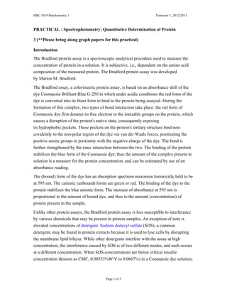
The Bradford protein assay is a spectroscopic analytical procedure used to measure the
concentration of protein in a solution. It is subjective, i.e., dependent on the amino acid
composition of the measured protein. The Bradford protein assay was developed
by Marion M. Bradford.
The Bradford assay, a colorimetric protein assay, is based on an absorbance shift of the
dye Coomassie Brilliant Blue G-250 in which under acidic conditions the red form of the
dye is converted into its bluer form to bind to the protein being assayed. During the
formation of this complex, two types of bond interaction take place: the red form of
Coomassie dye first donates its free electron to the ionizable groups on the protein, which
causes a disruption of the protein's native state, consequently exposing
its hydrophobic pockets. These pockets on the protein's tertiary structure bind non-
covalently to the non-polar region of the dye via van der Waals forces, positioning the
positive amine groups in proximity with the negative charge of the dye. The bond is
further strengthened by the ionic interaction between the two. The binding of the protein
stabilizes the blue form of the Coomassie dye; thus the amount of the complex present in
solution is a measure for the protein concentration, and can be estimated by use of an
absorbance reading.
The (bound) form of the dye has an absorption spectrum maximum historically held to be
at 595 nm. The cationic (unbound) forms are green or red. The binding of the dye to the
protein stabilizes the blue anionic form. The increase of absorbance at 595 nm is
proportional to the amount of bound dye, and thus to the amount (concentration) of
protein present in the sample.
Unlike other protein assays, the Bradford protein assay is less susceptible to interference
by various chemicals that may be present in protein samples. An exception of note is
elevated concentrations of detergent. Sodium dodecyl sulfate (SDS), a common
detergent, may be found in protein extracts because it is used to lyse cells by disrupting
the membrane lipid bilayer. While other detergents interfere with the assay at high
concentration, the interference caused by SDS is of two different modes, and each occurs
at a different concentration. When SDS concentrations are below critical micelle
concentration (known as CMC, 0.00333%W/V to 0.0667%) in a Coomassie dye solution,
Page 1 of 5
HBC 1019 Biochemistry 1 Trimester 1, 2012/2013
the detergent tends to bind strongly with the protein, inhibiting the protein binding sites
for the dye reagent. This can cause underestimations of protein concentration in solution.
When SDS concentrations are above CMC, the detergent associates strongly with the
green form of the Coomassie dye, causing the equilibrium to shift, thereby producing
more of the blue form. This causes an increase in the absorbance at 595 nm independent
of protein presence.
Materials, reagents and equipments:
Samples: low fat milk, soy milk and cultured-drink
Dye reagent,
Bovine γ-globulin standard set,
1X Phosphate buffered saline (PBS)
Spectrophotometer
Cuvettes
Microtubes
100 - 1000 ul adjustable-volume micropipette
10-100 ul adjustable-volume micropipette
2 – 20 ul adjustable-volume micropipette
Methods
I) Construction of linear graph from known protein standards
1. Get the set of data from your tutor and fill in the Table 1 as below.
2. Construct a standard curve linear by plotting the A595 values of the known
standards against concentration from the data in table below.
Table 1 Standard Curve Absorbance Values
Standard Standard (mg/ml) A595
#1 0.125 0.548
#2 0.250 0.010
#3 0.500 0.703
#4 0.750 0.897
#5 1.000 1.048
#6 1.500 1.222
#7 2.000 1.138
II) Quantitative determination of protein
1. Prepare a 1:50 dilution of the protein sample using PBS in two microtubes and
labeled them with Sample A, Sample B and Sample C.
2. Label 4 cuvettes as follows:
Label Standard (mg/ml)
Page 2 of 5
HBC 1019 Biochemistry 1 Trimester 1, 2012/2013
Blank PBS
A Sample A
B Sample B
C Sample C
3. Add 1 ml of dye reagent to each cuvette and 20 µl of PBS to the cuvette
labeled blank.
4. By using a fresh tip for each sample, pipette 20 µl of each standard into the
diluted sample into the appropriate cuvette.
5. Cover each cuvette with parafilm and invert each cuvette 3 times to mix.
6. Incubate at room temperature for a period of at least 5 minutes (but not to
exceed 60 minutes).
7. Visually compare the color of your unknown samples against the standards of
known concentrations. A representative set of standards and a typical color
spectrum are shown in figure below. Using the palette of standards, try to
qualitatively determine to which known standard your unknown sample
corresponds.
• Examine the color of the first unknown
• Compare it to Std. #1
• Is it lighter or darker?
• Compare it to Std. #2
• Is it lighter or darker, etc.
• Record your observations in Table 2 below.
Table 2 Unknown protein concentration
Sample Estimated Protein Concentration (mg/ml)
A 0.250
B 1.500
C 1.000
Page 3 of 5
HBC 1019 Biochemistry 1 Trimester 1, 2012/2013
8. Getting data from spectrophotometer and insert the absorbance data and the
concentration values for the unknown samples in Table 3. Read absorbance of
samples on spectrophotometer within one hour.
Table 3 Spectrophotometric Data for Unknown Samples
Sample A595 Protein Concentration
(mg/ml)
A 0.681
B 1.008
C 0.950
9. Compare your quantitative spectrophotometer determined concentrations with
qualitative estimates from table 3 to the table below. (Table 4)
Table 4 Estimated vs Quantitated Protein Concentrations
Sample Estimated Protein
Concentration (mg/ml) –
from table 2
Quantitated
Spectrophotometer
Protein (mg/ml) –from
table 3
A
B
C
10. Read the concentration of unknown samples by reading across from the
absorbance of unknown samples until meet the intersection with the standard
curve and then read the concentration. Record these data in table 5.
11. Adjust the final concentration of the unknown samples determined from
spectrophotometer before by multiplying the concentration by the dilution
factor used. For example, sample A is diluted 1:50 gives a reading of 0.224
absorbance units, which gives a concentration of M mg/ml. The final
concentration of milk is M x 50 = ___________mg/ml
Table 5 Final concentration of unknown samples
Sample A595 Concentration
read from
standard curve
(mg/ml)
Dilution
Factor
Final
Concentration
(mg/ml)
A
B
C
12. Find the protein content information for your samples on the package label
(look for ‘Nutrition Facts’). Convert this information into mg/ml of protein;
compare the values from the standard curve. Fill in table 5 below.
Page 4 of 5
HBC 1019 Biochemistry 1 Trimester 1, 2012/2013
Table 6 Comparing measured protein concentrations to the values found
on food labels
Sample Bradford Assay (mg/ml)
Final Concentration from
Table 5 (mg/ml)
Food Label (mg/ml)
A
B
C
Questions:
1. What are the functions of spectrophotometer?
spectrophotometer is a machine which tells about the growth of
microorganisms in culture by examining the colour change.
2. Why might the values you get be different from those of the manufacturer?
3. List the major sources of errors you can observe in this experiment.
A high concentration of buffer will cause an overestimated protein
concentration due to depletion of free protons from the solution by
conjugate base from the buffer. This will not be a problem if a low
concentration of protein (subsequently the buffer) is used.
4. How long can the samples sit before being read?
2 minute or less.
5. Do bubbles on the surface of the samples affect reading?
Yes, that’s why we put it carefully.
6. Do fingerprints on the cuvette affect the reading?
Yes, that’s why we wipe it before we put it.
Page 5 of 5

Más contenido relacionado

La actualidad más candente

serum protein estimation
serum protein estimationserum protein estimation
serum protein estimationtonnybite
 
Protein analysis
Protein analysisProtein analysis
Protein analysisLalit Singh
 
Kenyatta university biuret protein determination
Kenyatta university biuret protein determinationKenyatta university biuret protein determination
Kenyatta university biuret protein determinationLando Elvis
 
DIFFERENT METHODS OF PROTEIN ESTIMATION - PROTEINS AND ENZYMES ASSIGNMENT
DIFFERENT METHODS OF PROTEIN ESTIMATION - PROTEINS AND ENZYMES ASSIGNMENTDIFFERENT METHODS OF PROTEIN ESTIMATION - PROTEINS AND ENZYMES ASSIGNMENT
DIFFERENT METHODS OF PROTEIN ESTIMATION - PROTEINS AND ENZYMES ASSIGNMENTRishabh Sharma
 
Global analysis of protein
Global analysis of proteinGlobal analysis of protein
Global analysis of proteinKAUSHAL SAHU
 
Estimation of Serum Protien by Biuret method
Estimation of Serum Protien by Biuret methodEstimation of Serum Protien by Biuret method
Estimation of Serum Protien by Biuret methodDr. Almas A
 
Chitosen based Microsphere
Chitosen based MicrosphereChitosen based Microsphere
Chitosen based Microsphereomprakashsahu30
 
Protein estimation
Protein estimationProtein estimation
Protein estimationNitin Dudeja
 
Immunohistochemistry description of the Affinity method
Immunohistochemistry  description of the Affinity method Immunohistochemistry  description of the Affinity method
Immunohistochemistry description of the Affinity method HadeelAlboaklah
 
Analytical techniques for separation or purification of proteins
Analytical techniques for separation or purification of proteinsAnalytical techniques for separation or purification of proteins
Analytical techniques for separation or purification of proteinsrohini sane
 
Protein microarrays, ICAT, and HPLC protein purification
Protein microarrays, ICAT, and HPLC protein purificationProtein microarrays, ICAT, and HPLC protein purification
Protein microarrays, ICAT, and HPLC protein purificationRaul Soto
 
Two dimensional gel electrophoresis
Two dimensional gel electrophoresisTwo dimensional gel electrophoresis
Two dimensional gel electrophoresisShyam K Uthaman
 

La actualidad más candente (20)

serum protein estimation
serum protein estimationserum protein estimation
serum protein estimation
 
Bradford assay
Bradford assayBradford assay
Bradford assay
 
Protein analysis
Protein analysisProtein analysis
Protein analysis
 
Kenyatta university biuret protein determination
Kenyatta university biuret protein determinationKenyatta university biuret protein determination
Kenyatta university biuret protein determination
 
Protein analysis
Protein analysisProtein analysis
Protein analysis
 
Paragraph 10 rise
Paragraph 10 riseParagraph 10 rise
Paragraph 10 rise
 
DIFFERENT METHODS OF PROTEIN ESTIMATION - PROTEINS AND ENZYMES ASSIGNMENT
DIFFERENT METHODS OF PROTEIN ESTIMATION - PROTEINS AND ENZYMES ASSIGNMENTDIFFERENT METHODS OF PROTEIN ESTIMATION - PROTEINS AND ENZYMES ASSIGNMENT
DIFFERENT METHODS OF PROTEIN ESTIMATION - PROTEINS AND ENZYMES ASSIGNMENT
 
Protein analysis
Protein analysisProtein analysis
Protein analysis
 
Global analysis of protein
Global analysis of proteinGlobal analysis of protein
Global analysis of protein
 
Estimation of Serum Protien by Biuret method
Estimation of Serum Protien by Biuret methodEstimation of Serum Protien by Biuret method
Estimation of Serum Protien by Biuret method
 
Chitosen based Microsphere
Chitosen based MicrosphereChitosen based Microsphere
Chitosen based Microsphere
 
Protein estimation
Protein estimationProtein estimation
Protein estimation
 
Immunohistochemistry description of the Affinity method
Immunohistochemistry  description of the Affinity method Immunohistochemistry  description of the Affinity method
Immunohistochemistry description of the Affinity method
 
Protein purification
Protein purification Protein purification
Protein purification
 
Protein purification 2008
Protein purification  2008Protein purification  2008
Protein purification 2008
 
Analytical techniques for separation or purification of proteins
Analytical techniques for separation or purification of proteinsAnalytical techniques for separation or purification of proteins
Analytical techniques for separation or purification of proteins
 
Protein microarrays, ICAT, and HPLC protein purification
Protein microarrays, ICAT, and HPLC protein purificationProtein microarrays, ICAT, and HPLC protein purification
Protein microarrays, ICAT, and HPLC protein purification
 
Two dimensional gel electrophoresis
Two dimensional gel electrophoresisTwo dimensional gel electrophoresis
Two dimensional gel electrophoresis
 
New
NewNew
New
 
Clinical analysis
Clinical analysisClinical analysis
Clinical analysis
 

Similar a Practical 3 got

General techniques for identification and characterization of protein and Nuc...
General techniques for identification and characterization of protein and Nuc...General techniques for identification and characterization of protein and Nuc...
General techniques for identification and characterization of protein and Nuc...JohamSarfrazAli1
 
ICSA17 - Bula Dosagem Proteínas
 ICSA17 - Bula Dosagem Proteínas ICSA17 - Bula Dosagem Proteínas
ICSA17 - Bula Dosagem ProteínasRicardo Portela
 
Final submission –Pay attention to APA formatting, spelling, and
Final submission –Pay attention to APA formatting, spelling, andFinal submission –Pay attention to APA formatting, spelling, and
Final submission –Pay attention to APA formatting, spelling, andChereCheek752
 
PREPERATION F CELL EXTRACT
PREPERATION F CELL EXTRACTPREPERATION F CELL EXTRACT
PREPERATION F CELL EXTRACTYashwanth B S
 
Excipient combinations to manage protein viscosity for highly concentrated fo...
Excipient combinations to manage protein viscosity for highly concentrated fo...Excipient combinations to manage protein viscosity for highly concentrated fo...
Excipient combinations to manage protein viscosity for highly concentrated fo...MilliporeSigma
 
Microbial Examination of Milk .pptx
Microbial Examination of Milk .pptxMicrobial Examination of Milk .pptx
Microbial Examination of Milk .pptxS B_micro
 
Your guide to protein quantitation
Your guide to protein quantitationYour guide to protein quantitation
Your guide to protein quantitationCreative BioMart
 
Measurement growth in cell culture
Measurement growth in cell cultureMeasurement growth in cell culture
Measurement growth in cell cultureShailendra shera
 
The Viscosity Reduction Platform: Viscosity-reducing excipients for improveme...
The Viscosity Reduction Platform: Viscosity-reducing excipients for improveme...The Viscosity Reduction Platform: Viscosity-reducing excipients for improveme...
The Viscosity Reduction Platform: Viscosity-reducing excipients for improveme...MilliporeSigma
 
The Viscosity Reduction Platform: Viscosity Reducing Excipients for Protein F...
The Viscosity Reduction Platform: Viscosity Reducing Excipients for Protein F...The Viscosity Reduction Platform: Viscosity Reducing Excipients for Protein F...
The Viscosity Reduction Platform: Viscosity Reducing Excipients for Protein F...MilliporeSigma
 
The Viscosity Reduction Platform: Viscosity-reducing excipients for improveme...
The Viscosity Reduction Platform: Viscosity-reducing excipients for improveme...The Viscosity Reduction Platform: Viscosity-reducing excipients for improveme...
The Viscosity Reduction Platform: Viscosity-reducing excipients for improveme...Merck Life Sciences
 
The Viscosity Reduction Platform: Viscosity Reducing Excipients for Protein F...
The Viscosity Reduction Platform: Viscosity Reducing Excipients for Protein F...The Viscosity Reduction Platform: Viscosity Reducing Excipients for Protein F...
The Viscosity Reduction Platform: Viscosity Reducing Excipients for Protein F...Merck Life Sciences
 
Corvelva study-on-the-chemical-composition-profile-of-gardasil9
Corvelva study-on-the-chemical-composition-profile-of-gardasil9Corvelva study-on-the-chemical-composition-profile-of-gardasil9
Corvelva study-on-the-chemical-composition-profile-of-gardasil9ilfattoquotidiano.it
 
Assay fortification flour
Assay fortification flourAssay fortification flour
Assay fortification flourGonzalo Mulero
 
microbial assay antibiotics, vitamins, amino acids
microbial assay antibiotics,  vitamins,  amino acidsmicrobial assay antibiotics,  vitamins,  amino acids
microbial assay antibiotics, vitamins, amino acidsMicroShamim
 
IPQC Tests for Opthalmic Preparations.pptx
IPQC Tests for Opthalmic Preparations.pptxIPQC Tests for Opthalmic Preparations.pptx
IPQC Tests for Opthalmic Preparations.pptxSohailSheikh62
 
Estimation of milk protein
Estimation of milk proteinEstimation of milk protein
Estimation of milk proteinDeepanjan Basak
 
Estimation of milk protein
Estimation of milk proteinEstimation of milk protein
Estimation of milk proteinDeepanjan Basak
 

Similar a Practical 3 got (20)

Quantitative test for Protein by Bradford Assay.pptx
Quantitative test for Protein by Bradford Assay.pptxQuantitative test for Protein by Bradford Assay.pptx
Quantitative test for Protein by Bradford Assay.pptx
 
General techniques for identification and characterization of protein and Nuc...
General techniques for identification and characterization of protein and Nuc...General techniques for identification and characterization of protein and Nuc...
General techniques for identification and characterization of protein and Nuc...
 
ICSA17 - Bula Dosagem Proteínas
 ICSA17 - Bula Dosagem Proteínas ICSA17 - Bula Dosagem Proteínas
ICSA17 - Bula Dosagem Proteínas
 
Final submission –Pay attention to APA formatting, spelling, and
Final submission –Pay attention to APA formatting, spelling, andFinal submission –Pay attention to APA formatting, spelling, and
Final submission –Pay attention to APA formatting, spelling, and
 
6.pdf
6.pdf6.pdf
6.pdf
 
PREPERATION F CELL EXTRACT
PREPERATION F CELL EXTRACTPREPERATION F CELL EXTRACT
PREPERATION F CELL EXTRACT
 
Excipient combinations to manage protein viscosity for highly concentrated fo...
Excipient combinations to manage protein viscosity for highly concentrated fo...Excipient combinations to manage protein viscosity for highly concentrated fo...
Excipient combinations to manage protein viscosity for highly concentrated fo...
 
Microbial Examination of Milk .pptx
Microbial Examination of Milk .pptxMicrobial Examination of Milk .pptx
Microbial Examination of Milk .pptx
 
Your guide to protein quantitation
Your guide to protein quantitationYour guide to protein quantitation
Your guide to protein quantitation
 
Measurement growth in cell culture
Measurement growth in cell cultureMeasurement growth in cell culture
Measurement growth in cell culture
 
The Viscosity Reduction Platform: Viscosity-reducing excipients for improveme...
The Viscosity Reduction Platform: Viscosity-reducing excipients for improveme...The Viscosity Reduction Platform: Viscosity-reducing excipients for improveme...
The Viscosity Reduction Platform: Viscosity-reducing excipients for improveme...
 
The Viscosity Reduction Platform: Viscosity Reducing Excipients for Protein F...
The Viscosity Reduction Platform: Viscosity Reducing Excipients for Protein F...The Viscosity Reduction Platform: Viscosity Reducing Excipients for Protein F...
The Viscosity Reduction Platform: Viscosity Reducing Excipients for Protein F...
 
The Viscosity Reduction Platform: Viscosity-reducing excipients for improveme...
The Viscosity Reduction Platform: Viscosity-reducing excipients for improveme...The Viscosity Reduction Platform: Viscosity-reducing excipients for improveme...
The Viscosity Reduction Platform: Viscosity-reducing excipients for improveme...
 
The Viscosity Reduction Platform: Viscosity Reducing Excipients for Protein F...
The Viscosity Reduction Platform: Viscosity Reducing Excipients for Protein F...The Viscosity Reduction Platform: Viscosity Reducing Excipients for Protein F...
The Viscosity Reduction Platform: Viscosity Reducing Excipients for Protein F...
 
Corvelva study-on-the-chemical-composition-profile-of-gardasil9
Corvelva study-on-the-chemical-composition-profile-of-gardasil9Corvelva study-on-the-chemical-composition-profile-of-gardasil9
Corvelva study-on-the-chemical-composition-profile-of-gardasil9
 
Assay fortification flour
Assay fortification flourAssay fortification flour
Assay fortification flour
 
microbial assay antibiotics, vitamins, amino acids
microbial assay antibiotics,  vitamins,  amino acidsmicrobial assay antibiotics,  vitamins,  amino acids
microbial assay antibiotics, vitamins, amino acids
 
IPQC Tests for Opthalmic Preparations.pptx
IPQC Tests for Opthalmic Preparations.pptxIPQC Tests for Opthalmic Preparations.pptx
IPQC Tests for Opthalmic Preparations.pptx
 
Estimation of milk protein
Estimation of milk proteinEstimation of milk protein
Estimation of milk protein
 
Estimation of milk protein
Estimation of milk proteinEstimation of milk protein
Estimation of milk protein
 

Más de Osama Barayan

Estimating the-time-needed-for-mitosis
Estimating the-time-needed-for-mitosisEstimating the-time-needed-for-mitosis
Estimating the-time-needed-for-mitosisOsama Barayan
 
Practical 7 dna, rna and the flow of genetic information5
Practical 7 dna, rna and the flow of genetic information5Practical 7 dna, rna and the flow of genetic information5
Practical 7 dna, rna and the flow of genetic information5Osama Barayan
 
Practical 5 sds page
Practical 5 sds pagePractical 5 sds page
Practical 5 sds pageOsama Barayan
 
Practical 9 protein structure and function (3)
Practical 9 protein structure and function  (3)Practical 9 protein structure and function  (3)
Practical 9 protein structure and function (3)Osama Barayan
 

Más de Osama Barayan (11)

Estimating the-time-needed-for-mitosis
Estimating the-time-needed-for-mitosisEstimating the-time-needed-for-mitosis
Estimating the-time-needed-for-mitosis
 
Tutorial mitosis
Tutorial mitosisTutorial mitosis
Tutorial mitosis
 
Practical 7 dna, rna and the flow of genetic information5
Practical 7 dna, rna and the flow of genetic information5Practical 7 dna, rna and the flow of genetic information5
Practical 7 dna, rna and the flow of genetic information5
 
Practical 6 elisa
Practical 6 elisaPractical 6 elisa
Practical 6 elisa
 
Practical 5 sds page
Practical 5 sds pagePractical 5 sds page
Practical 5 sds page
 
Practical 4 sec
Practical 4 secPractical 4 sec
Practical 4 sec
 
Practical 2 tlc
Practical 2 tlcPractical 2 tlc
Practical 2 tlc
 
Lab8biochem
Lab8biochemLab8biochem
Lab8biochem
 
Hbc1019 tut4
Hbc1019 tut4Hbc1019 tut4
Hbc1019 tut4
 
Biochemistry I
Biochemistry IBiochemistry I
Biochemistry I
 
Practical 9 protein structure and function (3)
Practical 9 protein structure and function  (3)Practical 9 protein structure and function  (3)
Practical 9 protein structure and function (3)
 

Último

JORNADA 3 LIGA MURO 2024GHGHGHGHGHGH.pdf
JORNADA 3 LIGA MURO 2024GHGHGHGHGHGH.pdfJORNADA 3 LIGA MURO 2024GHGHGHGHGHGH.pdf
JORNADA 3 LIGA MURO 2024GHGHGHGHGHGH.pdfArturo Pacheco Alvarez
 
Benifits of Individual And Team Sports-Group 7.pptx
Benifits of Individual And Team Sports-Group 7.pptxBenifits of Individual And Team Sports-Group 7.pptx
Benifits of Individual And Team Sports-Group 7.pptxsherrymieg19
 
Italy Vs Albania Euro Cup 2024 Italy's Strategy for Success.docx
Italy Vs Albania Euro Cup 2024 Italy's Strategy for Success.docxItaly Vs Albania Euro Cup 2024 Italy's Strategy for Success.docx
Italy Vs Albania Euro Cup 2024 Italy's Strategy for Success.docxWorld Wide Tickets And Hospitality
 
Clash of Titans_ PSG vs Barcelona (1).pdf
Clash of Titans_ PSG vs Barcelona (1).pdfClash of Titans_ PSG vs Barcelona (1).pdf
Clash of Titans_ PSG vs Barcelona (1).pdfMuhammad Hashim
 
DONAL88 >LINK SLOT PG SOFT TERGACOR 2024
DONAL88 >LINK SLOT PG SOFT TERGACOR 2024DONAL88 >LINK SLOT PG SOFT TERGACOR 2024
DONAL88 >LINK SLOT PG SOFT TERGACOR 2024DONAL88 GACOR
 
Spain Vs Italy Showdown Between Italy and Spain Could Determine UEFA Euro 202...
Spain Vs Italy Showdown Between Italy and Spain Could Determine UEFA Euro 202...Spain Vs Italy Showdown Between Italy and Spain Could Determine UEFA Euro 202...
Spain Vs Italy Showdown Between Italy and Spain Could Determine UEFA Euro 202...World Wide Tickets And Hospitality
 
Introduction to Basketball-PowerPoint Presentation
Introduction to Basketball-PowerPoint PresentationIntroduction to Basketball-PowerPoint Presentation
Introduction to Basketball-PowerPoint PresentationJuliusMacaballug
 
PPT on INDIA VS PAKISTAN - A Sports Rivalry
PPT on INDIA VS PAKISTAN - A Sports RivalryPPT on INDIA VS PAKISTAN - A Sports Rivalry
PPT on INDIA VS PAKISTAN - A Sports Rivalryanirbannath184
 
Project & Portfolio, Market Analysis: WWE
Project & Portfolio, Market Analysis: WWEProject & Portfolio, Market Analysis: WWE
Project & Portfolio, Market Analysis: WWEDeShawn Ellis
 
JORNADA 2 LIGA MUROBASQUETBOL1 2024.docx
JORNADA 2 LIGA MUROBASQUETBOL1 2024.docxJORNADA 2 LIGA MUROBASQUETBOL1 2024.docx
JORNADA 2 LIGA MUROBASQUETBOL1 2024.docxArturo Pacheco Alvarez
 

Último (11)

NATIONAL SPORTS DAY WRITTEN QUIZ by QUI9
NATIONAL SPORTS DAY WRITTEN QUIZ by QUI9NATIONAL SPORTS DAY WRITTEN QUIZ by QUI9
NATIONAL SPORTS DAY WRITTEN QUIZ by QUI9
 
JORNADA 3 LIGA MURO 2024GHGHGHGHGHGH.pdf
JORNADA 3 LIGA MURO 2024GHGHGHGHGHGH.pdfJORNADA 3 LIGA MURO 2024GHGHGHGHGHGH.pdf
JORNADA 3 LIGA MURO 2024GHGHGHGHGHGH.pdf
 
Benifits of Individual And Team Sports-Group 7.pptx
Benifits of Individual And Team Sports-Group 7.pptxBenifits of Individual And Team Sports-Group 7.pptx
Benifits of Individual And Team Sports-Group 7.pptx
 
Italy Vs Albania Euro Cup 2024 Italy's Strategy for Success.docx
Italy Vs Albania Euro Cup 2024 Italy's Strategy for Success.docxItaly Vs Albania Euro Cup 2024 Italy's Strategy for Success.docx
Italy Vs Albania Euro Cup 2024 Italy's Strategy for Success.docx
 
Clash of Titans_ PSG vs Barcelona (1).pdf
Clash of Titans_ PSG vs Barcelona (1).pdfClash of Titans_ PSG vs Barcelona (1).pdf
Clash of Titans_ PSG vs Barcelona (1).pdf
 
DONAL88 >LINK SLOT PG SOFT TERGACOR 2024
DONAL88 >LINK SLOT PG SOFT TERGACOR 2024DONAL88 >LINK SLOT PG SOFT TERGACOR 2024
DONAL88 >LINK SLOT PG SOFT TERGACOR 2024
 
Spain Vs Italy Showdown Between Italy and Spain Could Determine UEFA Euro 202...
Spain Vs Italy Showdown Between Italy and Spain Could Determine UEFA Euro 202...Spain Vs Italy Showdown Between Italy and Spain Could Determine UEFA Euro 202...
Spain Vs Italy Showdown Between Italy and Spain Could Determine UEFA Euro 202...
 
Introduction to Basketball-PowerPoint Presentation
Introduction to Basketball-PowerPoint PresentationIntroduction to Basketball-PowerPoint Presentation
Introduction to Basketball-PowerPoint Presentation
 
PPT on INDIA VS PAKISTAN - A Sports Rivalry
PPT on INDIA VS PAKISTAN - A Sports RivalryPPT on INDIA VS PAKISTAN - A Sports Rivalry
PPT on INDIA VS PAKISTAN - A Sports Rivalry
 
Project & Portfolio, Market Analysis: WWE
Project & Portfolio, Market Analysis: WWEProject & Portfolio, Market Analysis: WWE
Project & Portfolio, Market Analysis: WWE
 
JORNADA 2 LIGA MUROBASQUETBOL1 2024.docx
JORNADA 2 LIGA MUROBASQUETBOL1 2024.docxJORNADA 2 LIGA MUROBASQUETBOL1 2024.docx
JORNADA 2 LIGA MUROBASQUETBOL1 2024.docx
 

Practical 3 got

  • 1. HBC 1019 Biochemistry 1 Trimester 1, 2012/2013 PRACTICAL : Spectrophotometry; Quantitative Determination of Protein 3 (**Please bring along graph papers for this practical) Introduction The Bradford protein assay is a spectroscopic analytical procedure used to measure the concentration of protein in a solution. It is subjective, i.e., dependent on the amino acid composition of the measured protein. The Bradford protein assay was developed by Marion M. Bradford. The Bradford assay, a colorimetric protein assay, is based on an absorbance shift of the dye Coomassie Brilliant Blue G-250 in which under acidic conditions the red form of the dye is converted into its bluer form to bind to the protein being assayed. During the formation of this complex, two types of bond interaction take place: the red form of Coomassie dye first donates its free electron to the ionizable groups on the protein, which causes a disruption of the protein's native state, consequently exposing its hydrophobic pockets. These pockets on the protein's tertiary structure bind non- covalently to the non-polar region of the dye via van der Waals forces, positioning the positive amine groups in proximity with the negative charge of the dye. The bond is further strengthened by the ionic interaction between the two. The binding of the protein stabilizes the blue form of the Coomassie dye; thus the amount of the complex present in solution is a measure for the protein concentration, and can be estimated by use of an absorbance reading. The (bound) form of the dye has an absorption spectrum maximum historically held to be at 595 nm. The cationic (unbound) forms are green or red. The binding of the dye to the protein stabilizes the blue anionic form. The increase of absorbance at 595 nm is proportional to the amount of bound dye, and thus to the amount (concentration) of protein present in the sample. Unlike other protein assays, the Bradford protein assay is less susceptible to interference by various chemicals that may be present in protein samples. An exception of note is elevated concentrations of detergent. Sodium dodecyl sulfate (SDS), a common detergent, may be found in protein extracts because it is used to lyse cells by disrupting the membrane lipid bilayer. While other detergents interfere with the assay at high concentration, the interference caused by SDS is of two different modes, and each occurs at a different concentration. When SDS concentrations are below critical micelle concentration (known as CMC, 0.00333%W/V to 0.0667%) in a Coomassie dye solution, Page 1 of 5
  • 2. HBC 1019 Biochemistry 1 Trimester 1, 2012/2013 the detergent tends to bind strongly with the protein, inhibiting the protein binding sites for the dye reagent. This can cause underestimations of protein concentration in solution. When SDS concentrations are above CMC, the detergent associates strongly with the green form of the Coomassie dye, causing the equilibrium to shift, thereby producing more of the blue form. This causes an increase in the absorbance at 595 nm independent of protein presence. Materials, reagents and equipments: Samples: low fat milk, soy milk and cultured-drink Dye reagent, Bovine γ-globulin standard set, 1X Phosphate buffered saline (PBS) Spectrophotometer Cuvettes Microtubes 100 - 1000 ul adjustable-volume micropipette 10-100 ul adjustable-volume micropipette 2 – 20 ul adjustable-volume micropipette Methods I) Construction of linear graph from known protein standards 1. Get the set of data from your tutor and fill in the Table 1 as below. 2. Construct a standard curve linear by plotting the A595 values of the known standards against concentration from the data in table below. Table 1 Standard Curve Absorbance Values Standard Standard (mg/ml) A595 #1 0.125 0.548 #2 0.250 0.010 #3 0.500 0.703 #4 0.750 0.897 #5 1.000 1.048 #6 1.500 1.222 #7 2.000 1.138 II) Quantitative determination of protein 1. Prepare a 1:50 dilution of the protein sample using PBS in two microtubes and labeled them with Sample A, Sample B and Sample C. 2. Label 4 cuvettes as follows: Label Standard (mg/ml) Page 2 of 5
  • 3. HBC 1019 Biochemistry 1 Trimester 1, 2012/2013 Blank PBS A Sample A B Sample B C Sample C 3. Add 1 ml of dye reagent to each cuvette and 20 µl of PBS to the cuvette labeled blank. 4. By using a fresh tip for each sample, pipette 20 µl of each standard into the diluted sample into the appropriate cuvette. 5. Cover each cuvette with parafilm and invert each cuvette 3 times to mix. 6. Incubate at room temperature for a period of at least 5 minutes (but not to exceed 60 minutes). 7. Visually compare the color of your unknown samples against the standards of known concentrations. A representative set of standards and a typical color spectrum are shown in figure below. Using the palette of standards, try to qualitatively determine to which known standard your unknown sample corresponds. • Examine the color of the first unknown • Compare it to Std. #1 • Is it lighter or darker? • Compare it to Std. #2 • Is it lighter or darker, etc. • Record your observations in Table 2 below. Table 2 Unknown protein concentration Sample Estimated Protein Concentration (mg/ml) A 0.250 B 1.500 C 1.000 Page 3 of 5
  • 4. HBC 1019 Biochemistry 1 Trimester 1, 2012/2013 8. Getting data from spectrophotometer and insert the absorbance data and the concentration values for the unknown samples in Table 3. Read absorbance of samples on spectrophotometer within one hour. Table 3 Spectrophotometric Data for Unknown Samples Sample A595 Protein Concentration (mg/ml) A 0.681 B 1.008 C 0.950 9. Compare your quantitative spectrophotometer determined concentrations with qualitative estimates from table 3 to the table below. (Table 4) Table 4 Estimated vs Quantitated Protein Concentrations Sample Estimated Protein Concentration (mg/ml) – from table 2 Quantitated Spectrophotometer Protein (mg/ml) –from table 3 A B C 10. Read the concentration of unknown samples by reading across from the absorbance of unknown samples until meet the intersection with the standard curve and then read the concentration. Record these data in table 5. 11. Adjust the final concentration of the unknown samples determined from spectrophotometer before by multiplying the concentration by the dilution factor used. For example, sample A is diluted 1:50 gives a reading of 0.224 absorbance units, which gives a concentration of M mg/ml. The final concentration of milk is M x 50 = ___________mg/ml Table 5 Final concentration of unknown samples Sample A595 Concentration read from standard curve (mg/ml) Dilution Factor Final Concentration (mg/ml) A B C 12. Find the protein content information for your samples on the package label (look for ‘Nutrition Facts’). Convert this information into mg/ml of protein; compare the values from the standard curve. Fill in table 5 below. Page 4 of 5
  • 5. HBC 1019 Biochemistry 1 Trimester 1, 2012/2013 Table 6 Comparing measured protein concentrations to the values found on food labels Sample Bradford Assay (mg/ml) Final Concentration from Table 5 (mg/ml) Food Label (mg/ml) A B C Questions: 1. What are the functions of spectrophotometer? spectrophotometer is a machine which tells about the growth of microorganisms in culture by examining the colour change. 2. Why might the values you get be different from those of the manufacturer? 3. List the major sources of errors you can observe in this experiment. A high concentration of buffer will cause an overestimated protein concentration due to depletion of free protons from the solution by conjugate base from the buffer. This will not be a problem if a low concentration of protein (subsequently the buffer) is used. 4. How long can the samples sit before being read? 2 minute or less. 5. Do bubbles on the surface of the samples affect reading? Yes, that’s why we put it carefully. 6. Do fingerprints on the cuvette affect the reading? Yes, that’s why we wipe it before we put it. Page 5 of 5
  • 6. HBC 1019 Biochemistry 1 Trimester 1, 2012/2013 Table 6 Comparing measured protein concentrations to the values found on food labels Sample Bradford Assay (mg/ml) Final Concentration from Table 5 (mg/ml) Food Label (mg/ml) A B C Questions: 1. What are the functions of spectrophotometer? spectrophotometer is a machine which tells about the growth of microorganisms in culture by examining the colour change. 2. Why might the values you get be different from those of the manufacturer? 3. List the major sources of errors you can observe in this experiment. A high concentration of buffer will cause an overestimated protein concentration due to depletion of free protons from the solution by conjugate base from the buffer. This will not be a problem if a low concentration of protein (subsequently the buffer) is used. 4. How long can the samples sit before being read? 2 minute or less. 5. Do bubbles on the surface of the samples affect reading? Yes, that’s why we put it carefully. 6. Do fingerprints on the cuvette affect the reading? Yes, that’s why we wipe it before we put it. Page 5 of 5