
Recommended
More Related Content
What's hot
What's hot (20)
Viewers also liked
Viewers also liked (20)
Similar to Circulatory System - The Heart
Similar to Circulatory System - The Heart (20)
Recently uploaded
Recently uploaded (20)
Circulatory System - The Heart
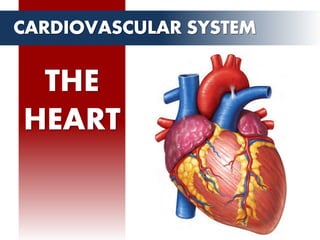
- 2. • muscular organ • size of a closed fist • weigh appr. 250 to 350 grams • BASE is attached to the aorta, pulmonary arteries and veins, and the vena cava. • APEX of the heart rests superior to the diaphragm. THE HEART OVERVIEW BASE APEX AORTA SUPERIOR VENA CAVA PULMONARY ARTERY INFERIOR VENA CAVA PULMONARY VEINS
- 3. • circulatory pump – takes in deoxygenated blood through the veins and delivers it to the lungs for oxygenation – blood is pumped into the various arteries (which provide oxygen and nutrients to body tissues by transporting the blood throughout the body). THE HEART OVERVIEW
- 4. THE HEART LOCATION LUNGS HEART LIVER DIAPHRAGM ESOPHAGUS T8 VERTEBRA STERNUM • Posterior to the sternum • Anterior to the esophagus and vertebra • Medial to the lungs • 2/3 left, 1/3 right
- 6. THE HEART PERICARDIUM • Prevent friction between beating heart and organs • Holds the heart in position and maintains a hollow space for expansion when full.
- 7. THE HEART HEART WALL • EPICARDIUM – outermost layer, same as visceral layer of pericardium. • MYOCARDIUM – muscular middle layer of the heart. • ENDOCARDIUM – simple squamous epithelium lining the inside of the heart.
- 8. • short, fat, branched and interconnected. • interlock at dark-staining junctions called intercalated discs. • gap junctions allow ions to pass • muscles behave like a single coordinated unit. THE HEART CARDIAC MUSCLES CARDIAC MUSCLE CELL INTERCALATED DISC DESMOSOME GAP JUNCTION
- 9. THE HEART CARDIAC MUSCLES CARDIAC MUSCLE SKELETAL MUSCLE Structure Short, fat, branched, Interconnected Long, cylindrical, multinucleate Means of Stimulation Nerve Stimulus Self-excitable Nerve stimulus Contraction Contract as a unit Individually activated Refractory period 200 ms 1-2 ms
- 10. MITRAL VALVE TRICUSPID VALVE AORTIC VALVE PULMONARY VALVE Chordae tendineae THE HEART VALVES • Prevents the blood from flowing backwards or regurgitating back into the heart. • Two types: – Atrioventricular (AV) valve – Semilunar valve
- 11. THE HEART VALVES Atrioventricular or AV Valve Tricuspid – right side Mitral – left side prevent backflow into the atria when the ventricles contract. MITRAL VALVE TRICUSPID VALVE AORTIC VALVE PULMONARY VALVE Chordae tendineae
- 12. THE HEART VALVES Semilunar Valve Aortic– left side Pulmonary – right side blood flows from ventricles to the arteries MITRAL VALVE TRICUSPID VALVE AORTIC VALVE PULMONARY VALVE Chordae tendineae
- 13. THE HEART CHAMBERS ATRIA VENTRICLES • receiving chambers for blood • smaller and less muscular than ventricles • larger, stronger • pumps blood out of the heart
- 14. THE HEART CHAMBERS RIGHT ATRIUM • deoxygenated blood(body) • FROM: superior/inferior vena cava coronary sinus • PUMPTS TO: right ventricle • THROUGH THE: tricuspid valve SUPERIOR VENA CAVA INFERIOR VENA CAVA TRICUSPID VALVE
- 15. THE HEART CHAMBERS RIGHT VENTRICLE • deoxygenated blood • FROM: right atrium • PUMPTS TO: pulmonary artery • THROUGH THE: tricuspid valve (in) pulmonary valve(out) PULMONARY ARTERY TRICUSPID VALVE
- 16. THE HEART CHAMBERS LEFT ATRIUM • oxygenated blood (lungs) • FROM: left /right pulmonary veins • PUMPTS TO: left ventricle • THROUGH THE: mitral valve MITRAL VALVE PULMONARY VEINS
- 17. THE HEART CHAMBERS LEFT VENTRICLE • oxygenated blood • FROM: left atrium • PUMPTS TO: aorta (to the body) • THROUGH THE: mitral valve (in) aortic valve(out) AORTA MITRAL VALVE
Editor's Notes
- The heart is a muscular organ about the size of a closed fist that functions as the body’s circulatory pump. 250 to 350 grams The heart is said to be in the shape of an inverted cone wherein the base is attached to the aorta, pulmonary arteries and veins, and the vena cava. The inferior tip of the heart, known as the apex, rests just superior to the diaphragm.
- It takes in deoxygenated blood through the veins and delivers it to the lungs for oxygenation. From the lungs, oxygenated blood goes back into the heart, and the heart pumps it into the various arteries (which provide oxygen and nutrients to body tissues by transporting the blood throughout the body).
- Where is the heart located and what are adjacent organs to it? LEFT: Transverse slice of the chest along the T8 Vertebra RIGHT: Frontal slice of the chest area The heart is located in the thoracic cavity medial to the lungs, posterior to the sternum and anterior to the vertebra and esophagus. The base of the heart is located along the body’s midline with the apex pointing toward the left side. Because the heart points to the left, about 2/3 of the heart’s mass is found on the left side of the body and the other 1/3 is on the right.
- The heart sits within a fluid-filled cavity called the pericardial cavity whose wall is lined with a special membrane known as the pericardium. Pericardium is a type of serous membrane that produces serous fluid to lubricate the heart and prevent friction between the ever beating heart and its surrounding organs. The pericardium also serves to hold the heart in position and maintain a hollow space for the heart to expand into when it is full. The pericardium has 2 layers—a visceral layer that covers the outside of the heart and a parietal layer that forms a sac around the outside of the pericardial cavity.
- The heart wall is made of 3 layers: epicardium, myocardium and endocardium. Epicardium. The epicardium is the outermost layer of the heart wall and is just another name for the visceral layer of the pericardium. The epicardium helps to lubricate and protect the outside of the heart. Below the epicardium is the second, thicker layer of the heart wall: the myocardium. Myocardium. The myocardium is the think layer containing the cardiac muscle tissue. Myocardium makes up the majority of the thickness and mass of the heart wall and is the part of the heart responsible for pumping blood. Below the myocardium is the thin endocardium layer. Endocardium. Endocardium is the simple squamous endothelium layer that lines the inside of the heart. This layer is very smooth and is responsible for keeping blood from sticking to the inside of the heart and forming potentially deadly blood clots.
- The circular and spiral arrangement of cardiac muscle bundles in the myocardium of the heart. In contrast to the long, cylindrical, multinucleate skeletal muscle fibers, cardiac cells are short, fat, branched, and interconnected. Each fiber contains one or at most two large, pale, centrally located nuclei In contrast to the structurally and functionally independent skeletal muscle fibers, the plasma mem- branes of adjacent cardiac cells interlock at dark-staining junctions called intercalated discs which contain anchoring desmosomes and gap junctions. The gap junctions allow ions to pass from cell to cell, transmitting current across the entire heart. Because the junctions electrically couple cardiac cells, the muscles in the heart be- haves as a single coordinated unit.
- To prevent blood from flowing backwards or “regurgitating” back into the heart, a system of one-way valves are present in the heart. The heart valves can be broken down into two types: atrioventricular and semilunar valves.
- Atrioventricular valves. The atrioventricular (AV) valves are located in each atrial ventricular junction and only allow blood to flow from the atria into the ventricles. The AV valve on the right side of the heart is called the tricuspid valve because it is made of three cusps (flaps) that separate to allow blood to pass through and connect to block regurgitation of blood. The AV valve on the left side of the heart is called the mitral valve or the bicuspid valve because it has two cusps. The AV valves are attached on the ventricular side to tough strings called chordae tendineae. The chordae tendineae pull on the AV valves to keep them from folding backwards and allowing blood to regurgitate past them. During the contraction of the ventricles, the AV valves look like domed parachutes with the chordae tendineae acting as the ropes holding the parachutes taut.
- Semilunar valves. The semilunar valves, so named for the crescent moon shape of their cusps, are located between the ventricles and the arteries that carry blood away from the heart. The semilunar valve on the right side of the heart is thepulmonary valve, so named because it prevents the backflow of blood from the pulmonary trunk into the right ventricle. The semilunar valve on the left side of the heart is the aortic valve, named for the fact that it prevents the aorta from regurgitating blood back into the left ventricle. The semilunar valves are smaller than the AV valves and do not have chordae tendineae to hold them in place. Instead, the cusps of the semilunar valves are cup shaped to catch regurgitating blood and use the blood’s pressure to snap shut.
- The heart contains 4 chambers: the right atrium, left atrium, right ventricle, andleft ventricle. The upper chambers, or atria act as receiving chambers for blood, so they are connected to the veins that carry blood to the heart. The ventricles are the larger, stronger pumping chambers that send blood out of the heart. Implication of the thickness of the wall. The chambers on the right side of the heart are smaller and have less myocardium in their heart wall when compared to the left side of the heart. This difference in size between the sides of the heart is related to their functions and the size of the 2 circulatory loops. The right side of the heart maintains pulmonary circulation to the nearby lungs while the left side of the heart pumps blood all the way to the extremities of the body in the systemic circulatory loop.