SlideShare una empresa de Scribd logo
1 de 10
Descargar para leer sin conexión
Section V – Gastrointestinal Radiology


                                                  Figure 1

186. You are shown a radiograph (Figure 1) from a small bowel follow-thru in a 32-year-old male with chronic
     abdominal pain. Which one of the following is the MOST likely diagnosis?
      A. Crohn’s disease
      B.   Sprue
      C. Midgut volvulus
      D. Paraduodenal hernia
      E.   Pseudomyxoma peritonei


                                  Diagnostic In-Training Exam 2002                                             1
Section V – Gastrointestinal Radiology
Question #186
Findings: The radiograph demonstrates encapsulation of jejunal loops in the left mid abdomen.

Rationales:
A. Incorrect. Crohn’s disease involves the small bowel in 75% of patients at the time of presentation. Affected
   segments show fold thickening and ulceration. Sinus tracts and fistulae are also noted. Although surrounding
   fibrofatty proliferation may produce a mass effect or displace bowel on SBFT, encapsulation of bowel loops is
   not noted with Crohn’s disease.
B. Incorrect. Sprue or gluten sensitive enteropathy produces villous atrophy and radiographically shows reversal of
   the normal fold pattern with loss of normal jejunal folds and an increased number of folds per inch in the
   ilium. Transient intussusceptions, hypomotility and flocculation of barium can also be seen. However, none of
   these findings are present on this film.
C. Incorrect. Midgut volvulus is produced by twisting of small bowel loops around the shortened mesenteric
   vascular pedicle in patients with congenital small bowel malrotation. A corkscrew deformity of the jejunal loops
   is noted on SBFT. Obstruction to venous return produces edema and hence thickened folds in the involved
   bowel. Such changes are not noted on this film.
D. Correct. Internal hernias are abnormal protrusions of intraabdominal structures through a normal opening or
   through a congenital or acquired defect in fascia or mesentery. Encapsulation of jejunal bowel loops in either
   the right or left mid abdomen is characteristic of paraduodenal hernia - the most common type of internal
   hernia. The amount of contained small bowel can vary from a few loops to the majority of the small bowel.
   Patient typically present with intermittent abdominal pain thought to be secondary to episodes of obstruction.
   Left sided paraduodenal hernia is more common than right sided paraduodenal hernia.
E. Incorrect. Pseudomyxoma peritonei results from seeding of the peritoneal cavity by benign or malignant mucin
   secreting cells. The cells typically come from a ruptured mucinous cystadenoma or cystadenocarcinoma of the
   appendix or ovary. Though bowel can be distorted, obstructed or tethered by tumor or mucin, encapsulation as
   noted here is not seen.

Citations:
Maglinte DDT, Bisset III GS, Congenital and Developmental Anomalies of the Small Bowel in Adolescents and Adults pp 247-251
in Clinical Imaging of the Small Intestine 2nd ed, eds Herlinger H, Maglinte DDT, and Birnbaum BA, Springer, New York, 1999.


2                                         American College of Radiology
Section V – Gastrointestinal Radiology


                                                  Figure 2

187. You are shown a contrast-enhanced CT (Figure 2) in an 84-year-old male with recent weight loss and guaiac
     positive stool. Which one of the following is the MOST likely diagnosis?
      A. Desmoid tumor
      B.   Intussusception
      C. Internal hernia
      D. Lipomatous ileocecal valve
      E.   Volvulus


                                  Diagnostic In-Training Exam 2002                                               3
Section V – Gastrointestinal Radiology
Question #187
Findings: The CT image demonstrates a long colo-colic intussusception involving the ascending and transverse
colon

Rationales:
A. Incorrect. Desmoid tumors are non metastasizing but locally invasive fibrous tumors. They can occur either in
   isolation or more commonly in association with Gardner’s syndrome, particularly following abdominal surgery.
   They may present as an abdominal mass or cause bowel obstruction. On CT they appear as a soft tissue mass in
   the mesentery either with well circumscribed or infiltrative margins. These findings are not present in this case.
B. Correct. The image demonstrates a long segment colo-colic intussusception. The vast majority of adult colo-
   colic intussusceptions have a malignant lead point with adenocarcinoma being the most common histologic
   type. The extensive edema seen with the intussusception often makes it difficult to delineate the precise lead
   point.
C. Incorrect. Internal hernias are abnormal protrusions of intraabdominal structures through a normal opening
   or through a congenital or acquired defect in fascia or mesentery An internal. The most common type is the
   paraduodenal hernia shown in the previous question. Although internal hernias can cause distortion of bowel
   loops they would not produce the bowel within bowel appearance noted here.
D. Incorrect. Lipomatous ileocecal valve is caused by submucosal infiltration of fat into the lips of the I-C valve. It
   is typically seen in older patients and is more common in women. Symptoms are usually absent. Although the
   mass can mimic an adenocarcinoma its fatty density on CT and smooth contour usually allow differentiation.
   The mass in this case is predominantly soft tissue density and much larger and more extensive and irregular
   than a lipomatous I-C valve.
E. Incorrect. A volvulus involving the bowel is produced by twisting of small bowel loops around a point of
   fixation. In midgut volvulus associated with malrotation the point of fixation is the shortened mesenteric
   vascular pedicle. Volvulus involving fewer bowel loops may also occur around an acquired adhesive band. On
   CT a swirling appearance of vessels is seen in conjunction with obstructed small bowel loops. Although arcing
   vessels are seen in this case, they are entering the ascending colon rather than twisting around a point of
   fixation.

Citations:
Gore RM, Eisenberg RL, Large Bowel Obstruction pp1247-1260 in Textbook of Gastrointestinal Radiology eds Gore RM, Levine
MS, Laufer I, W.B.Saunders, Philadelphia, 1994


4                                        American College of Radiology
Section V – Gastrointestinal Radiology


                                                 Figure 3

188. You are shown a radiograph (Figure 3) of the splenic flexure from a double contrast barium enema in a
     35-year-old female with diarrhea and abdominal pain. Which one of the following is the MOST likely diagno-
     sis?
      A. Typhlitis
      B.   Ulcerative colitis
      C. Toxic megacolon
      D. Crohn’s disease
      E.   Pseudomembranous colitis


                                 Diagnostic In-Training Exam 2002                                            5
Section V – Gastrointestinal Radiology
Question #188
Findings: The radiograph demonstrate areas of irregular colonic narrowing and ulceration. More normal caliber
bowel is also noted but demonstrates multiple small aphthous lesions.

Rationales:
A. Incorrect. Typhlitis refers to an acute enterocolitis associated with immunosuppression and neutropenia. The
   disease predominantly involves the cecum and right colon and is characterized by marked wall edema and
   inflammation. Diagnosis is usually suggested by CT or plain film in conjunction with the clinical setting.
   Barium enema or colonoscopy may place the patient at risk of perforation when inflammation is severe. The
   appearance of aphthous lesions and irregular areas of colonic narrowing and ulceration in the splenic flexure
   would not be typical for typhlitis.
B. Incorrect. Ulcerative colitis is a chronic inflammatory bowel disease, predominantly limited to the mucosa,
   with a peak age of onset of 15 to 25 years. The barium enema findings involve the rectum and extend
   proximally to a variable extent, often involving the entire colon. Mucosal abnormalities in acute active disease
   include granularity, mucosal stippling, collar button ulceration and inflammatory pseudopolyp formation. In
   patients with chronic disease, there can be narrowing of the colonic lumen, loss of haustration and shortening
   of the colon. The discontinuous irregular involvement noted in this case in addition to the presence of
   aphthous lesions are strongly against the diagnosis of UC.
C. Incorrect. Toxic megacolon is characterized by an ill, toxic appearing patient with diffuse colonic dilatation.
   The colon will also demonstrate loss of haustral folds and an irregular nodular mucosal surface corresponding
   to ulcerations and inflammatory pseudopolyps. Barium enema is contraindicated in patients with toxic
   megacolon, because of a risk of perforation. Causes include ulcerative colitis as well as infectious colitis.
D. Correct. Crohn’s disease is a chronic inflammatory bowel disease that, like UC has a peak age of onset of 15 to
   25 years. Unlike UC however, Crohn’s disease is characterized by transmural inflammation with aphthous
   lesions and discontinuous areas of mucosal ulceration and narrowing. Sinus tracts and fistulae are also common
   as is small bowel involvement which occurs in 75% of patients at presentation. The appearance in this case
   would be typical for colonic involvement with Crohn’s disease.
E. Incorrect. Pseudomembranous colitis is produced secondary to toxin producing Clostridium difficile infection.
   The disease usually follows the administration of broad spectrum antibiotics and produces watery diarrhea,
   fever, abdominal pain and leukocytosis. Moderate large bowel dilation with thumbprinting is noted on
   abdominal plain film exam. On barium enema small irregular plaque like filling defects or small nodules are
   noted. In severe cases the luminal margin may appear irregular from poor mucosal coating. These changes are
   not noted in this case.

Citations:
Gore RM, Laufer I, Ulcerative and Granulomatous Colitis: Idiopathic Inflammatory Bowel Disease in Textbook of Gastrointestinal
Radiology eds Gore RM, Levine MS, Laufer I, W.B.Saunders, Philadelphia, 1994


6                                         American College of Radiology
Section V – Gastrointestinal Radiology


                                                     Figure 4A


                                                     Figure 4B

189. You are shown a contrast-enhanced CT (Figure 4A and 4B) of a 25-year-old male with epigastric pain. Which
     one of the following is the MOST likely diagnosis?
      A. Mucinous ductectatic malignancy
      B.   Cystic fibrosis
      C. von Hipple Lindau disease
      D. Tuberous sclerosis
      E.   Solid and papillary epithelial neoplasm

                                  Diagnostic In-Training Exam 2002                                          7
Section V – Gastrointestinal Radiology
Question #189
Findings: Multiple cysts are identified in the pancreas. A solid enhancing lesion is also noted in the left kidney. In
addition an enhancing mass is noted involving the spinal cord.

Rationales:
A. Incorrect. Mucinous ductectatic tumor is a pancreatic mucinous tumor with an intraductal growth pattern.
   Both benign and malignant variants are reported. The median age of patients is in the 7th decade but unlike
   the usual mucinous cystic tumor, the lesion is more common in men than women. On CT cystic areas are noted
   along with distension of the pancreatic ducts with mucin. The appearance can mimic the changes of chronic
   pancreatitis. Duct distension is not noted in this case, nor would this entity explain the spinal and renal
   abnormalities noted above.
B. Incorrect. Cystic fibrosis affects multiple epithelial tissues including sweat gland, lung, pancreas, and bowel.
   It is inherited as an autosomal recessive trait and occurs in 1 in 3000 Caucasian live births in the US. Pancreatic
   secretions in cystic fibrosis patients have increased viscosity and are thought to cause inspissation and resulting
   pancreatic atrophy. By 2 years of age more than 80% of patients will have evidence of pancreatic insufficiency.
   On CT the pancreas often demonstrates diffuse fatty replacement. Less commonly replacement of the gland by
   multiple macroscopic cysts occurs. Although multiple cysts are seen in this patient, cystic fibrosis would not
   explain the renal or CNS masses.
C. Correct. von Hippel Lindau disease is characterized by cysts and or neoplasm in multiple organs including
   the pancreas, kidney, liver, epididymis and CNS. It is inherited in an autosomal dominant fashion. Onset of
   symptoms is typically in the third to fifth decade. Commonly associated lesions include hemangioblastomas
   (seen in retina, cerebrum, cerebellum and spinal cord) renal carcinoma and hemangioblastomas, pancreatic cysts
   and cystadenomas, and epididymal cysts. The pancreatic cysts, renal carcinoma and spinal hemangioblastoma
   seen in this case would be typical for von Hippel Lindau disease.
D. Incorrect. Tuberous sclerosis is an autosomal dominant syndrome which has skin, CNS, cardiovascular renal, and
   pulmonary manifestations. Renal lesions including intrarenal aneurysms, carcinoma, cysts and angiomyolipomas,
   are reported. The latter occur in 40-80% of patients and can cause spontaneous hemorrhage. Diagnosis of AMLs
   can be made by showing intratumoral fat on CT or MRI. Although CNS lesions occur, tuberous sclerosis would
   not be expected to present the pancreatic or spinal cord findings noted in this case.
E. Incorrect. Solid and papillary epithelial neoplasm is an uncommon pancreatic tumor of young females. More
   than 95% of cases are found in adolescent or postadolescent girls and young women. The tumor is generally
   a large cystic and solid mass that frequently contains areas of necrosis and hemorrhage with fluid-debris levels.
   The sex of the patient and the morphologic appearance of the tumor in the test patient make solid and papillar y
   epithelial neoplasm an unlikely diagnosis.

Citations:
Stanley RJ, Semelka RC, Pancreas in Computed Body Tomography with MRI Correlation third ed., eds Lee JKTL, Sagl SS, Stanley
RJ, and Heiken JR, Lippincott-Raven, New York 1998.


8                                        American College of Radiology
Section V – Gastrointestinal Radiology


                                                 Figure 5

190. You are shown an image from an endoscopic retrograde cholangiogram (Figure 5) in a 24-year-old female
     with painless jaundice. Which one of the following is the MOST likely diagnosis?
      A. Adenomyomatosis
      B.   Mirizzi syndrome
      C. Sclerosing cholangitis
      D. Caroli’s disease
      E.   Klatskin tumor


                                  Diagnostic In-Training Exam 2002                                           9
Section V – Gastrointestinal Radiology
Question         #190
Findings: The image demonstrates obstruction and dilation of the intra and extrahepatic biliary tree down to a
point just above the juncture with the cystic duct. At this point there is thinning of the contrast column and an
apparent filling defect outlined in the cystic duct. The CBD is normal in size.

Rationales:
A. Incorrect. Adenomyomatosis is characterized by hyperplastic changes in the wall of the gallbladder with
   formation of intramural diverticuli or Aschoff-Rokitansky sinuses. This may occur in a diffuse or segmental
   fashion. The clinical significance of this finding is uncertain, although it is important to differentiate it from
   other causes of gallbladder wall thickening such as carcinoma and cholecystitis. The intramural diverticuli do
   not typically involve the distal cystic duct and would not be responsible for the findings shown here.
B. Correct. Mirizzi syndrome is partial or complete obstruction of the common hepatic duct associated with a
   stone lodged in the distal cystic duct. It occurs because the distal cystic duct and common hepatic duct are
   often bound together in a common sheath. The clinical presentation is often progressive jaundice and
   abdominal pain. The findings depicted here are typical for Mirizzi syndrome.
C. Incorrect. Primary sclerosing cholangitis is an idiopathic disorder characterized by inflammation, fibrosis and
   strictures involving the intra and extrahepatic biliary tree. Ulcerative colitis is the most frequent associated
   condition and is present in 50-75% of cases. Men are affected twice as often as women. Symptoms are usually
   insidious in onset and consist of fatigue, right upper quadrant pain, jaundice and pruritus. Multiple short
   irregular strictures are seen at cholangiography diffusely distributed throughout the biliary tree. A beaded
   appearance of the ducts is often noted with bandlike strictures and small diverticulum like outpouchings
   occasionally seen. Such findings are not noted in this case.
D. Incorrect. Caroli’s disease is a congenital disorder characterized by diffuse or segmental dilatation of the
   intrahepatic biliary tree. Two types have been characterized, a simple and a periportal fibrosis type. The latter is
   more common and is associated with congenital hepatic fibrosis, cirrhosis and portal hypertension. The former
   is associated with medullary sponge kidney. Patients usually present in early adulthood with symptoms of
   cholangitis (fever, chills, abdominal pain). An increased incidence of malignant transformation is also noted.
   The uniform dilation of the biliary tree down to the point of obstruction noted here would not be seen in
   Caroli’s disease.
E. Incorrect. Klatskin tumor refers to a cholangiocarcinoma occurring at the confluence of the right and left bile
   ducts and common hepatic duct. An increased incidence of cholangiocarcinoma has been noted in patients
   with PSC and choledochal cyst as well as patients infested with Clonorchis sinensis or Opisthorchis viverrini.
   Patients usually present with painless jaundice. The peak incidence is around 65 years of age. The tumor
   typically appears as a short segmental stricture at the bifurcation and would not be consistent with the
   appearance shown here.

Citations:
Zeman RK, Cholelithiasis and Cholecystitis in Textbook of Gastrointestinal Radiology eds Gore RM, Levine MS,
Laufer I, W.B.Saunders, Philadelphia, 1994


10                                      American College of Radiology

Más contenido relacionado

La actualidad más candente

Imaging in Inflammatory Bowel Disease
Imaging in Inflammatory Bowel DiseaseImaging in Inflammatory Bowel Disease
Imaging in Inflammatory Bowel DiseaseI A Shad
 
L13 inflammatory bowel disease f
L13 inflammatory bowel disease fL13 inflammatory bowel disease f
L13 inflammatory bowel disease fMohammad Manzoor
 
Acs0502 Abdominal Mass 2006
Acs0502 Abdominal Mass 2006Acs0502 Abdominal Mass 2006
Acs0502 Abdominal Mass 2006medbookonline
 
Gastric Cancer ( stomach tumor )
Gastric Cancer ( stomach tumor )Gastric Cancer ( stomach tumor )
Gastric Cancer ( stomach tumor )D.A.B.M
 
Surgical Management Of Diverticular Disease
Surgical Management Of Diverticular DiseaseSurgical Management Of Diverticular Disease
Surgical Management Of Diverticular DiseaseReda Hussein
 
Early Detection and Management of Oesophageal and Gastric Tumours
Early Detection and Management of Oesophageal and Gastric TumoursEarly Detection and Management of Oesophageal and Gastric Tumours
Early Detection and Management of Oesophageal and Gastric TumoursHossam Ghoneim
 
Carcinoma colon
Carcinoma colonCarcinoma colon
Carcinoma colonAgasya raj
 
Gastric polyps & tumors by Dr. Karan Arora
Gastric polyps & tumors by Dr. Karan AroraGastric polyps & tumors by Dr. Karan Arora
Gastric polyps & tumors by Dr. Karan AroraKaran Arora
 
Prenatal imaging of git & Liver
Prenatal imaging of git &  LiverPrenatal imaging of git &  Liver
Prenatal imaging of git & Livernidhi aggarwal
 
Barrett's Oesophagus - Treatment and Management
Barrett's Oesophagus - Treatment and ManagementBarrett's Oesophagus - Treatment and Management
Barrett's Oesophagus - Treatment and Managementmeducationdotnet
 
Barret's Esophagus
Barret's EsophagusBarret's Esophagus
Barret's Esophagusfitango
 
Surgical Management of Ulcerative Colitis
Surgical Management of Ulcerative ColitisSurgical Management of Ulcerative Colitis
Surgical Management of Ulcerative ColitisHappykumar Kagathara
 

La actualidad más candente (20)

Imaging in Inflammatory Bowel Disease
Imaging in Inflammatory Bowel DiseaseImaging in Inflammatory Bowel Disease
Imaging in Inflammatory Bowel Disease
 
Diverticular disease
Diverticular diseaseDiverticular disease
Diverticular disease
 
Gastric carcinoma radiology ppt
Gastric carcinoma radiology  ppt Gastric carcinoma radiology  ppt
Gastric carcinoma radiology ppt
 
265-1738-1-PB APPENDIX CYSTADENOMA
265-1738-1-PB APPENDIX CYSTADENOMA265-1738-1-PB APPENDIX CYSTADENOMA
265-1738-1-PB APPENDIX CYSTADENOMA
 
Choledochalcyst
CholedochalcystCholedochalcyst
Choledochalcyst
 
L13 inflammatory bowel disease f
L13 inflammatory bowel disease fL13 inflammatory bowel disease f
L13 inflammatory bowel disease f
 
Acs0502 Abdominal Mass 2006
Acs0502 Abdominal Mass 2006Acs0502 Abdominal Mass 2006
Acs0502 Abdominal Mass 2006
 
Gastric polyps
Gastric polypsGastric polyps
Gastric polyps
 
Gastric Cancer ( stomach tumor )
Gastric Cancer ( stomach tumor )Gastric Cancer ( stomach tumor )
Gastric Cancer ( stomach tumor )
 
Gallbladder
GallbladderGallbladder
Gallbladder
 
Surgical Management Of Diverticular Disease
Surgical Management Of Diverticular DiseaseSurgical Management Of Diverticular Disease
Surgical Management Of Diverticular Disease
 
Early Detection and Management of Oesophageal and Gastric Tumours
Early Detection and Management of Oesophageal and Gastric TumoursEarly Detection and Management of Oesophageal and Gastric Tumours
Early Detection and Management of Oesophageal and Gastric Tumours
 
Acute ependicite!
Acute ependicite!Acute ependicite!
Acute ependicite!
 
Carcinoma colon
Carcinoma colonCarcinoma colon
Carcinoma colon
 
Gastric polyps & tumors by Dr. Karan Arora
Gastric polyps & tumors by Dr. Karan AroraGastric polyps & tumors by Dr. Karan Arora
Gastric polyps & tumors by Dr. Karan Arora
 
Identifying and Treating Abdominal Lump in Children
Identifying and Treating Abdominal Lump in ChildrenIdentifying and Treating Abdominal Lump in Children
Identifying and Treating Abdominal Lump in Children
 
Prenatal imaging of git & Liver
Prenatal imaging of git &  LiverPrenatal imaging of git &  Liver
Prenatal imaging of git & Liver
 
Barrett's Oesophagus - Treatment and Management
Barrett's Oesophagus - Treatment and ManagementBarrett's Oesophagus - Treatment and Management
Barrett's Oesophagus - Treatment and Management
 
Barret's Esophagus
Barret's EsophagusBarret's Esophagus
Barret's Esophagus
 
Surgical Management of Ulcerative Colitis
Surgical Management of Ulcerative ColitisSurgical Management of Ulcerative Colitis
Surgical Management of Ulcerative Colitis
 

Destacado

Research, Development, Test, and Evaluation: A Defensible Process for Federal...
Research, Development, Test, and Evaluation: A Defensible Process for Federal...Research, Development, Test, and Evaluation: A Defensible Process for Federal...
Research, Development, Test, and Evaluation: A Defensible Process for Federal...Duane Blackburn
 
Financing Solutions From PAETEC
Financing Solutions From PAETECFinancing Solutions From PAETEC
Financing Solutions From PAETECTracey Gaffney
 
Logika Informatika
Logika InformatikaLogika Informatika
Logika Informatikaandrewaja
 
Koncernpresentation 23 April 09
Koncernpresentation 23 April 09Koncernpresentation 23 April 09
Koncernpresentation 23 April 09OEMInternational
 
Hartford Meridian Event Photos
Hartford Meridian Event PhotosHartford Meridian Event Photos
Hartford Meridian Event Photosguest5b2bec
 
Change Management (In Malay Language)
Change Management (In Malay Language)Change Management (In Malay Language)
Change Management (In Malay Language)ewan shah
 
Building research student communities: is there a role for library and learni...
Building research student communities: is there a role for library and learni...Building research student communities: is there a role for library and learni...
Building research student communities: is there a role for library and learni...Jo Webb
 
In library school or job hunting: tips & tricks to build up your professional...
In library school or job hunting: tips & tricks to build up your professional...In library school or job hunting: tips & tricks to build up your professional...
In library school or job hunting: tips & tricks to build up your professional...Lisa Chow
 
Where We Are Today – The Current Landscape of Identity Management
Where We Are Today – The Current Landscape of Identity ManagementWhere We Are Today – The Current Landscape of Identity Management
Where We Are Today – The Current Landscape of Identity ManagementDuane Blackburn
 
A Brief Word From Graham Richards
A Brief Word From Graham RichardsA Brief Word From Graham Richards
A Brief Word From Graham RichardsGraham Richards
 
Marking a mark: assessment for learning
Marking a mark: assessment for learningMarking a mark: assessment for learning
Marking a mark: assessment for learningJo Webb
 
Transit of Venus Presentation
Transit of Venus PresentationTransit of Venus Presentation
Transit of Venus Presentationbecnicholas
 
Sign Career Portfolio
Sign Career PortfolioSign Career Portfolio
Sign Career Portfoliomegapixeldi
 

Destacado (20)

Llistat llibres
Llistat llibresLlistat llibres
Llistat llibres
 
Research, Development, Test, and Evaluation: A Defensible Process for Federal...
Research, Development, Test, and Evaluation: A Defensible Process for Federal...Research, Development, Test, and Evaluation: A Defensible Process for Federal...
Research, Development, Test, and Evaluation: A Defensible Process for Federal...
 
Financing Solutions From PAETEC
Financing Solutions From PAETECFinancing Solutions From PAETEC
Financing Solutions From PAETEC
 
Logika Informatika
Logika InformatikaLogika Informatika
Logika Informatika
 
Mama
MamaMama
Mama
 
Virginia Empowerment Zone
Virginia Empowerment ZoneVirginia Empowerment Zone
Virginia Empowerment Zone
 
Choose a craftsman
Choose a craftsmanChoose a craftsman
Choose a craftsman
 
Environmental Group Of Support Sulkowice
Environmental Group Of Support  SulkowiceEnvironmental Group Of Support  Sulkowice
Environmental Group Of Support Sulkowice
 
Koncernpresentation 23 April 09
Koncernpresentation 23 April 09Koncernpresentation 23 April 09
Koncernpresentation 23 April 09
 
Hartford Meridian Event Photos
Hartford Meridian Event PhotosHartford Meridian Event Photos
Hartford Meridian Event Photos
 
Change Management (In Malay Language)
Change Management (In Malay Language)Change Management (In Malay Language)
Change Management (In Malay Language)
 
Building research student communities: is there a role for library and learni...
Building research student communities: is there a role for library and learni...Building research student communities: is there a role for library and learni...
Building research student communities: is there a role for library and learni...
 
The Starbucks Experience Principle 4
The Starbucks Experience Principle 4The Starbucks Experience Principle 4
The Starbucks Experience Principle 4
 
In library school or job hunting: tips & tricks to build up your professional...
In library school or job hunting: tips & tricks to build up your professional...In library school or job hunting: tips & tricks to build up your professional...
In library school or job hunting: tips & tricks to build up your professional...
 
Where We Are Today – The Current Landscape of Identity Management
Where We Are Today – The Current Landscape of Identity ManagementWhere We Are Today – The Current Landscape of Identity Management
Where We Are Today – The Current Landscape of Identity Management
 
A Brief Word From Graham Richards
A Brief Word From Graham RichardsA Brief Word From Graham Richards
A Brief Word From Graham Richards
 
Marking a mark: assessment for learning
Marking a mark: assessment for learningMarking a mark: assessment for learning
Marking a mark: assessment for learning
 
Transit of Venus Presentation
Transit of Venus PresentationTransit of Venus Presentation
Transit of Venus Presentation
 
European Union
European UnionEuropean Union
European Union
 
Sign Career Portfolio
Sign Career PortfolioSign Career Portfolio
Sign Career Portfolio
 

Similar a 23204904

23204995
2320499523204995
23204995radgirl
 
23204943
2320494323204943
23204943radgirl
 
23205039
2320503923205039
23205039radgirl
 
Ulcerative colitis
Ulcerative colitisUlcerative colitis
Ulcerative colitisSabonaBulto
 
Laparoscopic Excision of Foregut Duplication Cyst of Stomach
Laparoscopic Excision of Foregut Duplication Cyst of StomachLaparoscopic Excision of Foregut Duplication Cyst of Stomach
Laparoscopic Excision of Foregut Duplication Cyst of StomachApollo Hospitals
 
Combined 03 clinical training--pathology benign_diverticular disease
Combined 03 clinical training--pathology benign_diverticular diseaseCombined 03 clinical training--pathology benign_diverticular disease
Combined 03 clinical training--pathology benign_diverticular diseaseIknifem
 
23205045
2320504523205045
23205045radgirl
 
Choledochal Cyst.pptx
Choledochal Cyst.pptxCholedochal Cyst.pptx
Choledochal Cyst.pptxEetaJain1
 
Inflammatory bowel disease imaging
Inflammatory bowel disease imagingInflammatory bowel disease imaging
Inflammatory bowel disease imagingKaram Manzalawi
 
Inflammatory bowel disease- Lower gi hemorrhage
Inflammatory bowel disease-  Lower gi hemorrhageInflammatory bowel disease-  Lower gi hemorrhage
Inflammatory bowel disease- Lower gi hemorrhageSelvaraj Balasubramani
 
84984907 case-analysis-final
84984907 case-analysis-final84984907 case-analysis-final
84984907 case-analysis-finalhomeworkping3
 
Management of Perforated Duodenal Diverticulum: Case Report
Management of Perforated Duodenal Diverticulum: Case ReportManagement of Perforated Duodenal Diverticulum: Case Report
Management of Perforated Duodenal Diverticulum: Case Reportasclepiuspdfs
 
MIDGUT VOLVULUS AND MALROTATION : AN UNUSUAL CAUSE OF INTESTINAL OBSTRUCTION ...
MIDGUT VOLVULUS AND MALROTATION : AN UNUSUAL CAUSE OF INTESTINAL OBSTRUCTION ...MIDGUT VOLVULUS AND MALROTATION : AN UNUSUAL CAUSE OF INTESTINAL OBSTRUCTION ...
MIDGUT VOLVULUS AND MALROTATION : AN UNUSUAL CAUSE OF INTESTINAL OBSTRUCTION ...WCER 2021
 
Imaging of Acute Diverticulitis
Imaging of Acute Diverticulitis  Imaging of Acute Diverticulitis
Imaging of Acute Diverticulitis Sakher Alkhaderi
 
Inflammatory bowel disease
Inflammatory bowel diseaseInflammatory bowel disease
Inflammatory bowel diseaseDrPoojaPandey4
 
Mesenteric cyst - Journal club
Mesenteric cyst - Journal clubMesenteric cyst - Journal club
Mesenteric cyst - Journal clubPriyadarshan Konar
 
Neonatal Necrotizing Enterocolitis
Neonatal Necrotizing EnterocolitisNeonatal Necrotizing Enterocolitis
Neonatal Necrotizing Enterocolitissumona keya
 
Neonatal Necrotizing Enterocolitis
Neonatal Necrotizing EnterocolitisNeonatal Necrotizing Enterocolitis
Neonatal Necrotizing Enterocolitissumona keya
 
Lymphangioma in small bowel disease
Lymphangioma in small bowel diseaseLymphangioma in small bowel disease
Lymphangioma in small bowel diseaseShikha Saxena
 

Similar a 23204904 (20)

23204995
2320499523204995
23204995
 
23204943
2320494323204943
23204943
 
23205039
2320503923205039
23205039
 
Ulcerative colitis
Ulcerative colitisUlcerative colitis
Ulcerative colitis
 
Laparoscopic Excision of Foregut Duplication Cyst of Stomach
Laparoscopic Excision of Foregut Duplication Cyst of StomachLaparoscopic Excision of Foregut Duplication Cyst of Stomach
Laparoscopic Excision of Foregut Duplication Cyst of Stomach
 
Combined 03 clinical training--pathology benign_diverticular disease
Combined 03 clinical training--pathology benign_diverticular diseaseCombined 03 clinical training--pathology benign_diverticular disease
Combined 03 clinical training--pathology benign_diverticular disease
 
23205045
2320504523205045
23205045
 
Choledochal Cyst.pptx
Choledochal Cyst.pptxCholedochal Cyst.pptx
Choledochal Cyst.pptx
 
Inflammatory bowel disease imaging
Inflammatory bowel disease imagingInflammatory bowel disease imaging
Inflammatory bowel disease imaging
 
Inflammatory bowel disease- Lower gi hemorrhage
Inflammatory bowel disease-  Lower gi hemorrhageInflammatory bowel disease-  Lower gi hemorrhage
Inflammatory bowel disease- Lower gi hemorrhage
 
84984907 case-analysis-final
84984907 case-analysis-final84984907 case-analysis-final
84984907 case-analysis-final
 
Management of Perforated Duodenal Diverticulum: Case Report
Management of Perforated Duodenal Diverticulum: Case ReportManagement of Perforated Duodenal Diverticulum: Case Report
Management of Perforated Duodenal Diverticulum: Case Report
 
MIDGUT VOLVULUS AND MALROTATION : AN UNUSUAL CAUSE OF INTESTINAL OBSTRUCTION ...
MIDGUT VOLVULUS AND MALROTATION : AN UNUSUAL CAUSE OF INTESTINAL OBSTRUCTION ...MIDGUT VOLVULUS AND MALROTATION : AN UNUSUAL CAUSE OF INTESTINAL OBSTRUCTION ...
MIDGUT VOLVULUS AND MALROTATION : AN UNUSUAL CAUSE OF INTESTINAL OBSTRUCTION ...
 
Imaging of Acute Diverticulitis
Imaging of Acute Diverticulitis  Imaging of Acute Diverticulitis
Imaging of Acute Diverticulitis
 
Inflammatory bowel disease
Inflammatory bowel diseaseInflammatory bowel disease
Inflammatory bowel disease
 
Mesenteric cyst - Journal club
Mesenteric cyst - Journal clubMesenteric cyst - Journal club
Mesenteric cyst - Journal club
 
Neonatal Necrotizing Enterocolitis
Neonatal Necrotizing EnterocolitisNeonatal Necrotizing Enterocolitis
Neonatal Necrotizing Enterocolitis
 
Neonatal Necrotizing Enterocolitis
Neonatal Necrotizing EnterocolitisNeonatal Necrotizing Enterocolitis
Neonatal Necrotizing Enterocolitis
 
Choledochal cyst
Choledochal cyst Choledochal cyst
Choledochal cyst
 
Lymphangioma in small bowel disease
Lymphangioma in small bowel diseaseLymphangioma in small bowel disease
Lymphangioma in small bowel disease
 

Más de radgirl

23205065
2320506523205065
23205065radgirl
 
23205062
2320506223205062
23205062radgirl
 
23205059
2320505923205059
23205059radgirl
 
23205056
2320505623205056
23205056radgirl
 
23205042
2320504223205042
23205042radgirl
 
23205052
2320505223205052
23205052radgirl
 
23205048
2320504823205048
23205048radgirl
 
23205036
2320503623205036
23205036radgirl
 
23205032
2320503223205032
23205032radgirl
 
23205029
2320502923205029
23205029radgirl
 
23205025
2320502523205025
23205025radgirl
 
23205022
2320502223205022
23205022radgirl
 
23205019
2320501923205019
23205019radgirl
 
23205016
2320501623205016
23205016radgirl
 
23205011
2320501123205011
23205011radgirl
 
23204998
2320499823204998
23204998radgirl
 
23205008
2320500823205008
23205008radgirl
 
23205004
2320500423205004
23205004radgirl
 
23205001
2320500123205001
23205001radgirl
 
23204989
2320498923204989
23204989radgirl
 

Más de radgirl (20)

23205065
2320506523205065
23205065
 
23205062
2320506223205062
23205062
 
23205059
2320505923205059
23205059
 
23205056
2320505623205056
23205056
 
23205042
2320504223205042
23205042
 
23205052
2320505223205052
23205052
 
23205048
2320504823205048
23205048
 
23205036
2320503623205036
23205036
 
23205032
2320503223205032
23205032
 
23205029
2320502923205029
23205029
 
23205025
2320502523205025
23205025
 
23205022
2320502223205022
23205022
 
23205019
2320501923205019
23205019
 
23205016
2320501623205016
23205016
 
23205011
2320501123205011
23205011
 
23204998
2320499823204998
23204998
 
23205008
2320500823205008
23205008
 
23205004
2320500423205004
23205004
 
23205001
2320500123205001
23205001
 
23204989
2320498923204989
23204989
 

Último

Statistical modeling in pharmaceutical research and development.
Statistical modeling in pharmaceutical research and development.Statistical modeling in pharmaceutical research and development.
Statistical modeling in pharmaceutical research and development.ANJALI
 
LUNG TUMORS AND ITS CLASSIFICATIONS.pdf
LUNG TUMORS AND ITS  CLASSIFICATIONS.pdfLUNG TUMORS AND ITS  CLASSIFICATIONS.pdf
LUNG TUMORS AND ITS CLASSIFICATIONS.pdfDolisha Warbi
 
Informed Consent Empowering Healthcare Decision-Making.pptx
Informed Consent Empowering Healthcare Decision-Making.pptxInformed Consent Empowering Healthcare Decision-Making.pptx
Informed Consent Empowering Healthcare Decision-Making.pptxSasikiranMarri
 
Nutrition of OCD for my Nutritional Neuroscience Class
Nutrition of OCD for my Nutritional Neuroscience ClassNutrition of OCD for my Nutritional Neuroscience Class
Nutrition of OCD for my Nutritional Neuroscience Classmanuelazg2001
 
Hematology and Immunology - Leukocytes Functions
Hematology and Immunology - Leukocytes FunctionsHematology and Immunology - Leukocytes Functions
Hematology and Immunology - Leukocytes FunctionsMedicoseAcademics
 
Radiation Dosimetry Parameters and Isodose Curves.pptx
Radiation Dosimetry Parameters and Isodose Curves.pptxRadiation Dosimetry Parameters and Isodose Curves.pptx
Radiation Dosimetry Parameters and Isodose Curves.pptxDr. Dheeraj Kumar
 
VarSeq 2.6.0: Advancing Pharmacogenomics and Genomic Analysis
VarSeq 2.6.0: Advancing Pharmacogenomics and Genomic AnalysisVarSeq 2.6.0: Advancing Pharmacogenomics and Genomic Analysis
VarSeq 2.6.0: Advancing Pharmacogenomics and Genomic AnalysisGolden Helix
 
Monoclonal antibody production by hybridoma technology
Monoclonal antibody production by hybridoma technologyMonoclonal antibody production by hybridoma technology
Monoclonal antibody production by hybridoma technologyHasnat Tariq
 
Giftedness: Understanding Everyday Neurobiology for Self-Knowledge
Giftedness: Understanding Everyday Neurobiology for Self-KnowledgeGiftedness: Understanding Everyday Neurobiology for Self-Knowledge
Giftedness: Understanding Everyday Neurobiology for Self-Knowledgeassessoriafabianodea
 
Biomechanics- Shoulder Joint!!!!!!!!!!!!
Biomechanics- Shoulder Joint!!!!!!!!!!!!Biomechanics- Shoulder Joint!!!!!!!!!!!!
Biomechanics- Shoulder Joint!!!!!!!!!!!!ibtesaam huma
 
The next social challenge to public health: the information environment.pptx
The next social challenge to public health:  the information environment.pptxThe next social challenge to public health:  the information environment.pptx
The next social challenge to public health: the information environment.pptxTina Purnat
 
medico legal aspects of wound - forensic medicine
medico legal aspects of wound - forensic medicinemedico legal aspects of wound - forensic medicine
medico legal aspects of wound - forensic medicinethanaram patel
 
Clinical Pharmacotherapy of Scabies Disease
Clinical Pharmacotherapy of Scabies DiseaseClinical Pharmacotherapy of Scabies Disease
Clinical Pharmacotherapy of Scabies DiseaseSreenivasa Reddy Thalla
 
Big Data Analysis Suggests COVID Vaccination Increases Excess Mortality Of ...
Big Data Analysis Suggests COVID  Vaccination Increases Excess Mortality Of  ...Big Data Analysis Suggests COVID  Vaccination Increases Excess Mortality Of  ...
Big Data Analysis Suggests COVID Vaccination Increases Excess Mortality Of ...sdateam0
 
COVID-19 (NOVEL CORONA VIRUS DISEASE PANDEMIC ).pptx
COVID-19  (NOVEL CORONA  VIRUS DISEASE PANDEMIC ).pptxCOVID-19  (NOVEL CORONA  VIRUS DISEASE PANDEMIC ).pptx
COVID-19 (NOVEL CORONA VIRUS DISEASE PANDEMIC ).pptxBibekananda shah
 
Primary headache and facial pain. (2024)
Primary headache and facial pain. (2024)Primary headache and facial pain. (2024)
Primary headache and facial pain. (2024)Mohamed Rizk Khodair
 
METHODS OF ACQUIRING KNOWLEDGE IN NURSING.pptx by navdeep kaur
METHODS OF ACQUIRING KNOWLEDGE IN NURSING.pptx by navdeep kaurMETHODS OF ACQUIRING KNOWLEDGE IN NURSING.pptx by navdeep kaur
METHODS OF ACQUIRING KNOWLEDGE IN NURSING.pptx by navdeep kaurNavdeep Kaur
 
Culture and Health Disorders Social change.pptx
Culture and Health Disorders Social change.pptxCulture and Health Disorders Social change.pptx
Culture and Health Disorders Social change.pptxDr. Dheeraj Kumar
 
PHYSIOTHERAPY IN HEART TRANSPLANTATION..
PHYSIOTHERAPY IN HEART TRANSPLANTATION..PHYSIOTHERAPY IN HEART TRANSPLANTATION..
PHYSIOTHERAPY IN HEART TRANSPLANTATION..AneriPatwari
 
Presentation on Parasympathetic Nervous System
Presentation on Parasympathetic Nervous SystemPresentation on Parasympathetic Nervous System
Presentation on Parasympathetic Nervous SystemPrerana Jadhav
 

Último (20)

Statistical modeling in pharmaceutical research and development.
Statistical modeling in pharmaceutical research and development.Statistical modeling in pharmaceutical research and development.
Statistical modeling in pharmaceutical research and development.
 
LUNG TUMORS AND ITS CLASSIFICATIONS.pdf
LUNG TUMORS AND ITS  CLASSIFICATIONS.pdfLUNG TUMORS AND ITS  CLASSIFICATIONS.pdf
LUNG TUMORS AND ITS CLASSIFICATIONS.pdf
 
Informed Consent Empowering Healthcare Decision-Making.pptx
Informed Consent Empowering Healthcare Decision-Making.pptxInformed Consent Empowering Healthcare Decision-Making.pptx
Informed Consent Empowering Healthcare Decision-Making.pptx
 
Nutrition of OCD for my Nutritional Neuroscience Class
Nutrition of OCD for my Nutritional Neuroscience ClassNutrition of OCD for my Nutritional Neuroscience Class
Nutrition of OCD for my Nutritional Neuroscience Class
 
Hematology and Immunology - Leukocytes Functions
Hematology and Immunology - Leukocytes FunctionsHematology and Immunology - Leukocytes Functions
Hematology and Immunology - Leukocytes Functions
 
Radiation Dosimetry Parameters and Isodose Curves.pptx
Radiation Dosimetry Parameters and Isodose Curves.pptxRadiation Dosimetry Parameters and Isodose Curves.pptx
Radiation Dosimetry Parameters and Isodose Curves.pptx
 
VarSeq 2.6.0: Advancing Pharmacogenomics and Genomic Analysis
VarSeq 2.6.0: Advancing Pharmacogenomics and Genomic AnalysisVarSeq 2.6.0: Advancing Pharmacogenomics and Genomic Analysis
VarSeq 2.6.0: Advancing Pharmacogenomics and Genomic Analysis
 
Monoclonal antibody production by hybridoma technology
Monoclonal antibody production by hybridoma technologyMonoclonal antibody production by hybridoma technology
Monoclonal antibody production by hybridoma technology
 
Giftedness: Understanding Everyday Neurobiology for Self-Knowledge
Giftedness: Understanding Everyday Neurobiology for Self-KnowledgeGiftedness: Understanding Everyday Neurobiology for Self-Knowledge
Giftedness: Understanding Everyday Neurobiology for Self-Knowledge
 
Biomechanics- Shoulder Joint!!!!!!!!!!!!
Biomechanics- Shoulder Joint!!!!!!!!!!!!Biomechanics- Shoulder Joint!!!!!!!!!!!!
Biomechanics- Shoulder Joint!!!!!!!!!!!!
 
The next social challenge to public health: the information environment.pptx
The next social challenge to public health:  the information environment.pptxThe next social challenge to public health:  the information environment.pptx
The next social challenge to public health: the information environment.pptx
 
medico legal aspects of wound - forensic medicine
medico legal aspects of wound - forensic medicinemedico legal aspects of wound - forensic medicine
medico legal aspects of wound - forensic medicine
 
Clinical Pharmacotherapy of Scabies Disease
Clinical Pharmacotherapy of Scabies DiseaseClinical Pharmacotherapy of Scabies Disease
Clinical Pharmacotherapy of Scabies Disease
 
Big Data Analysis Suggests COVID Vaccination Increases Excess Mortality Of ...
Big Data Analysis Suggests COVID  Vaccination Increases Excess Mortality Of  ...Big Data Analysis Suggests COVID  Vaccination Increases Excess Mortality Of  ...
Big Data Analysis Suggests COVID Vaccination Increases Excess Mortality Of ...
 
COVID-19 (NOVEL CORONA VIRUS DISEASE PANDEMIC ).pptx
COVID-19  (NOVEL CORONA  VIRUS DISEASE PANDEMIC ).pptxCOVID-19  (NOVEL CORONA  VIRUS DISEASE PANDEMIC ).pptx
COVID-19 (NOVEL CORONA VIRUS DISEASE PANDEMIC ).pptx
 
Primary headache and facial pain. (2024)
Primary headache and facial pain. (2024)Primary headache and facial pain. (2024)
Primary headache and facial pain. (2024)
 
METHODS OF ACQUIRING KNOWLEDGE IN NURSING.pptx by navdeep kaur
METHODS OF ACQUIRING KNOWLEDGE IN NURSING.pptx by navdeep kaurMETHODS OF ACQUIRING KNOWLEDGE IN NURSING.pptx by navdeep kaur
METHODS OF ACQUIRING KNOWLEDGE IN NURSING.pptx by navdeep kaur
 
Culture and Health Disorders Social change.pptx
Culture and Health Disorders Social change.pptxCulture and Health Disorders Social change.pptx
Culture and Health Disorders Social change.pptx
 
PHYSIOTHERAPY IN HEART TRANSPLANTATION..
PHYSIOTHERAPY IN HEART TRANSPLANTATION..PHYSIOTHERAPY IN HEART TRANSPLANTATION..
PHYSIOTHERAPY IN HEART TRANSPLANTATION..
 
Presentation on Parasympathetic Nervous System
Presentation on Parasympathetic Nervous SystemPresentation on Parasympathetic Nervous System
Presentation on Parasympathetic Nervous System
 

23204904

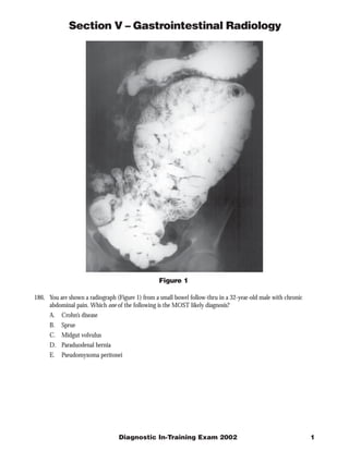
  • 1. Section V – Gastrointestinal Radiology Figure 1 186. You are shown a radiograph (Figure 1) from a small bowel follow-thru in a 32-year-old male with chronic abdominal pain. Which one of the following is the MOST likely diagnosis? A. Crohn’s disease B. Sprue C. Midgut volvulus D. Paraduodenal hernia E. Pseudomyxoma peritonei Diagnostic In-Training Exam 2002 1
  • 2. Section V – Gastrointestinal Radiology Question #186 Findings: The radiograph demonstrates encapsulation of jejunal loops in the left mid abdomen. Rationales: A. Incorrect. Crohn’s disease involves the small bowel in 75% of patients at the time of presentation. Affected segments show fold thickening and ulceration. Sinus tracts and fistulae are also noted. Although surrounding fibrofatty proliferation may produce a mass effect or displace bowel on SBFT, encapsulation of bowel loops is not noted with Crohn’s disease. B. Incorrect. Sprue or gluten sensitive enteropathy produces villous atrophy and radiographically shows reversal of the normal fold pattern with loss of normal jejunal folds and an increased number of folds per inch in the ilium. Transient intussusceptions, hypomotility and flocculation of barium can also be seen. However, none of these findings are present on this film. C. Incorrect. Midgut volvulus is produced by twisting of small bowel loops around the shortened mesenteric vascular pedicle in patients with congenital small bowel malrotation. A corkscrew deformity of the jejunal loops is noted on SBFT. Obstruction to venous return produces edema and hence thickened folds in the involved bowel. Such changes are not noted on this film. D. Correct. Internal hernias are abnormal protrusions of intraabdominal structures through a normal opening or through a congenital or acquired defect in fascia or mesentery. Encapsulation of jejunal bowel loops in either the right or left mid abdomen is characteristic of paraduodenal hernia - the most common type of internal hernia. The amount of contained small bowel can vary from a few loops to the majority of the small bowel. Patient typically present with intermittent abdominal pain thought to be secondary to episodes of obstruction. Left sided paraduodenal hernia is more common than right sided paraduodenal hernia. E. Incorrect. Pseudomyxoma peritonei results from seeding of the peritoneal cavity by benign or malignant mucin secreting cells. The cells typically come from a ruptured mucinous cystadenoma or cystadenocarcinoma of the appendix or ovary. Though bowel can be distorted, obstructed or tethered by tumor or mucin, encapsulation as noted here is not seen. Citations: Maglinte DDT, Bisset III GS, Congenital and Developmental Anomalies of the Small Bowel in Adolescents and Adults pp 247-251 in Clinical Imaging of the Small Intestine 2nd ed, eds Herlinger H, Maglinte DDT, and Birnbaum BA, Springer, New York, 1999. 2 American College of Radiology
  • 3. Section V – Gastrointestinal Radiology Figure 2 187. You are shown a contrast-enhanced CT (Figure 2) in an 84-year-old male with recent weight loss and guaiac positive stool. Which one of the following is the MOST likely diagnosis? A. Desmoid tumor B. Intussusception C. Internal hernia D. Lipomatous ileocecal valve E. Volvulus Diagnostic In-Training Exam 2002 3
  • 4. Section V – Gastrointestinal Radiology Question #187 Findings: The CT image demonstrates a long colo-colic intussusception involving the ascending and transverse colon Rationales: A. Incorrect. Desmoid tumors are non metastasizing but locally invasive fibrous tumors. They can occur either in isolation or more commonly in association with Gardner’s syndrome, particularly following abdominal surgery. They may present as an abdominal mass or cause bowel obstruction. On CT they appear as a soft tissue mass in the mesentery either with well circumscribed or infiltrative margins. These findings are not present in this case. B. Correct. The image demonstrates a long segment colo-colic intussusception. The vast majority of adult colo- colic intussusceptions have a malignant lead point with adenocarcinoma being the most common histologic type. The extensive edema seen with the intussusception often makes it difficult to delineate the precise lead point. C. Incorrect. Internal hernias are abnormal protrusions of intraabdominal structures through a normal opening or through a congenital or acquired defect in fascia or mesentery An internal. The most common type is the paraduodenal hernia shown in the previous question. Although internal hernias can cause distortion of bowel loops they would not produce the bowel within bowel appearance noted here. D. Incorrect. Lipomatous ileocecal valve is caused by submucosal infiltration of fat into the lips of the I-C valve. It is typically seen in older patients and is more common in women. Symptoms are usually absent. Although the mass can mimic an adenocarcinoma its fatty density on CT and smooth contour usually allow differentiation. The mass in this case is predominantly soft tissue density and much larger and more extensive and irregular than a lipomatous I-C valve. E. Incorrect. A volvulus involving the bowel is produced by twisting of small bowel loops around a point of fixation. In midgut volvulus associated with malrotation the point of fixation is the shortened mesenteric vascular pedicle. Volvulus involving fewer bowel loops may also occur around an acquired adhesive band. On CT a swirling appearance of vessels is seen in conjunction with obstructed small bowel loops. Although arcing vessels are seen in this case, they are entering the ascending colon rather than twisting around a point of fixation. Citations: Gore RM, Eisenberg RL, Large Bowel Obstruction pp1247-1260 in Textbook of Gastrointestinal Radiology eds Gore RM, Levine MS, Laufer I, W.B.Saunders, Philadelphia, 1994 4 American College of Radiology
  • 5. Section V – Gastrointestinal Radiology Figure 3 188. You are shown a radiograph (Figure 3) of the splenic flexure from a double contrast barium enema in a 35-year-old female with diarrhea and abdominal pain. Which one of the following is the MOST likely diagno- sis? A. Typhlitis B. Ulcerative colitis C. Toxic megacolon D. Crohn’s disease E. Pseudomembranous colitis Diagnostic In-Training Exam 2002 5
  • 6. Section V – Gastrointestinal Radiology Question #188 Findings: The radiograph demonstrate areas of irregular colonic narrowing and ulceration. More normal caliber bowel is also noted but demonstrates multiple small aphthous lesions. Rationales: A. Incorrect. Typhlitis refers to an acute enterocolitis associated with immunosuppression and neutropenia. The disease predominantly involves the cecum and right colon and is characterized by marked wall edema and inflammation. Diagnosis is usually suggested by CT or plain film in conjunction with the clinical setting. Barium enema or colonoscopy may place the patient at risk of perforation when inflammation is severe. The appearance of aphthous lesions and irregular areas of colonic narrowing and ulceration in the splenic flexure would not be typical for typhlitis. B. Incorrect. Ulcerative colitis is a chronic inflammatory bowel disease, predominantly limited to the mucosa, with a peak age of onset of 15 to 25 years. The barium enema findings involve the rectum and extend proximally to a variable extent, often involving the entire colon. Mucosal abnormalities in acute active disease include granularity, mucosal stippling, collar button ulceration and inflammatory pseudopolyp formation. In patients with chronic disease, there can be narrowing of the colonic lumen, loss of haustration and shortening of the colon. The discontinuous irregular involvement noted in this case in addition to the presence of aphthous lesions are strongly against the diagnosis of UC. C. Incorrect. Toxic megacolon is characterized by an ill, toxic appearing patient with diffuse colonic dilatation. The colon will also demonstrate loss of haustral folds and an irregular nodular mucosal surface corresponding to ulcerations and inflammatory pseudopolyps. Barium enema is contraindicated in patients with toxic megacolon, because of a risk of perforation. Causes include ulcerative colitis as well as infectious colitis. D. Correct. Crohn’s disease is a chronic inflammatory bowel disease that, like UC has a peak age of onset of 15 to 25 years. Unlike UC however, Crohn’s disease is characterized by transmural inflammation with aphthous lesions and discontinuous areas of mucosal ulceration and narrowing. Sinus tracts and fistulae are also common as is small bowel involvement which occurs in 75% of patients at presentation. The appearance in this case would be typical for colonic involvement with Crohn’s disease. E. Incorrect. Pseudomembranous colitis is produced secondary to toxin producing Clostridium difficile infection. The disease usually follows the administration of broad spectrum antibiotics and produces watery diarrhea, fever, abdominal pain and leukocytosis. Moderate large bowel dilation with thumbprinting is noted on abdominal plain film exam. On barium enema small irregular plaque like filling defects or small nodules are noted. In severe cases the luminal margin may appear irregular from poor mucosal coating. These changes are not noted in this case. Citations: Gore RM, Laufer I, Ulcerative and Granulomatous Colitis: Idiopathic Inflammatory Bowel Disease in Textbook of Gastrointestinal Radiology eds Gore RM, Levine MS, Laufer I, W.B.Saunders, Philadelphia, 1994 6 American College of Radiology
  • 7. Section V – Gastrointestinal Radiology Figure 4A Figure 4B 189. You are shown a contrast-enhanced CT (Figure 4A and 4B) of a 25-year-old male with epigastric pain. Which one of the following is the MOST likely diagnosis? A. Mucinous ductectatic malignancy B. Cystic fibrosis C. von Hipple Lindau disease D. Tuberous sclerosis E. Solid and papillary epithelial neoplasm Diagnostic In-Training Exam 2002 7
  • 8. Section V – Gastrointestinal Radiology Question #189 Findings: Multiple cysts are identified in the pancreas. A solid enhancing lesion is also noted in the left kidney. In addition an enhancing mass is noted involving the spinal cord. Rationales: A. Incorrect. Mucinous ductectatic tumor is a pancreatic mucinous tumor with an intraductal growth pattern. Both benign and malignant variants are reported. The median age of patients is in the 7th decade but unlike the usual mucinous cystic tumor, the lesion is more common in men than women. On CT cystic areas are noted along with distension of the pancreatic ducts with mucin. The appearance can mimic the changes of chronic pancreatitis. Duct distension is not noted in this case, nor would this entity explain the spinal and renal abnormalities noted above. B. Incorrect. Cystic fibrosis affects multiple epithelial tissues including sweat gland, lung, pancreas, and bowel. It is inherited as an autosomal recessive trait and occurs in 1 in 3000 Caucasian live births in the US. Pancreatic secretions in cystic fibrosis patients have increased viscosity and are thought to cause inspissation and resulting pancreatic atrophy. By 2 years of age more than 80% of patients will have evidence of pancreatic insufficiency. On CT the pancreas often demonstrates diffuse fatty replacement. Less commonly replacement of the gland by multiple macroscopic cysts occurs. Although multiple cysts are seen in this patient, cystic fibrosis would not explain the renal or CNS masses. C. Correct. von Hippel Lindau disease is characterized by cysts and or neoplasm in multiple organs including the pancreas, kidney, liver, epididymis and CNS. It is inherited in an autosomal dominant fashion. Onset of symptoms is typically in the third to fifth decade. Commonly associated lesions include hemangioblastomas (seen in retina, cerebrum, cerebellum and spinal cord) renal carcinoma and hemangioblastomas, pancreatic cysts and cystadenomas, and epididymal cysts. The pancreatic cysts, renal carcinoma and spinal hemangioblastoma seen in this case would be typical for von Hippel Lindau disease. D. Incorrect. Tuberous sclerosis is an autosomal dominant syndrome which has skin, CNS, cardiovascular renal, and pulmonary manifestations. Renal lesions including intrarenal aneurysms, carcinoma, cysts and angiomyolipomas, are reported. The latter occur in 40-80% of patients and can cause spontaneous hemorrhage. Diagnosis of AMLs can be made by showing intratumoral fat on CT or MRI. Although CNS lesions occur, tuberous sclerosis would not be expected to present the pancreatic or spinal cord findings noted in this case. E. Incorrect. Solid and papillary epithelial neoplasm is an uncommon pancreatic tumor of young females. More than 95% of cases are found in adolescent or postadolescent girls and young women. The tumor is generally a large cystic and solid mass that frequently contains areas of necrosis and hemorrhage with fluid-debris levels. The sex of the patient and the morphologic appearance of the tumor in the test patient make solid and papillar y epithelial neoplasm an unlikely diagnosis. Citations: Stanley RJ, Semelka RC, Pancreas in Computed Body Tomography with MRI Correlation third ed., eds Lee JKTL, Sagl SS, Stanley RJ, and Heiken JR, Lippincott-Raven, New York 1998. 8 American College of Radiology
  • 9. Section V – Gastrointestinal Radiology Figure 5 190. You are shown an image from an endoscopic retrograde cholangiogram (Figure 5) in a 24-year-old female with painless jaundice. Which one of the following is the MOST likely diagnosis? A. Adenomyomatosis B. Mirizzi syndrome C. Sclerosing cholangitis D. Caroli’s disease E. Klatskin tumor Diagnostic In-Training Exam 2002 9
  • 10. Section V – Gastrointestinal Radiology Question #190 Findings: The image demonstrates obstruction and dilation of the intra and extrahepatic biliary tree down to a point just above the juncture with the cystic duct. At this point there is thinning of the contrast column and an apparent filling defect outlined in the cystic duct. The CBD is normal in size. Rationales: A. Incorrect. Adenomyomatosis is characterized by hyperplastic changes in the wall of the gallbladder with formation of intramural diverticuli or Aschoff-Rokitansky sinuses. This may occur in a diffuse or segmental fashion. The clinical significance of this finding is uncertain, although it is important to differentiate it from other causes of gallbladder wall thickening such as carcinoma and cholecystitis. The intramural diverticuli do not typically involve the distal cystic duct and would not be responsible for the findings shown here. B. Correct. Mirizzi syndrome is partial or complete obstruction of the common hepatic duct associated with a stone lodged in the distal cystic duct. It occurs because the distal cystic duct and common hepatic duct are often bound together in a common sheath. The clinical presentation is often progressive jaundice and abdominal pain. The findings depicted here are typical for Mirizzi syndrome. C. Incorrect. Primary sclerosing cholangitis is an idiopathic disorder characterized by inflammation, fibrosis and strictures involving the intra and extrahepatic biliary tree. Ulcerative colitis is the most frequent associated condition and is present in 50-75% of cases. Men are affected twice as often as women. Symptoms are usually insidious in onset and consist of fatigue, right upper quadrant pain, jaundice and pruritus. Multiple short irregular strictures are seen at cholangiography diffusely distributed throughout the biliary tree. A beaded appearance of the ducts is often noted with bandlike strictures and small diverticulum like outpouchings occasionally seen. Such findings are not noted in this case. D. Incorrect. Caroli’s disease is a congenital disorder characterized by diffuse or segmental dilatation of the intrahepatic biliary tree. Two types have been characterized, a simple and a periportal fibrosis type. The latter is more common and is associated with congenital hepatic fibrosis, cirrhosis and portal hypertension. The former is associated with medullary sponge kidney. Patients usually present in early adulthood with symptoms of cholangitis (fever, chills, abdominal pain). An increased incidence of malignant transformation is also noted. The uniform dilation of the biliary tree down to the point of obstruction noted here would not be seen in Caroli’s disease. E. Incorrect. Klatskin tumor refers to a cholangiocarcinoma occurring at the confluence of the right and left bile ducts and common hepatic duct. An increased incidence of cholangiocarcinoma has been noted in patients with PSC and choledochal cyst as well as patients infested with Clonorchis sinensis or Opisthorchis viverrini. Patients usually present with painless jaundice. The peak incidence is around 65 years of age. The tumor typically appears as a short segmental stricture at the bifurcation and would not be consistent with the appearance shown here. Citations: Zeman RK, Cholelithiasis and Cholecystitis in Textbook of Gastrointestinal Radiology eds Gore RM, Levine MS, Laufer I, W.B.Saunders, Philadelphia, 1994 10 American College of Radiology