SlideShare a Scribd company logo
1 of 7
Download to read offline
287
Management of Diabetic Foot
Sunil Gupta, Nagpur
Introduction
The management of diabetic foot disease may seem poorly defined by comparison with complications
such as nephropathy, hyperlipidaemia and retinopathy, for which clear guidelines exist. A
multidisciplinary team, approach, particularly in specific diabetic foot clinics, is very successful in
avoiding and treating foot complications, This strategy has been shown to reduce both the incidence
of major leg amputation (by 40% or more), and the duration of in-patient admissions for the treatment
of diabetic foot ulceration.1,2
The major challenges relating to diabetes foot are:-
1.	 Foot ulceration is common, affecting up to 25% of patients with diabetes during their lifetime.3
2.	 Over 85% of lower limb amputations are preceded by foot ulcers and Diabetes remains a major
cause of non-traumatic amputation across the world with rates being as much as 15 times higher
than in the non-diabetic population.
3.	 Prevention is the first step towards solving diabetic foot problems. Although it was estimated
that an ankle is lost to diabetes somewhere in the world every 30 seconds, a more important fact
is that up to 85% of all amputations in diabetes should be preventable.4
4.	 Strategies aimed at preventing foot ulcers are cost-effective and can even be cost-saving if
increase education and effort are focused on those patients with recognized risk factors for the
development of foot problem.5
5.	 Diabetes is now the most common cause of Charcot neuro-arthropathy in Western countries,
another condition that should be generally preventable.6
Epidemiology of Diabetic foot Disease:
One-third of all diabetic patients have significant peripheral neuropathy and/or peripheral vascular
disease (PVD). Diabetic foot problems are the commonest reason for hospitalization of diabetic
patients (about 30% of admissions) and absorb some 20% of the total health-care costs of the disease-
more than all other diabetic complication.2,7
In India prevalence of foot ulcers in diabetic patients in
clinic population is 3% , which is much lower than reported in the western world.Apossible reasoning
for the low prevalence in Indians is younger age and shorter duration of diabetes.8,9
PVD has been
reported to be low amongAsians10-13
ranging between 3-6% as against 25-45% inWestern patients.14-16
The prevalence of PVD increases with advancing age and is 3.2% below 50 years of age and rises to
55% in those above 80 years of age17
Similarly it also increases with increased duration of diabetes,
15% at 10 years and 45% after 20 years.18
Etiopathogenesis of diabetic foot lesions:
The breakdown of the diabetic foot does not occur spontaneously, and there are many warning signs
that may be used to predict those at risk. Dr. Elliott Joslin recognised this more than 75 years ago,
when he stated that ā€œDiabetic gangrene is not heaven-sent but is earth-bornā€.19
Ulcers invariably
occur as a consequence of an interaction between environmental hazards and specific pathologies
in the lower limb.
5 : 10
Medicine Update 2012 ļ® Vol. 22
288
Diabetic Neuropathy: More than 60% of diabetic foot
ulcers are the result of underlying neuropathy.20, 21
The more
commonly described mechanisms of action is the polyol
pathway.22
The hyperglycaemic state leads to an increase
in action of the enzymes aldose reductase and sorbitol
dehydrogenase. This results in the conversion of intracellular
glucose to sorbitol and fructose. The accumulation of these
sugar products results in a decrease in the synthesis of nerve
cell myoinositol, required for normal neuron conduction.
Additionally, the chemical conversion of glucose results in
a depletion of nicotinamide adenine dinucleotide phosphate
(NADP) stores, which are necessary for the detoxification of
reactive oxygen species (ROS) and for the synthesis of the
vasodilator nitric oxide (NO). There is a resultant increase
in oxidative stress on the nerve cell and an increase in
vasoconstriction leading to ischemia, which will promote
nerve cell injury and death. Hyperglycemia and oxidative
stress also contribute to the abnormal glycation of nerve cell
proteins and activation of protein kinase C, (PK-C) resulting
in further nerve dysfunction and ischemia.
Neuropathy in diabetic patients is manifested in the motor,
autonomic,andsensorycomponentsofthenervoussystem.20
Damage to the innervations of the intrinsic foot muscles leads
to an imbalance between flexion and extension of the affected
footcausing.Autonomicneuropathyleadstoadiminutionin
sweat and oil gland functionality.As a result, the foot loses its
natural ability to moisturize the overlying skin and becomes
dry and increasingly susceptible to tears and the subsequent
development of infection.
Peripheral Vascular Disease in Diabetes
Peripheral arterial disease (PAD) is a contributing factor to
the development of foot ulcers in up to 50% of cases.23,24
It
commonly affects the tibial and peroneal arteries of the calf.
Endothelial cell dysfunction and smooth cell abnormalities
developinperipheralarteriesasaconsequenceofthepersistent
hyperglycaemic state.25
Moreover, smoking, hypertension,
and hyperlipidemia are other factors that are common in
diabetic patients and contribute to the development of PAD
Plantar callus:
Callus forms under weight-bearing areas as a consequence of
dry skin (autonomic dysfunction), insensitivity and repetitive
moderate stress from high foot pressure. It acts as a foreign
body and cause ulceration.29
Callus should be removed by the
podiatrist or other trained health care professional.
Foot deformity
A combination of motor neuropathy, cheiroarthropathy
and altered gait patterns are thought to result in the ā€œhigh
riskā€ neuropathic foot with clawing of the toes, prominent
metatarsal heads, high arch and small muscle wasting.28
Assessment of Diabetic Foot
A task force of the Foot Care Interest Group of the American
Diabetes Association (ADA) released a 2008 report that
specifies recommended components of foot examinations for
patients with diabetes.24
Providers should take a history of
all risk factors given in table 3. The foot should be examined
for deformities. Hyperextension of the metatarsal-phalangeal
joint with interphalangeal or distal phalangeal joint flexion
leads to hammer toes. In examining for PAD, the dorsalis
pedis and posterior tibial pulses should be palpated and
characterized as present or absent.30
Claudication, loss of hair,
and the presence of pale, thin, shiny, or cool skin are physical
	
Table 1: Limb Amputation in Diabetes: Differences- Western
vs Indian Experience 8
Limb Amputation Western Indian
Mean age at amputation 75 years 61.25 years
Mortality at 2 years 50% 14.28%
Contra lateral limb amp. At 2 years 30-50% 8.92%
AK:BK amputation 1:2 1:6
Indication: Neuropathic 10% 76.78%
Ischemic 90% 23.21%
Table 2 : Economics of Treatment of Diabetic Foot: Cost of
Treatment of Complete Healing (In India )26
Type of lesion Treatment Direct cost (US$)
Neuropathic Ulcer Ambulatory 56
Infected neuropathic foot Ambulatory 165
Advanced Diabetic foot Salvage 1080
Advanced Diabetic foot Limb amputation 960
Advanced Diabetic foot Salvage then Amputation 2650
Neuroischemic foot Bypass 1960
Table 327
Factors increasing the Risk of Diabetic Foot Ulceration:
ā€¢	 Peripheral neuropathy : somatic or autonomic
ā€¢	 Peripheral vascular disease
ā€¢	 Past foot ulcer history (The annual risk of re-ulceration is found to
be up to 50%)
ā€¢	 Plantar callus and elevated foot pressure
ā€¢	 Foot deformity, Nail abnormalities,
ā€¢	 Psychosocial factors (Anxiety, Depression, non-compliance)
ā€¢	 Other microvascular complication, especially chronic renal failure
ā€¢	 Diabetic Nephropathy, patients with end stage Renal Disease on di-
alysis subjects with renal or pancreas- renal transplants.
ā€¢	 Interdigital Infection in feet. Temperature difference between feet.
ā€¢	 Edema
ā€¢	 Ethnic background
ā€¢	 Living alone
ā€¢	 Poor social background
ā€¢	 History of Smoking
289
Management of Diabetic Foot
findings suggestive of potential ischemia. Measuring the
Ankle Brachial Index (ABI) can be used for determining the
extent of vascular disease. The ABI is obtained by measuring
the systolic blood pressures in the ankles (dorsalis pedis and
posterior tibial arteries) and arms (brachial artery) using a
handheld Doppler and then calculating a ratio. Ratios below
0.91 are suggestive of obstruction. However, in patients with
calcified, poorly compressible vessels or aorto-iliac stenosis,
the results of theABI can be complicated.31
If there is a strong
suspicionofPAD,thepatientshouldundergovascularimaging
/ peripheral arterial angiogram.
Thelossofpressuresensationinthefoothasbeenidentifiedas
a significant predictive factor for the likelihood of ulceration.
A screening can be done by the diabetic foot is the 10-gauge
monofilament. The monofilament is tested on various sites
along the plantar aspect of the toes, the ball of the foot, and
between the great and second toe. The test is considered
reflective of an ulcer risk if the patient is unable to sense the
monofilament when it is pressed against the foot with enough
pressure to bend it.32
Areas of callus should not be tested24
Wound healing in Diabetic foot:
Diabetes may influence foot wound healing by an impairment
of the peripheral circulation, altered leukocyte function,
disturbedbalanceofcytokinesandproteasesandevenchronic
hyperglycemia itself.6,33
Recently, it has been suggested that
levels of matrix metalloproteinases (MMP) are important in
predicting the likelihood of wound healing and a high level
of MMP-1 seems essential to wound healing.34
Offloading
The neuropathic plantar foot wounds will heal satisfactorily
whenoffloadedinaTotalContactCast(TCC).6
ThePrincipleof
TCCmanagementisthatpressureismitigatedbut,inaddition,
the device is irremovable thus enforcing compliance with
therapy. The Removable Cast Walkers (RCW) redistribute
pressure in a similar manner to the TCC, but itā€™s results are
inferior to TCC due to patientā€™s non-compliance.Appropriate
offloading result in angiogenesis, fibroblast proliferation and
presence of granulation tissue. Offloading is an essential
component to the management of predominantly neuropathic
plantar foot ulcers (UT 1A and 2A) ulcers. For those patients
treated with irremovable cast walkers, it is recommended that
the cast be removed initially on a weekly basis for wound
assessment,debridementandcleansing.Healingcangenerally
be achieved in a period of 6-12 weeks in a cast. It is strongly
recommended that after the plantar wound has healed, that the
cast be worn for a further 4 weeks to permit the scar tissue to
firm up. Thereafter, the patient may be gradually transferred
to appropriate footwear which may need extra depth or in the
case of severe deformity, custom moulded.
Management of Infection:
Clinically non- infected ulcers:
Where ulcers are not infected and predominantly neuropathic
(UT grade 1A, 2A), the use of antibiotics may be withheld
as Chantelau et al35
have shown that with appropriate wound
management,patientsdoequallywellwithorwithoutsystemic
antibiotics in a randomized controlled trial. Nevertheless,
frequentreview,debridementandcallusremovaltogetherwith
offloading are essential parts of management of neuropathic
footulcersandifsignsofinfectiondevelop,antibioticsmaybe
needed. For those ulcers with an ischemic component which
do not have gross signs of infection (UT 1C, 2C) antibiotics
should probably be given in most cases as the combination
of infection and ischemia in the diabetic foot are a common
cause of ultimate lower extremity amputation.
Clinically infected ulcers
Non- limb- threatening infected ulcers (UT 1B, 1D,2B,2D)
can generally be treated on an outpatient basis, and oral
broad- spectrum antibiotics should be used initially until
results of sensitivities are obtained. Generally, mild infections
are relatively superficial and limited, moderate infections
involve deeper tissues.36
Any ulcer with clinical evidence
of infection should have tissue taken and sent for culture
and sensitivity. Although superficial swabs are commonly
taken, deep (preferably tissue) specimens are preferable in
terms of accuracy of diagnosis.36
Most infective ulcers are
polymicrobial, often with a mixture of anaerobes and aerobes.
Ifthereisanysuspicionofosteomyelitis,furtherinvestigations
Table 428
Traditional Meggitt-Wagner Ulcer Classification System
Grade 0 : No ulcer, but high-risk foot (bony prominences, callus, deformi-
ties, etc)
Grade 1 : Superficial, full-thickness ulcer
Grade 2 : Deep ulcer, may involve tendons, but without bone involvement
Grade 3: Deep ulcer with osteomyelitis
Grade 4 : Local gangrene (toes or forefoot)
Grade 5: Gangrene of whole foot
Table 5 28
University of Texas wound classifi-
cation system	
Grade 0: Pre- or post-ulcerative
lesion, completely epithelialized
Stage A: without infection or is-
chemia
Stage B: with infection
Stage C: with ischemia
Stage D: with infection and ischemia
Grade1:Superficial wound not in-
volving tendon, capsule, or bone
Stage A: without infection or is-
chemia
Stage B: with infection
Stage C: with ischemia
Stage D: with infection and ischemia
Grade2:Wound penetrating to
tendon or capsule
Stage A: without infection or is-
chemia
Stage B: with infection
Stage C: with ischemia
Stage D: with infection and ischemia
Grade 3:Wound penetrating to
bone or joint
Stage A: without infection or is-
chemia
Stage B: with infection
Stage C: with ischemia
Stage D: with infection and ischemia
Medicine Update 2012 ļ® Vol. 22
290
should be done. Suitable broad- spectrum antibiotics such as
clindamycin or the amoxicillin- clavulanate combination36
may be started till the C & S report is available.
Limb- threatening infection
Patients with limb-threatening infection usually have
systemic symptoms and signs and require hospitalization
with parental antibiotics. Deep wound and blood cultures
should be taken, the circulation assessed with non- invasive
studies initially, and metabolic control is usually achieved by
intravenous insulin infusion. Early surgical debridement is
often indicated in such cases, and initial antibiotic regimens
should be broad- spectrum until sensitivities are determined
fromcultures.Examplesofinitialantibioticregimensinclude:
clindamycin and ciprofloxacin, or flucloxacillin, ampicillin
and metronidazole. The Polymerase chain reaction (PCR)
assay has been shown to be effective at identifying many
virulent organisms.36
A recent study from France37
showed
the potential advantages of using this new technique in the
rapid distinction between colonizing and virulent infecting
organisms.
An increasing problem in diabetic foot clinics is the
antibiotic- resistant pathogens such as methicillin- resistant
staphylococcus aureus (MRSA). If MRSA is felt to be an
infecting organism, there are useful new agents such as
linezolid,36
which can be given parentally or orally.
Osteomyelitis:
Contrary to traditional teaching, it is increasingly recognized
that some cases of localized osteomyelitis can be managed
by long- term (10-12weeks) antibiotic therapy38
; However,
localized bony resection after appropriate antibiotic therapy
remains a common approach. Those cases with osteomyelitis
confined to one bone without involvement of a joint are most
likely to respond to antibiotic therapy particularly in the
absence of PVD.
Adjunctive Therapy
Growth factors
The recombinant platelet derived growth factor (PDGF)
Becaplermin was the first growth factor, approved for treating
diabeticneuropathicfootulcer.Thereissomesupportfortheir
use from randomized clinical studies,39
their expense together
with the fact that most neuropathic ulcers can be healed with
appropriate offloading, have limited their use and there is still
no consensus as their place in day ā€“ to ā€“ day clinical practice.40
Hyperbaric oxygen
Hyperbaric oxygen (HBO) has been promoted for the
management of non- healing diabetic foot ulcers in ischemic
diabetic foot wounds. Though the systematic review of the
International working group considered HBO accepted, that
there was some evidence to support its use, it is clear that
more data are required from larger controlled trials not only
to confirm efficacy but also to clarify which wounds might
best benefit from this expensive treatment.40,41
Negative pressure wound therapy
Over the past several years negative pressure wound therapy
(NPWT) using vacuum- assisted closure has emerged as
the treatment of complex wounds of the diabetic foot.42
It
decreases local tissue edema and removes excessive fluid and
pro-inflammatory exudates from the wound bed.There is now
controlled trial evidence for the use of NPWT in both local
postoperativewoundsinthediabeticfoot43
and,morerecently,
in the management of complex but non- surgical diabetic
foot ulcers.44
It is clear that this treatment helps promote the
formation of granulation tissue, but its cost will limit its use.
Bioengineered skin substitutes
Tissue engineered skin,Apligruf comprises of cultured living
dermis and sequentially cultured epidermis. Dermagraft is
dermis derived from human fibroblasts used for treatment of
non-infected neuropathic ulcers. A systematic review on this
topic concluded that the trials assessed were of questionable
quality and until high quality studies were performed,
recommendations for the use of these skin substitutes could
not be made.45
Larval Therapy
The use of sterile maggots, the larvae of the common green
bottlefly,isnotnew.Sterilemaggotsareusefulindesloughing
woundsthatareresistanttosurgicaldebridement.Itisbelieved
that they secrete a broad spectrum of powerful enzymes that
break down dead tissue. Limited evidence also suggests that
they do not harm healthy tissue because the enzymes are
inactivated by inhibitors present in normal skin.27
Charcot neuroarthropathy
Charcotneuroarthropathy(CN)isanon-infectivearthropathy
whichoccursduetoresultofacombinationofmotor,autonomic,
and sensory neuropathies in which there is muscle and joint
laxity that lead to changes in the arches of the foot. Further,
the autonomic denervation leads to bone demineralization via
the impairment of vascular smooth muscle, which leads to
an increase in blood flow to the bone with a consequential
osteolysis.The exact mechanism underlying the development
of CN remains unclear. Patient may have history of trauma
and may present with a warm, swollen foot and may be
accompanied by pain or at least discomfort. The treatment
of CN in acute phase is offloading of the affected foot by use
of a plaster cast. The cast should continue until the swelling
and hyperemia have resolved and then custom moulded shoes
with appropriate insoles are indicated.46
Bisphosphonates are
potent inhibitors of osteoclast activation and intravenous
Pamidronate has been shown to be useful in reducing disease
activity in acute CN47
. Larger randomized controlled trials
291
Management of Diabetic Foot
Charcotā€™s Arthropathy
Planter Infected Ulcer
Self Inspection
Cut the nails straight
Take care of Dry skin
to see feet in mirror for redness,
Swelling, ulcer or crack foot
Hammer Toes
Foot Care
Tinea Interdigitalies
Gangrene Bunion with Nail Changes
Medicine Update 2012 ļ® Vol. 22
292
are required to confirm these preliminary observations. The
management of advanced CN with bone deformity requires
reconstructive surgery.
Footwear, Orthoses, and Hosiery
Inappropriate (Often tight) footwear is a major cause of
ulceration. Studies have suggested that specialized foot wear
with padded hosiery reduces high foot pressures and gives
all- around protection to the high-risk diabetic foot provided
that the shoes are fitted to accommodate the padded socks.48
Injected Liquid Silicone
A randomized, double-blind trial of injected liquid silicone
in the diabetic foot has confirmed that silicone injections
were associated with increased soft-tissue thickness under
the metatarsal head, decreases foot pressure, and reduced
callus formation. Such an ā€œinjectable orthosisā€ might well be
beneficial in high-risk patients. Subsequent follow-up studies
confirmedthatpatientsatgreatestriskofulcerationweremost
likely to benefit from silicone injections but that after two
years of follow-up, the benefits of injections, though still
demonstrable,werereducedcomparedtobaseline,suggesting
that booster injections may periodically be needed. This is an
area of ongoing research49, 50
Patient Education
Patients need to be informed of the risk of having sensory
loss and the need for regular self-inspection, foot hygiene,
and podiatry treatment as required. They must also be told
what action to take in the event of injury or the discovery of
a foot ulcer.51
The Diabetic foot: Need For A Team Ap-
proach
A number of reports have shown the benefits of the
multidisciplinary approach to diabetic foot care. The team
might include diabetologists, surgeons (both orthopaedic and
vascular), specialist nurses, diabetes educators, podiatrists,
orthotists, pedorthotists and patient or a care taker of patient.27
Conclusion
Patients with diabetes are at an increased risk for developing
foot ulcerations. The consequences of persistent and poorly
controlled hyperglycemia lead to neuropathic and vascular
abnormalities that cause foot deformities and ulceration.
The feet of diabetic patients should be examined at least
annually to determine predisposing conditions to ulceration.
Treatment plans should be based on examination findings and
the individual risk for ulceration. If ulcers are present, the
treatment strategy should include offloading, debridement,
and appropriate dressings. Further, the presence of infections
should be determined by clinical findings and appropriate
wound cultures and treated based on the culture results. If
evidence for ischemia is present, revascularization may be
indicatedtorestorearterialbloodflowandincreasethechance
for limb salvage. There are adjunctive therapies available
that can also contribute to the overall healing process of
the wounds in affected patients. Patient education and team
approach towards management plays the key role to words
the success. .
References
1.	 Assal JP, et al. Patient education in diabetes. In: Recent Trends in
Diabetic Research, Stockholm: Almqvist and Wiksel International
1982:276-89.
2.	 Thomson FJ, et al. A team approach to diabetic foot care: the Man-
chester experience. Foot 1991;1:75-82.
3.	 Singh N, Armstrong DG, Lipsky BA. Preventing foot ulcer in pa-
tients with diabetes. JAMA 2005;293:217-228
4.	 Boulton AJM, Vileikyte L, Ragnarson-Tennvall G, Apel-
qvist J:Ā  The global burden of diabetic foot disease. Lan-
cetĀ 2005;Ā 366:1719-1724.
5.	 Ragnarson- Tennvall G, Apelqvist J. Prevalence of diabetes -re-
lated foot ulcers and amputations: a cost-utility analysis based on
Markov model simulations. Diabetologia 2001;44:2077-2087.
6.	 Boulton AJM. The diabetic foot: from art to science: the 18th
Ca-
millow Golgi lecture. Diabetologia 2004; 47:1343-1353.
7.	 Williams R, Airey M. The size of the problem: economic aspects
of foot problems in diabetes. In: Boulton AJM, Connor H, Cava-
nagh PR, eds. The Foot in Diabetes, 3rd
edn. Chichester: Wiley,
2000:3-17
8.	 Pendsey SP, Epidemiological aspects of Diabetic Foot. Int. J Diab.
Dev Countries 1994; 14:37-38
9.	 International consensus on the Diabetic Foot, by the International
working group on the Diabetic Foot, 1999.
10.	 Mohan V, Premlatha G, Shastry NG. Peripheral vascular disease
in non-insulin dependent diabetes mellitus in South India; Diab
Res Clin Pract 1995; 27:235-240.
11. 	 De silva D. The prevalence of macrovascular disease and lipid ab-
normalities amongst diabetic patient in Sri Lanka. Postgrad Med
J 1993; 69:557-561
12.	 Pendsey SP. Peripheral vascular disease (PVD): An Indian Scena-
rio, Dibetologia Croatia1998;27-4: 153-156.
13. 	 Pendsey SP. Diabetic Foot Syndrome. RSSDI Text book of Diabe-
tes Mellitus, 2nd
Edition, 2008;2:959-972.
14. 	 Migdalis IN, et al. peripheral vascular disease in newly diagnosed
non insulin dependent diabetes. Int Angiol 1992; 11 : 230-232.
15. 	 Marinelli Mr. Beach KW, Glass MJ et al. Non invasive testing vs,
clinical evaluation of arterial disease: a prospective study. J AM
Med Assoc 1979;2031.
16. 	 Walters DP. Et al. the prevalence, detection and epidemiological
correlates of peripheral vascular disease : a comparison of diabe-
tic and non diabetic subjects in an English community. Diab Med
1992; 9: 710-5.
17. 	 Janka H U, StandI E and Mehnert H. Peripheral vascular disease
in diabetes mellitus and its relation to cardiovascular risk factor :
screening with Doppler ultrasonic technique. Diabetes Care 1980;
3: 207.
293
Management of Diabetic Foot
18. 	 Palumbo PJ, Melton LJ. Peripheral vascular disease and diabetes
in M. I. Harris and R. F. Hamman (Eds), Diabetes in America,
NIH 1985; publ no.85 ā€“ 1468. Washington:US Government Prin-
ting Office, 1985;XVI-21.
19. 	Joslin EP: The Menace of diabetic gangrene. N Engl J Med
211:16-20, 1934
20. 	 Bowering CK: Diabetic foot ulcers: pathophysiology, assessment,
and therapy. Can Fam Phys 47:1007-1016, 2001
21. 	 Dyck PJ, Davies JL, et al : Risk factors for severity of diabetic
polyneuropathy. Diabetes Care 1999;22:1479-1486
22. 	 Feldman EL, Russell JW,et al: New insights into the pathogenesis
of diabetic neuropathy. Curr Opin Neurol 1999;5:553-563.
23. 	 Huijberts MS et al,: Advanced glycation end products and diabetic
foot disease. Diabetes
24. 	 Boulton AJ et al , : Comprehensive foot examination and risk as-
sessment. Diabetes Care 2008;31:1679-1685.
25. 	 Zochodone DW: Diabetic polyneuropathy: an update. Curr Opin
Neurol 2008;21:527-533.
26. 	 Pendsey S P. Indian Scenario; the Diabetic Foot in complications
of Diabetes in Indian Scenarion; Nidus 99 Diabetology Initiative
in Diabetology: Proceedings; 1.
27. 	 Andrew J.M.Ā Boulton and David G. Armstrong, The diabetic foot,
Clinical Diabetes, Translating research into practice. Edi. Vivian
A. Fonseca. Publisher Elsevier, 2006;179-195.
28. 	Andrew J.M.Ā Boulton, Foot Problems in Patients With Diabetes.
Textbook of Diabetes, 4th
edition, Editor- Richard I.G. Holt, Cli-
ve S. Cockram , Allan Flyvbjerg, Barry J. Goldstein, publisher
Wiley-Blackwell, 2010;727-742.
29. 	 Murray HJ, Young MJ, Boulton AJM. The relationship between
callus formation, high foot pressures and neuropathy in diabetic
foot ulceration. Diabet Med 1996;13:979-982.
30. 	 Khan NA et al , : Does the clinical examination predict lower ex-
tremity peripheral arterial disease? JAMA 2006;295:536-546
31. 	 American Diabetes Association: Peripheral arterial disease in
people with diabetes. Diabetes Care 2003;26:3333-3341,
32.	 Armstrong DG et al,: Choosing a practical screening instrument
to identify patients at risk for diabetic foot ulceration. Arch Intern
Med 1998;158:289-292.
33. 	 Sibbald,Ā RG & Woo,Ā KY. The biology of chronic foot ulcers in
persons with diabetes.Ā Diabet Metab Res Rev 2008;Ā 24(Suppl
1):25ā€“30.
34. 	 MĆ¼ller,Ā M, Trocme,Ā et al, Matrix metalloproteinases and diabetic
foot ulcers: the ratio of MMP-1 to TIMP-1 is a predictor of wound
healing.Ā Diabet MedĀ 2008;Ā 25:419ā€“426.
35.	 Chantelau,Ā EA, Tanudjaja,Ā T, et al, Antibiotic treatment for un-
complicated neuropathic forefoot ulcers in diabetes: a controlled
trial.Ā Diabet MedĀ 1996;Ā 26:267ā€“276.68Ā Lipsky,Ā BA. New develop-
ments in diagnosing and treating diabetic foot infections.Ā Diabet
Metab Res Rev2008;Ā 24(Suppl 1):S66ā€“S71.
36.	 Lipsky,Ā BA. New developments in diagnosing and treating diabet-
ic foot infections.Ā Diabet Metab Res Rev 2008;Ā 24(Suppl 1):S66ā€“
S71.
37.	 SottoĀ AĀ ,Ā RichardĀ J-LĀ ,Ā JourdanĀ NĀ ,Ā CombescureĀ CĀ ,Ā BouzigesĀ NĀ ,
LavigneĀ J-PĀ .Miniaturised oligonucleotide arrays: a new tool
for discriminating colonisation from infection due to Staphy-
lococcus aureusĀ in diabetic foot ulcers. Diabetes CareĀ 2007;
30Ā :2819Ā ā€“Ā 2828. Ā 
38.	 GameĀ FLĀ ,Ā JeffcoateĀ WJĀ .Primarily non-surgical management
ofĀ osteomyelitis of the foot in diabetes. DiabetologiaĀ 2008;
51Ā :962Ā ā€“Ā 967.Ā 
39.	 WiemanĀ TJĀ ,Ā SmiellĀ JMĀ ,Ā YachinĀ SĀ .Efļ¬Ā cacy and safety ofĀ a topi-
cal gel formulation of recombinant human platelet-derived grow-
th factor-Ā BB (Becaplermin) in patients with chronic neuropathic
diabetic foot ulcers. Diabetes CareĀ 1998; 21Ā :822Ā ā€“Ā 827.
40.	 Jeffcoate WJ, Lipsky BA,Ā BerendtĀ AR, CavanaghĀ PRĀ ,Ā BusĀ SAĀ ,Ā Pe-
ters EJĀ ,et al. Unresolved issues in the management of ulcers of the
foot in diabetes. Diabet MedĀ 2008; 25Ā :1380Ā ā€“1389.
41.	 HinchliffeĀ RJĀ ,Ā ValkĀ GDĀ ,Ā ApelqvistĀ J,Ā ArmstrongĀ DG, Bakker
K,Ā Game FLĀ ,et alĀ .A systematic review of the effectiveness of
interventions to enhance the healing of chronic ulcers of the foot
in diabetes. DiabetĀ Metab Res RevĀ 2008; 24Ā (Suppl 1):119Ā ā€“Ā 144.Ā 
42.	 ArmstrongĀ DGĀ ,Ā BoultonĀ AJMĀ .Negative pressure wound thera-
pyĀ (VAC). In:Ā BoultonĀ AJM,Ā CavanaghĀ PRĀ ,Ā RaymanĀ GĀ , eds.The
FootĀ in Diabetes, 4th edn.Ā Chichester: John Wiley & Sons Ltd,
2006:Ā 360Ā ā€“Ā 364.Ā 
43. 	ArmstrongĀ DG,Ā Lavery LA; Diabetic Foot Study Consorti-
um.Ā Negative pressure wound therapy after partial diabeticĀ foot
amputation: a multicentre randomised controlled trial. Lan-
cetĀ 2005;Ā 366:1704-1710. Ā 
44.	 BlumeĀ PA,Ā WaltersĀ J,Ā PayneĀ W,Ā AyalaĀ J,Ā LantisĀ J. Comparison
ofĀ negative pressure wound therapy using vacuum-assisted closu-
re with advanced moist wound therapy in the treatment of diabetic
foot ulcers: a multicentre randomised controlled trial. Diabetes
CareĀ 2008; 31:631Ā ā€“Ā 636.Ā 
45.	 BlozikĀ E,Ā SchererĀ MĀ .Skin replacement therapies for diabetic foot
ulcers: systematic review and meta- analysis. Diabetes CareĀ 2008;
31:693Ā ā€“Ā 694.
46.	 RathurĀ HĀ ,Ā BoultonĀ AJMĀ .The neuropathic diabetic foot. Natl Clin
Pract Endocrinol MetabĀ 2007; 3:14Ā ā€“Ā 25. Ā 
47.	JudeĀ EB,Ā Selby PLĀ ,Ā BurgessĀ J,Ā LilleystoneĀ P, Mawer
EB,Ā PageĀ SR,Ā et al.Ā Bisphosphonates in the treatment of Char-
cot neuroarthropathy: a double-blind randomised controlled trial.
DiabetologiaĀ 2001; 44:2032Ā ā€“Ā 2037. Ā 
48.	 Murray HJ, Veves A, Young MJ, et al: Role of experimental socks
in the care of the high-risk diabetic foot. A multicentre evaluation
study. Diabetes Care 1993;16:1190-1192.
49.	 Van Schie CHM, Whalley A, Vileikyte L, et al: Efficacy of in-
jected liquid silicone in the diabetic foot to reduce risk factors
for ulceration: a randomized double-blind placebo controlled trial.
Diabetes CareĀ 2000;23:634-638.
50.	 Van Schie CHM, Whalley A, Vileikyte L, BoultonĀ AJM: Efficacy
of injected liquid silicone is related to peak plantar foot pressures
in the neuropathic diabetic foot. Wounds 2002;14:26-30.
51. 	 Mason J, Oā€™Keefe C, Mclntosh A, et al: A systematic review of
foot ulcer in patient with type 2 diabetics: I. Prevention. Diabeties
Med 1999;16:801-812.

More Related Content

What's hot

Diabetic foot ortho view
Diabetic foot ortho viewDiabetic foot ortho view
Diabetic foot ortho viewMEEQAT HOSPITAL
Ā 
Diabetic foot lecture - 4rth year Medical Students 2017(Revision )
Diabetic foot lecture  - 4rth year Medical Students  2017(Revision )Diabetic foot lecture  - 4rth year Medical Students  2017(Revision )
Diabetic foot lecture - 4rth year Medical Students 2017(Revision )Abdulgadir Almograby
Ā 
Diabetic foot vinay 1
Diabetic foot vinay 1Diabetic foot vinay 1
Diabetic foot vinay 1Vinay Jain
Ā 
Year 3 Rheumatology Course
Year 3 Rheumatology CourseYear 3 Rheumatology Course
Year 3 Rheumatology Coursemeducationdotnet
Ā 
Cerasola iperten rischio cardio-renale_trani_2013
Cerasola iperten rischio cardio-renale_trani_2013Cerasola iperten rischio cardio-renale_trani_2013
Cerasola iperten rischio cardio-renale_trani_2013Giuseppe Quintaliani
Ā 
Cardiovascular diseased and Rheumatoid arthritis
Cardiovascular diseased and Rheumatoid arthritisCardiovascular diseased and Rheumatoid arthritis
Cardiovascular diseased and Rheumatoid arthritisDiana Girnita
Ā 
ICD-10 Codes for Documenting the Chronic Symptoms of Gout
ICD-10 Codes for Documenting the Chronic Symptoms of GoutICD-10 Codes for Documenting the Chronic Symptoms of Gout
ICD-10 Codes for Documenting the Chronic Symptoms of GoutOutsource Strategies International
Ā 
ueda2012 reducing risk of cardiovascular diseases-d.nabil
ueda2012 reducing risk of cardiovascular diseases-d.nabilueda2012 reducing risk of cardiovascular diseases-d.nabil
ueda2012 reducing risk of cardiovascular diseases-d.nabilueda2015
Ā 
Diabetic Foot Assessment
Diabetic Foot Assessment Diabetic Foot Assessment
Diabetic Foot Assessment Cameron Kippen
Ā 
Diabetic foot by Dr. Basil Tumaini and Dr. May Shoo
Diabetic foot by Dr. Basil Tumaini and Dr. May ShooDiabetic foot by Dr. Basil Tumaini and Dr. May Shoo
Diabetic foot by Dr. Basil Tumaini and Dr. May ShooBasil Tumaini
Ā 
Ing.2015. kongre sepsis değiştirilmiş
Ing.2015. kongre sepsis değiştirilmişIng.2015. kongre sepsis değiştirilmiş
Ing.2015. kongre sepsis değiştirilmiştyfngnc
Ā 
Diabetic foot Wilson N. M.D. MBBS
Diabetic foot    Wilson N.  M.D. MBBSDiabetic foot    Wilson N.  M.D. MBBS
Diabetic foot Wilson N. M.D. MBBSM.D. Njeru
Ā 
The diabetic foot
The diabetic footThe diabetic foot
The diabetic footDavid Lewis
Ā 
Diabetic foot + gangrene
Diabetic foot + gangreneDiabetic foot + gangrene
Diabetic foot + gangrenegroup7usmkk
Ā 
Crystal deposition diseases
Crystal deposition diseasesCrystal deposition diseases
Crystal deposition diseasesMuhammad Eimaduddin
Ā 

What's hot (20)

Diabetic foot
Diabetic footDiabetic foot
Diabetic foot
Ā 
Diabetic foot ortho view
Diabetic foot ortho viewDiabetic foot ortho view
Diabetic foot ortho view
Ā 
Diabetic foot lecture - 4rth year Medical Students 2017(Revision )
Diabetic foot lecture  - 4rth year Medical Students  2017(Revision )Diabetic foot lecture  - 4rth year Medical Students  2017(Revision )
Diabetic foot lecture - 4rth year Medical Students 2017(Revision )
Ā 
Diabetic foot vinay 1
Diabetic foot vinay 1Diabetic foot vinay 1
Diabetic foot vinay 1
Ā 
Year 3 Rheumatology Course
Year 3 Rheumatology CourseYear 3 Rheumatology Course
Year 3 Rheumatology Course
Ā 
Cerasola iperten rischio cardio-renale_trani_2013
Cerasola iperten rischio cardio-renale_trani_2013Cerasola iperten rischio cardio-renale_trani_2013
Cerasola iperten rischio cardio-renale_trani_2013
Ā 
Cardiovascular diseased and Rheumatoid arthritis
Cardiovascular diseased and Rheumatoid arthritisCardiovascular diseased and Rheumatoid arthritis
Cardiovascular diseased and Rheumatoid arthritis
Ā 
ICD-10 Codes for Documenting the Chronic Symptoms of Gout
ICD-10 Codes for Documenting the Chronic Symptoms of GoutICD-10 Codes for Documenting the Chronic Symptoms of Gout
ICD-10 Codes for Documenting the Chronic Symptoms of Gout
Ā 
Dyslipidemia managment samir rafla2
Dyslipidemia managment samir rafla2Dyslipidemia managment samir rafla2
Dyslipidemia managment samir rafla2
Ā 
ueda2012 reducing risk of cardiovascular diseases-d.nabil
ueda2012 reducing risk of cardiovascular diseases-d.nabilueda2012 reducing risk of cardiovascular diseases-d.nabil
ueda2012 reducing risk of cardiovascular diseases-d.nabil
Ā 
Diabetic Foot Assessment
Diabetic Foot Assessment Diabetic Foot Assessment
Diabetic Foot Assessment
Ā 
Diabetic foot by Dr. Basil Tumaini and Dr. May Shoo
Diabetic foot by Dr. Basil Tumaini and Dr. May ShooDiabetic foot by Dr. Basil Tumaini and Dr. May Shoo
Diabetic foot by Dr. Basil Tumaini and Dr. May Shoo
Ā 
Diabetic foot
Diabetic footDiabetic foot
Diabetic foot
Ā 
Ing.2015. kongre sepsis değiştirilmiş
Ing.2015. kongre sepsis değiştirilmişIng.2015. kongre sepsis değiştirilmiş
Ing.2015. kongre sepsis değiştirilmiş
Ā 
Diabetic foot Wilson N. M.D. MBBS
Diabetic foot    Wilson N.  M.D. MBBSDiabetic foot    Wilson N.  M.D. MBBS
Diabetic foot Wilson N. M.D. MBBS
Ā 
The diabetic foot
The diabetic footThe diabetic foot
The diabetic foot
Ā 
Diabetic neuropathy
Diabetic neuropathyDiabetic neuropathy
Diabetic neuropathy
Ā 
Diabetic foot + gangrene
Diabetic foot + gangreneDiabetic foot + gangrene
Diabetic foot + gangrene
Ā 
Crystal deposition diseases
Crystal deposition diseasesCrystal deposition diseases
Crystal deposition diseases
Ā 
diabetic foot
diabetic footdiabetic foot
diabetic foot
Ā 

Similar to Diabetic Foot Clinic

Diabetic foot all you need to know
Diabetic foot   all you need to knowDiabetic foot   all you need to know
Diabetic foot all you need to knowAnkit Sharma
Ā 
Diabetic Foot Ulcer.pptx
Diabetic Foot Ulcer.pptxDiabetic Foot Ulcer.pptx
Diabetic Foot Ulcer.pptxGaniDwiCahya2
Ā 
Diabetic Foot slide show vascular surgery
Diabetic Foot slide show vascular surgeryDiabetic Foot slide show vascular surgery
Diabetic Foot slide show vascular surgerydrmetwally7
Ā 
Diabetic foot
Diabetic foot Diabetic foot
Diabetic foot PayalSindel
Ā 
diabetes Orientation Talk The dealing with diabetic complications pptx
diabetes Orientation Talk The dealing with diabetic complications pptxdiabetes Orientation Talk The dealing with diabetic complications pptx
diabetes Orientation Talk The dealing with diabetic complications pptxGovindRankawat1
Ā 
A Basic Review on Diabetic Neuropathy
A Basic Review on Diabetic NeuropathyA Basic Review on Diabetic Neuropathy
A Basic Review on Diabetic Neuropathyijtsrd
Ā 
IRJET- Foot Care Sandal for Diabetic Patients Monitoring by using Lab View So...
IRJET- Foot Care Sandal for Diabetic Patients Monitoring by using Lab View So...IRJET- Foot Care Sandal for Diabetic Patients Monitoring by using Lab View So...
IRJET- Foot Care Sandal for Diabetic Patients Monitoring by using Lab View So...IRJET Journal
Ā 
Diabetes-related Clinical Complications: Novel Approaches for Diagnosis and M...
Diabetes-related Clinical Complications: Novel Approaches for Diagnosis and M...Diabetes-related Clinical Complications: Novel Approaches for Diagnosis and M...
Diabetes-related Clinical Complications: Novel Approaches for Diagnosis and M...asclepiuspdfs
Ā 
Biomarkers of diabetic retinopathy
Biomarkers of diabetic retinopathyBiomarkers of diabetic retinopathy
Biomarkers of diabetic retinopathyrx_sonali
Ā 
Diabetes Presentation Nosscr 52010
Diabetes Presentation Nosscr 52010Diabetes Presentation Nosscr 52010
Diabetes Presentation Nosscr 52010Law Firm
Ā 
The Diabetic Foot: What You Need to Know
The Diabetic Foot: What You Need to KnowThe Diabetic Foot: What You Need to Know
The Diabetic Foot: What You Need to KnowOmar Haqqani
Ā 
IWGDF-2023-05-PAD-Guideline.pdf
IWGDF-2023-05-PAD-Guideline.pdfIWGDF-2023-05-PAD-Guideline.pdf
IWGDF-2023-05-PAD-Guideline.pdfssuser844039
Ā 
Segmentation and Classification for Hyperspectral Imaging of Foot Inspection ...
Segmentation and Classification for Hyperspectral Imaging of Foot Inspection ...Segmentation and Classification for Hyperspectral Imaging of Foot Inspection ...
Segmentation and Classification for Hyperspectral Imaging of Foot Inspection ...ijtsrd
Ā 
PAD & Lower Extremity Interventions
PAD & Lower Extremity InterventionsPAD & Lower Extremity Interventions
PAD & Lower Extremity InterventionsNAJEEB ULLAH SOFI
Ā 
pathology and Complications of type 2 diabetes mellitus
pathology and Complications of type 2 diabetes mellituspathology and Complications of type 2 diabetes mellitus
pathology and Complications of type 2 diabetes mellitusAiswarya Thomas
Ā 
Diagnosing peripheral arterial disease and assessing degree of ischemia
Diagnosing peripheral arterial disease and assessing degree of ischemia Diagnosing peripheral arterial disease and assessing degree of ischemia
Diagnosing peripheral arterial disease and assessing degree of ischemia Perimed
Ā 

Similar to Diabetic Foot Clinic (20)

Diabetic foot all you need to know
Diabetic foot   all you need to knowDiabetic foot   all you need to know
Diabetic foot all you need to know
Ā 
Diabetic Foot Ulcer.pptx
Diabetic Foot Ulcer.pptxDiabetic Foot Ulcer.pptx
Diabetic Foot Ulcer.pptx
Ā 
Diabetic Foot slide show vascular surgery
Diabetic Foot slide show vascular surgeryDiabetic Foot slide show vascular surgery
Diabetic Foot slide show vascular surgery
Ā 
Diabetic foot
Diabetic foot Diabetic foot
Diabetic foot
Ā 
diabetes Orientation Talk The dealing with diabetic complications pptx
diabetes Orientation Talk The dealing with diabetic complications pptxdiabetes Orientation Talk The dealing with diabetic complications pptx
diabetes Orientation Talk The dealing with diabetic complications pptx
Ā 
A Basic Review on Diabetic Neuropathy
A Basic Review on Diabetic NeuropathyA Basic Review on Diabetic Neuropathy
A Basic Review on Diabetic Neuropathy
Ā 
Piediabetico
PiediabeticoPiediabetico
Piediabetico
Ā 
IRJET- Foot Care Sandal for Diabetic Patients Monitoring by using Lab View So...
IRJET- Foot Care Sandal for Diabetic Patients Monitoring by using Lab View So...IRJET- Foot Care Sandal for Diabetic Patients Monitoring by using Lab View So...
IRJET- Foot Care Sandal for Diabetic Patients Monitoring by using Lab View So...
Ā 
6571552380502.ppt
6571552380502.ppt6571552380502.ppt
6571552380502.ppt
Ā 
Diabetes-related Clinical Complications: Novel Approaches for Diagnosis and M...
Diabetes-related Clinical Complications: Novel Approaches for Diagnosis and M...Diabetes-related Clinical Complications: Novel Approaches for Diagnosis and M...
Diabetes-related Clinical Complications: Novel Approaches for Diagnosis and M...
Ā 
Group 6 diabetes
Group 6 diabetesGroup 6 diabetes
Group 6 diabetes
Ā 
Biomarkers of diabetic retinopathy
Biomarkers of diabetic retinopathyBiomarkers of diabetic retinopathy
Biomarkers of diabetic retinopathy
Ā 
Diabetes Presentation Nosscr 52010
Diabetes Presentation Nosscr 52010Diabetes Presentation Nosscr 52010
Diabetes Presentation Nosscr 52010
Ā 
The Diabetic Foot: What You Need to Know
The Diabetic Foot: What You Need to KnowThe Diabetic Foot: What You Need to Know
The Diabetic Foot: What You Need to Know
Ā 
IWGDF-2023-05-PAD-Guideline.pdf
IWGDF-2023-05-PAD-Guideline.pdfIWGDF-2023-05-PAD-Guideline.pdf
IWGDF-2023-05-PAD-Guideline.pdf
Ā 
Segmentation and Classification for Hyperspectral Imaging of Foot Inspection ...
Segmentation and Classification for Hyperspectral Imaging of Foot Inspection ...Segmentation and Classification for Hyperspectral Imaging of Foot Inspection ...
Segmentation and Classification for Hyperspectral Imaging of Foot Inspection ...
Ā 
PAD & Lower Extremity Interventions
PAD & Lower Extremity InterventionsPAD & Lower Extremity Interventions
PAD & Lower Extremity Interventions
Ā 
pathology and Complications of type 2 diabetes mellitus
pathology and Complications of type 2 diabetes mellituspathology and Complications of type 2 diabetes mellitus
pathology and Complications of type 2 diabetes mellitus
Ā 
Diabetic foot
Diabetic    footDiabetic    foot
Diabetic foot
Ā 
Diagnosing peripheral arterial disease and assessing degree of ischemia
Diagnosing peripheral arterial disease and assessing degree of ischemia Diagnosing peripheral arterial disease and assessing degree of ischemia
Diagnosing peripheral arterial disease and assessing degree of ischemia
Ā 

Recently uploaded

Book Call Girls in Hosur - 7001305949 | 24x7 Service Available Near Me
Book Call Girls in Hosur - 7001305949 | 24x7 Service Available Near MeBook Call Girls in Hosur - 7001305949 | 24x7 Service Available Near Me
Book Call Girls in Hosur - 7001305949 | 24x7 Service Available Near Menarwatsonia7
Ā 
Low Rate Call Girls In Bommanahalli Just Call 7001305949
Low Rate Call Girls In Bommanahalli Just Call 7001305949Low Rate Call Girls In Bommanahalli Just Call 7001305949
Low Rate Call Girls In Bommanahalli Just Call 7001305949ps5894268
Ā 
Russian Call Girls Hyderabad Saloni 9907093804 Independent Escort Service Hyd...
Russian Call Girls Hyderabad Saloni 9907093804 Independent Escort Service Hyd...Russian Call Girls Hyderabad Saloni 9907093804 Independent Escort Service Hyd...
Russian Call Girls Hyderabad Saloni 9907093804 Independent Escort Service Hyd...delhimodelshub1
Ā 
VIP Call Girls Hyderabad Megha 9907093804 Independent Escort Service Hyderabad
VIP Call Girls Hyderabad Megha 9907093804 Independent Escort Service HyderabadVIP Call Girls Hyderabad Megha 9907093804 Independent Escort Service Hyderabad
VIP Call Girls Hyderabad Megha 9907093804 Independent Escort Service Hyderabaddelhimodelshub1
Ā 
Russian Escorts Delhi | 9711199171 | all area service available
Russian Escorts Delhi | 9711199171 | all area service availableRussian Escorts Delhi | 9711199171 | all area service available
Russian Escorts Delhi | 9711199171 | all area service availablesandeepkumar69420
Ā 
Kukatpally Call Girls Services 9907093804 High Class Babes Here Call Now
Kukatpally Call Girls Services 9907093804 High Class Babes Here Call NowKukatpally Call Girls Services 9907093804 High Class Babes Here Call Now
Kukatpally Call Girls Services 9907093804 High Class Babes Here Call NowHyderabad Call Girls Services
Ā 
Gurgaon Sector 68 Call Girls ( 9873940964 ) Book Hot And Sexy Girls In A Few ...
Gurgaon Sector 68 Call Girls ( 9873940964 ) Book Hot And Sexy Girls In A Few ...Gurgaon Sector 68 Call Girls ( 9873940964 ) Book Hot And Sexy Girls In A Few ...
Gurgaon Sector 68 Call Girls ( 9873940964 ) Book Hot And Sexy Girls In A Few ...ggsonu500
Ā 
Call Girls Service Chandigarh Grishma ā¤ļøšŸ‘ 9907093804 šŸ‘„šŸ«¦ Independent Escort Se...
Call Girls Service Chandigarh Grishma ā¤ļøšŸ‘ 9907093804 šŸ‘„šŸ«¦ Independent Escort Se...Call Girls Service Chandigarh Grishma ā¤ļøšŸ‘ 9907093804 šŸ‘„šŸ«¦ Independent Escort Se...
Call Girls Service Chandigarh Grishma ā¤ļøšŸ‘ 9907093804 šŸ‘„šŸ«¦ Independent Escort Se...High Profile Call Girls Chandigarh Aarushi
Ā 
Book Call Girls in Noida Pick Up Drop With Cash Payment 9711199171 Call Girls
Book Call Girls in Noida Pick Up Drop With Cash Payment 9711199171 Call GirlsBook Call Girls in Noida Pick Up Drop With Cash Payment 9711199171 Call Girls
Book Call Girls in Noida Pick Up Drop With Cash Payment 9711199171 Call GirlsCall Girls Noida
Ā 
Gurgaon iffco chowk šŸ” Call Girls Service šŸ” ( 8264348440 ) unlimited hard sex ...
Gurgaon iffco chowk šŸ” Call Girls Service šŸ” ( 8264348440 ) unlimited hard sex ...Gurgaon iffco chowk šŸ” Call Girls Service šŸ” ( 8264348440 ) unlimited hard sex ...
Gurgaon iffco chowk šŸ” Call Girls Service šŸ” ( 8264348440 ) unlimited hard sex ...soniya singh
Ā 
Hi,Fi Call Girl In Whitefield - [ Cash on Delivery ] Contact 7001305949 Escor...
Hi,Fi Call Girl In Whitefield - [ Cash on Delivery ] Contact 7001305949 Escor...Hi,Fi Call Girl In Whitefield - [ Cash on Delivery ] Contact 7001305949 Escor...
Hi,Fi Call Girl In Whitefield - [ Cash on Delivery ] Contact 7001305949 Escor...narwatsonia7
Ā 
Call Girls Secunderabad 7001305949 all area service COD available Any Time
Call Girls Secunderabad 7001305949 all area service COD available Any TimeCall Girls Secunderabad 7001305949 all area service COD available Any Time
Call Girls Secunderabad 7001305949 all area service COD available Any Timedelhimodelshub1
Ā 
Single Assessment Framework - What We Know So Far
Single Assessment Framework - What We Know So FarSingle Assessment Framework - What We Know So Far
Single Assessment Framework - What We Know So FarCareLineLive
Ā 
Leading transformational change: inner and outer skills
Leading transformational change: inner and outer skillsLeading transformational change: inner and outer skills
Leading transformational change: inner and outer skillsHelenBevan4
Ā 
Russian Call Girls in Hyderabad Ishita 9907093804 Independent Escort Service ...
Russian Call Girls in Hyderabad Ishita 9907093804 Independent Escort Service ...Russian Call Girls in Hyderabad Ishita 9907093804 Independent Escort Service ...
Russian Call Girls in Hyderabad Ishita 9907093804 Independent Escort Service ...delhimodelshub1
Ā 

Recently uploaded (20)

College Call Girls Dehradun Kavya šŸ” 7001305949 šŸ” šŸ“ Independent Escort Service...
College Call Girls Dehradun Kavya šŸ” 7001305949 šŸ” šŸ“ Independent Escort Service...College Call Girls Dehradun Kavya šŸ” 7001305949 šŸ” šŸ“ Independent Escort Service...
College Call Girls Dehradun Kavya šŸ” 7001305949 šŸ” šŸ“ Independent Escort Service...
Ā 
Book Call Girls in Hosur - 7001305949 | 24x7 Service Available Near Me
Book Call Girls in Hosur - 7001305949 | 24x7 Service Available Near MeBook Call Girls in Hosur - 7001305949 | 24x7 Service Available Near Me
Book Call Girls in Hosur - 7001305949 | 24x7 Service Available Near Me
Ā 
Low Rate Call Girls In Bommanahalli Just Call 7001305949
Low Rate Call Girls In Bommanahalli Just Call 7001305949Low Rate Call Girls In Bommanahalli Just Call 7001305949
Low Rate Call Girls In Bommanahalli Just Call 7001305949
Ā 
VIP Call Girls Lucknow Isha šŸ” 9719455033 šŸ” šŸŽ¶ Independent Escort Service Lucknow
VIP Call Girls Lucknow Isha šŸ” 9719455033 šŸ” šŸŽ¶ Independent Escort Service LucknowVIP Call Girls Lucknow Isha šŸ” 9719455033 šŸ” šŸŽ¶ Independent Escort Service Lucknow
VIP Call Girls Lucknow Isha šŸ” 9719455033 šŸ” šŸŽ¶ Independent Escort Service Lucknow
Ā 
Call Girl Dehradun Aashi šŸ” 7001305949 šŸ” šŸ’ƒ Independent Escort Service Dehradun
Call Girl Dehradun Aashi šŸ” 7001305949 šŸ” šŸ’ƒ Independent Escort Service DehradunCall Girl Dehradun Aashi šŸ” 7001305949 šŸ” šŸ’ƒ Independent Escort Service Dehradun
Call Girl Dehradun Aashi šŸ” 7001305949 šŸ” šŸ’ƒ Independent Escort Service Dehradun
Ā 
Call Girls in Lucknow Esha šŸ” 8923113531 šŸ” šŸŽ¶ Independent Escort Service Lucknow
Call Girls in Lucknow Esha šŸ” 8923113531  šŸ” šŸŽ¶ Independent Escort Service LucknowCall Girls in Lucknow Esha šŸ” 8923113531  šŸ” šŸŽ¶ Independent Escort Service Lucknow
Call Girls in Lucknow Esha šŸ” 8923113531 šŸ” šŸŽ¶ Independent Escort Service Lucknow
Ā 
Russian Call Girls Hyderabad Saloni 9907093804 Independent Escort Service Hyd...
Russian Call Girls Hyderabad Saloni 9907093804 Independent Escort Service Hyd...Russian Call Girls Hyderabad Saloni 9907093804 Independent Escort Service Hyd...
Russian Call Girls Hyderabad Saloni 9907093804 Independent Escort Service Hyd...
Ā 
VIP Call Girls Hyderabad Megha 9907093804 Independent Escort Service Hyderabad
VIP Call Girls Hyderabad Megha 9907093804 Independent Escort Service HyderabadVIP Call Girls Hyderabad Megha 9907093804 Independent Escort Service Hyderabad
VIP Call Girls Hyderabad Megha 9907093804 Independent Escort Service Hyderabad
Ā 
Russian Escorts Delhi | 9711199171 | all area service available
Russian Escorts Delhi | 9711199171 | all area service availableRussian Escorts Delhi | 9711199171 | all area service available
Russian Escorts Delhi | 9711199171 | all area service available
Ā 
Kukatpally Call Girls Services 9907093804 High Class Babes Here Call Now
Kukatpally Call Girls Services 9907093804 High Class Babes Here Call NowKukatpally Call Girls Services 9907093804 High Class Babes Here Call Now
Kukatpally Call Girls Services 9907093804 High Class Babes Here Call Now
Ā 
Gurgaon Sector 68 Call Girls ( 9873940964 ) Book Hot And Sexy Girls In A Few ...
Gurgaon Sector 68 Call Girls ( 9873940964 ) Book Hot And Sexy Girls In A Few ...Gurgaon Sector 68 Call Girls ( 9873940964 ) Book Hot And Sexy Girls In A Few ...
Gurgaon Sector 68 Call Girls ( 9873940964 ) Book Hot And Sexy Girls In A Few ...
Ā 
Call Girl Guwahati Aashi šŸ‘‰ 7001305949 šŸ‘ˆ šŸ” Independent Escort Service Guwahati
Call Girl Guwahati Aashi šŸ‘‰ 7001305949 šŸ‘ˆ šŸ” Independent Escort Service GuwahatiCall Girl Guwahati Aashi šŸ‘‰ 7001305949 šŸ‘ˆ šŸ” Independent Escort Service Guwahati
Call Girl Guwahati Aashi šŸ‘‰ 7001305949 šŸ‘ˆ šŸ” Independent Escort Service Guwahati
Ā 
Call Girls Service Chandigarh Grishma ā¤ļøšŸ‘ 9907093804 šŸ‘„šŸ«¦ Independent Escort Se...
Call Girls Service Chandigarh Grishma ā¤ļøšŸ‘ 9907093804 šŸ‘„šŸ«¦ Independent Escort Se...Call Girls Service Chandigarh Grishma ā¤ļøšŸ‘ 9907093804 šŸ‘„šŸ«¦ Independent Escort Se...
Call Girls Service Chandigarh Grishma ā¤ļøšŸ‘ 9907093804 šŸ‘„šŸ«¦ Independent Escort Se...
Ā 
Book Call Girls in Noida Pick Up Drop With Cash Payment 9711199171 Call Girls
Book Call Girls in Noida Pick Up Drop With Cash Payment 9711199171 Call GirlsBook Call Girls in Noida Pick Up Drop With Cash Payment 9711199171 Call Girls
Book Call Girls in Noida Pick Up Drop With Cash Payment 9711199171 Call Girls
Ā 
Gurgaon iffco chowk šŸ” Call Girls Service šŸ” ( 8264348440 ) unlimited hard sex ...
Gurgaon iffco chowk šŸ” Call Girls Service šŸ” ( 8264348440 ) unlimited hard sex ...Gurgaon iffco chowk šŸ” Call Girls Service šŸ” ( 8264348440 ) unlimited hard sex ...
Gurgaon iffco chowk šŸ” Call Girls Service šŸ” ( 8264348440 ) unlimited hard sex ...
Ā 
Hi,Fi Call Girl In Whitefield - [ Cash on Delivery ] Contact 7001305949 Escor...
Hi,Fi Call Girl In Whitefield - [ Cash on Delivery ] Contact 7001305949 Escor...Hi,Fi Call Girl In Whitefield - [ Cash on Delivery ] Contact 7001305949 Escor...
Hi,Fi Call Girl In Whitefield - [ Cash on Delivery ] Contact 7001305949 Escor...
Ā 
Call Girls Secunderabad 7001305949 all area service COD available Any Time
Call Girls Secunderabad 7001305949 all area service COD available Any TimeCall Girls Secunderabad 7001305949 all area service COD available Any Time
Call Girls Secunderabad 7001305949 all area service COD available Any Time
Ā 
Single Assessment Framework - What We Know So Far
Single Assessment Framework - What We Know So FarSingle Assessment Framework - What We Know So Far
Single Assessment Framework - What We Know So Far
Ā 
Leading transformational change: inner and outer skills
Leading transformational change: inner and outer skillsLeading transformational change: inner and outer skills
Leading transformational change: inner and outer skills
Ā 
Russian Call Girls in Hyderabad Ishita 9907093804 Independent Escort Service ...
Russian Call Girls in Hyderabad Ishita 9907093804 Independent Escort Service ...Russian Call Girls in Hyderabad Ishita 9907093804 Independent Escort Service ...
Russian Call Girls in Hyderabad Ishita 9907093804 Independent Escort Service ...
Ā 

Diabetic Foot Clinic

  • 1. 287 Management of Diabetic Foot Sunil Gupta, Nagpur Introduction The management of diabetic foot disease may seem poorly defined by comparison with complications such as nephropathy, hyperlipidaemia and retinopathy, for which clear guidelines exist. A multidisciplinary team, approach, particularly in specific diabetic foot clinics, is very successful in avoiding and treating foot complications, This strategy has been shown to reduce both the incidence of major leg amputation (by 40% or more), and the duration of in-patient admissions for the treatment of diabetic foot ulceration.1,2 The major challenges relating to diabetes foot are:- 1. Foot ulceration is common, affecting up to 25% of patients with diabetes during their lifetime.3 2. Over 85% of lower limb amputations are preceded by foot ulcers and Diabetes remains a major cause of non-traumatic amputation across the world with rates being as much as 15 times higher than in the non-diabetic population. 3. Prevention is the first step towards solving diabetic foot problems. Although it was estimated that an ankle is lost to diabetes somewhere in the world every 30 seconds, a more important fact is that up to 85% of all amputations in diabetes should be preventable.4 4. Strategies aimed at preventing foot ulcers are cost-effective and can even be cost-saving if increase education and effort are focused on those patients with recognized risk factors for the development of foot problem.5 5. Diabetes is now the most common cause of Charcot neuro-arthropathy in Western countries, another condition that should be generally preventable.6 Epidemiology of Diabetic foot Disease: One-third of all diabetic patients have significant peripheral neuropathy and/or peripheral vascular disease (PVD). Diabetic foot problems are the commonest reason for hospitalization of diabetic patients (about 30% of admissions) and absorb some 20% of the total health-care costs of the disease- more than all other diabetic complication.2,7 In India prevalence of foot ulcers in diabetic patients in clinic population is 3% , which is much lower than reported in the western world.Apossible reasoning for the low prevalence in Indians is younger age and shorter duration of diabetes.8,9 PVD has been reported to be low amongAsians10-13 ranging between 3-6% as against 25-45% inWestern patients.14-16 The prevalence of PVD increases with advancing age and is 3.2% below 50 years of age and rises to 55% in those above 80 years of age17 Similarly it also increases with increased duration of diabetes, 15% at 10 years and 45% after 20 years.18 Etiopathogenesis of diabetic foot lesions: The breakdown of the diabetic foot does not occur spontaneously, and there are many warning signs that may be used to predict those at risk. Dr. Elliott Joslin recognised this more than 75 years ago, when he stated that ā€œDiabetic gangrene is not heaven-sent but is earth-bornā€.19 Ulcers invariably occur as a consequence of an interaction between environmental hazards and specific pathologies in the lower limb. 5 : 10
  • 2. Medicine Update 2012 ļ® Vol. 22 288 Diabetic Neuropathy: More than 60% of diabetic foot ulcers are the result of underlying neuropathy.20, 21 The more commonly described mechanisms of action is the polyol pathway.22 The hyperglycaemic state leads to an increase in action of the enzymes aldose reductase and sorbitol dehydrogenase. This results in the conversion of intracellular glucose to sorbitol and fructose. The accumulation of these sugar products results in a decrease in the synthesis of nerve cell myoinositol, required for normal neuron conduction. Additionally, the chemical conversion of glucose results in a depletion of nicotinamide adenine dinucleotide phosphate (NADP) stores, which are necessary for the detoxification of reactive oxygen species (ROS) and for the synthesis of the vasodilator nitric oxide (NO). There is a resultant increase in oxidative stress on the nerve cell and an increase in vasoconstriction leading to ischemia, which will promote nerve cell injury and death. Hyperglycemia and oxidative stress also contribute to the abnormal glycation of nerve cell proteins and activation of protein kinase C, (PK-C) resulting in further nerve dysfunction and ischemia. Neuropathy in diabetic patients is manifested in the motor, autonomic,andsensorycomponentsofthenervoussystem.20 Damage to the innervations of the intrinsic foot muscles leads to an imbalance between flexion and extension of the affected footcausing.Autonomicneuropathyleadstoadiminutionin sweat and oil gland functionality.As a result, the foot loses its natural ability to moisturize the overlying skin and becomes dry and increasingly susceptible to tears and the subsequent development of infection. Peripheral Vascular Disease in Diabetes Peripheral arterial disease (PAD) is a contributing factor to the development of foot ulcers in up to 50% of cases.23,24 It commonly affects the tibial and peroneal arteries of the calf. Endothelial cell dysfunction and smooth cell abnormalities developinperipheralarteriesasaconsequenceofthepersistent hyperglycaemic state.25 Moreover, smoking, hypertension, and hyperlipidemia are other factors that are common in diabetic patients and contribute to the development of PAD Plantar callus: Callus forms under weight-bearing areas as a consequence of dry skin (autonomic dysfunction), insensitivity and repetitive moderate stress from high foot pressure. It acts as a foreign body and cause ulceration.29 Callus should be removed by the podiatrist or other trained health care professional. Foot deformity A combination of motor neuropathy, cheiroarthropathy and altered gait patterns are thought to result in the ā€œhigh riskā€ neuropathic foot with clawing of the toes, prominent metatarsal heads, high arch and small muscle wasting.28 Assessment of Diabetic Foot A task force of the Foot Care Interest Group of the American Diabetes Association (ADA) released a 2008 report that specifies recommended components of foot examinations for patients with diabetes.24 Providers should take a history of all risk factors given in table 3. The foot should be examined for deformities. Hyperextension of the metatarsal-phalangeal joint with interphalangeal or distal phalangeal joint flexion leads to hammer toes. In examining for PAD, the dorsalis pedis and posterior tibial pulses should be palpated and characterized as present or absent.30 Claudication, loss of hair, and the presence of pale, thin, shiny, or cool skin are physical Table 1: Limb Amputation in Diabetes: Differences- Western vs Indian Experience 8 Limb Amputation Western Indian Mean age at amputation 75 years 61.25 years Mortality at 2 years 50% 14.28% Contra lateral limb amp. At 2 years 30-50% 8.92% AK:BK amputation 1:2 1:6 Indication: Neuropathic 10% 76.78% Ischemic 90% 23.21% Table 2 : Economics of Treatment of Diabetic Foot: Cost of Treatment of Complete Healing (In India )26 Type of lesion Treatment Direct cost (US$) Neuropathic Ulcer Ambulatory 56 Infected neuropathic foot Ambulatory 165 Advanced Diabetic foot Salvage 1080 Advanced Diabetic foot Limb amputation 960 Advanced Diabetic foot Salvage then Amputation 2650 Neuroischemic foot Bypass 1960 Table 327 Factors increasing the Risk of Diabetic Foot Ulceration: ā€¢ Peripheral neuropathy : somatic or autonomic ā€¢ Peripheral vascular disease ā€¢ Past foot ulcer history (The annual risk of re-ulceration is found to be up to 50%) ā€¢ Plantar callus and elevated foot pressure ā€¢ Foot deformity, Nail abnormalities, ā€¢ Psychosocial factors (Anxiety, Depression, non-compliance) ā€¢ Other microvascular complication, especially chronic renal failure ā€¢ Diabetic Nephropathy, patients with end stage Renal Disease on di- alysis subjects with renal or pancreas- renal transplants. ā€¢ Interdigital Infection in feet. Temperature difference between feet. ā€¢ Edema ā€¢ Ethnic background ā€¢ Living alone ā€¢ Poor social background ā€¢ History of Smoking
  • 3. 289 Management of Diabetic Foot findings suggestive of potential ischemia. Measuring the Ankle Brachial Index (ABI) can be used for determining the extent of vascular disease. The ABI is obtained by measuring the systolic blood pressures in the ankles (dorsalis pedis and posterior tibial arteries) and arms (brachial artery) using a handheld Doppler and then calculating a ratio. Ratios below 0.91 are suggestive of obstruction. However, in patients with calcified, poorly compressible vessels or aorto-iliac stenosis, the results of theABI can be complicated.31 If there is a strong suspicionofPAD,thepatientshouldundergovascularimaging / peripheral arterial angiogram. Thelossofpressuresensationinthefoothasbeenidentifiedas a significant predictive factor for the likelihood of ulceration. A screening can be done by the diabetic foot is the 10-gauge monofilament. The monofilament is tested on various sites along the plantar aspect of the toes, the ball of the foot, and between the great and second toe. The test is considered reflective of an ulcer risk if the patient is unable to sense the monofilament when it is pressed against the foot with enough pressure to bend it.32 Areas of callus should not be tested24 Wound healing in Diabetic foot: Diabetes may influence foot wound healing by an impairment of the peripheral circulation, altered leukocyte function, disturbedbalanceofcytokinesandproteasesandevenchronic hyperglycemia itself.6,33 Recently, it has been suggested that levels of matrix metalloproteinases (MMP) are important in predicting the likelihood of wound healing and a high level of MMP-1 seems essential to wound healing.34 Offloading The neuropathic plantar foot wounds will heal satisfactorily whenoffloadedinaTotalContactCast(TCC).6 ThePrincipleof TCCmanagementisthatpressureismitigatedbut,inaddition, the device is irremovable thus enforcing compliance with therapy. The Removable Cast Walkers (RCW) redistribute pressure in a similar manner to the TCC, but itā€™s results are inferior to TCC due to patientā€™s non-compliance.Appropriate offloading result in angiogenesis, fibroblast proliferation and presence of granulation tissue. Offloading is an essential component to the management of predominantly neuropathic plantar foot ulcers (UT 1A and 2A) ulcers. For those patients treated with irremovable cast walkers, it is recommended that the cast be removed initially on a weekly basis for wound assessment,debridementandcleansing.Healingcangenerally be achieved in a period of 6-12 weeks in a cast. It is strongly recommended that after the plantar wound has healed, that the cast be worn for a further 4 weeks to permit the scar tissue to firm up. Thereafter, the patient may be gradually transferred to appropriate footwear which may need extra depth or in the case of severe deformity, custom moulded. Management of Infection: Clinically non- infected ulcers: Where ulcers are not infected and predominantly neuropathic (UT grade 1A, 2A), the use of antibiotics may be withheld as Chantelau et al35 have shown that with appropriate wound management,patientsdoequallywellwithorwithoutsystemic antibiotics in a randomized controlled trial. Nevertheless, frequentreview,debridementandcallusremovaltogetherwith offloading are essential parts of management of neuropathic footulcersandifsignsofinfectiondevelop,antibioticsmaybe needed. For those ulcers with an ischemic component which do not have gross signs of infection (UT 1C, 2C) antibiotics should probably be given in most cases as the combination of infection and ischemia in the diabetic foot are a common cause of ultimate lower extremity amputation. Clinically infected ulcers Non- limb- threatening infected ulcers (UT 1B, 1D,2B,2D) can generally be treated on an outpatient basis, and oral broad- spectrum antibiotics should be used initially until results of sensitivities are obtained. Generally, mild infections are relatively superficial and limited, moderate infections involve deeper tissues.36 Any ulcer with clinical evidence of infection should have tissue taken and sent for culture and sensitivity. Although superficial swabs are commonly taken, deep (preferably tissue) specimens are preferable in terms of accuracy of diagnosis.36 Most infective ulcers are polymicrobial, often with a mixture of anaerobes and aerobes. Ifthereisanysuspicionofosteomyelitis,furtherinvestigations Table 428 Traditional Meggitt-Wagner Ulcer Classification System Grade 0 : No ulcer, but high-risk foot (bony prominences, callus, deformi- ties, etc) Grade 1 : Superficial, full-thickness ulcer Grade 2 : Deep ulcer, may involve tendons, but without bone involvement Grade 3: Deep ulcer with osteomyelitis Grade 4 : Local gangrene (toes or forefoot) Grade 5: Gangrene of whole foot Table 5 28 University of Texas wound classifi- cation system Grade 0: Pre- or post-ulcerative lesion, completely epithelialized Stage A: without infection or is- chemia Stage B: with infection Stage C: with ischemia Stage D: with infection and ischemia Grade1:Superficial wound not in- volving tendon, capsule, or bone Stage A: without infection or is- chemia Stage B: with infection Stage C: with ischemia Stage D: with infection and ischemia Grade2:Wound penetrating to tendon or capsule Stage A: without infection or is- chemia Stage B: with infection Stage C: with ischemia Stage D: with infection and ischemia Grade 3:Wound penetrating to bone or joint Stage A: without infection or is- chemia Stage B: with infection Stage C: with ischemia Stage D: with infection and ischemia
  • 4. Medicine Update 2012 ļ® Vol. 22 290 should be done. Suitable broad- spectrum antibiotics such as clindamycin or the amoxicillin- clavulanate combination36 may be started till the C & S report is available. Limb- threatening infection Patients with limb-threatening infection usually have systemic symptoms and signs and require hospitalization with parental antibiotics. Deep wound and blood cultures should be taken, the circulation assessed with non- invasive studies initially, and metabolic control is usually achieved by intravenous insulin infusion. Early surgical debridement is often indicated in such cases, and initial antibiotic regimens should be broad- spectrum until sensitivities are determined fromcultures.Examplesofinitialantibioticregimensinclude: clindamycin and ciprofloxacin, or flucloxacillin, ampicillin and metronidazole. The Polymerase chain reaction (PCR) assay has been shown to be effective at identifying many virulent organisms.36 A recent study from France37 showed the potential advantages of using this new technique in the rapid distinction between colonizing and virulent infecting organisms. An increasing problem in diabetic foot clinics is the antibiotic- resistant pathogens such as methicillin- resistant staphylococcus aureus (MRSA). If MRSA is felt to be an infecting organism, there are useful new agents such as linezolid,36 which can be given parentally or orally. Osteomyelitis: Contrary to traditional teaching, it is increasingly recognized that some cases of localized osteomyelitis can be managed by long- term (10-12weeks) antibiotic therapy38 ; However, localized bony resection after appropriate antibiotic therapy remains a common approach. Those cases with osteomyelitis confined to one bone without involvement of a joint are most likely to respond to antibiotic therapy particularly in the absence of PVD. Adjunctive Therapy Growth factors The recombinant platelet derived growth factor (PDGF) Becaplermin was the first growth factor, approved for treating diabeticneuropathicfootulcer.Thereissomesupportfortheir use from randomized clinical studies,39 their expense together with the fact that most neuropathic ulcers can be healed with appropriate offloading, have limited their use and there is still no consensus as their place in day ā€“ to ā€“ day clinical practice.40 Hyperbaric oxygen Hyperbaric oxygen (HBO) has been promoted for the management of non- healing diabetic foot ulcers in ischemic diabetic foot wounds. Though the systematic review of the International working group considered HBO accepted, that there was some evidence to support its use, it is clear that more data are required from larger controlled trials not only to confirm efficacy but also to clarify which wounds might best benefit from this expensive treatment.40,41 Negative pressure wound therapy Over the past several years negative pressure wound therapy (NPWT) using vacuum- assisted closure has emerged as the treatment of complex wounds of the diabetic foot.42 It decreases local tissue edema and removes excessive fluid and pro-inflammatory exudates from the wound bed.There is now controlled trial evidence for the use of NPWT in both local postoperativewoundsinthediabeticfoot43 and,morerecently, in the management of complex but non- surgical diabetic foot ulcers.44 It is clear that this treatment helps promote the formation of granulation tissue, but its cost will limit its use. Bioengineered skin substitutes Tissue engineered skin,Apligruf comprises of cultured living dermis and sequentially cultured epidermis. Dermagraft is dermis derived from human fibroblasts used for treatment of non-infected neuropathic ulcers. A systematic review on this topic concluded that the trials assessed were of questionable quality and until high quality studies were performed, recommendations for the use of these skin substitutes could not be made.45 Larval Therapy The use of sterile maggots, the larvae of the common green bottlefly,isnotnew.Sterilemaggotsareusefulindesloughing woundsthatareresistanttosurgicaldebridement.Itisbelieved that they secrete a broad spectrum of powerful enzymes that break down dead tissue. Limited evidence also suggests that they do not harm healthy tissue because the enzymes are inactivated by inhibitors present in normal skin.27 Charcot neuroarthropathy Charcotneuroarthropathy(CN)isanon-infectivearthropathy whichoccursduetoresultofacombinationofmotor,autonomic, and sensory neuropathies in which there is muscle and joint laxity that lead to changes in the arches of the foot. Further, the autonomic denervation leads to bone demineralization via the impairment of vascular smooth muscle, which leads to an increase in blood flow to the bone with a consequential osteolysis.The exact mechanism underlying the development of CN remains unclear. Patient may have history of trauma and may present with a warm, swollen foot and may be accompanied by pain or at least discomfort. The treatment of CN in acute phase is offloading of the affected foot by use of a plaster cast. The cast should continue until the swelling and hyperemia have resolved and then custom moulded shoes with appropriate insoles are indicated.46 Bisphosphonates are potent inhibitors of osteoclast activation and intravenous Pamidronate has been shown to be useful in reducing disease activity in acute CN47 . Larger randomized controlled trials
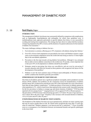
  • 5. 291 Management of Diabetic Foot Charcotā€™s Arthropathy Planter Infected Ulcer Self Inspection Cut the nails straight Take care of Dry skin to see feet in mirror for redness, Swelling, ulcer or crack foot Hammer Toes Foot Care Tinea Interdigitalies Gangrene Bunion with Nail Changes
  • 6. Medicine Update 2012 ļ® Vol. 22 292 are required to confirm these preliminary observations. The management of advanced CN with bone deformity requires reconstructive surgery. Footwear, Orthoses, and Hosiery Inappropriate (Often tight) footwear is a major cause of ulceration. Studies have suggested that specialized foot wear with padded hosiery reduces high foot pressures and gives all- around protection to the high-risk diabetic foot provided that the shoes are fitted to accommodate the padded socks.48 Injected Liquid Silicone A randomized, double-blind trial of injected liquid silicone in the diabetic foot has confirmed that silicone injections were associated with increased soft-tissue thickness under the metatarsal head, decreases foot pressure, and reduced callus formation. Such an ā€œinjectable orthosisā€ might well be beneficial in high-risk patients. Subsequent follow-up studies confirmedthatpatientsatgreatestriskofulcerationweremost likely to benefit from silicone injections but that after two years of follow-up, the benefits of injections, though still demonstrable,werereducedcomparedtobaseline,suggesting that booster injections may periodically be needed. This is an area of ongoing research49, 50 Patient Education Patients need to be informed of the risk of having sensory loss and the need for regular self-inspection, foot hygiene, and podiatry treatment as required. They must also be told what action to take in the event of injury or the discovery of a foot ulcer.51 The Diabetic foot: Need For A Team Ap- proach A number of reports have shown the benefits of the multidisciplinary approach to diabetic foot care. The team might include diabetologists, surgeons (both orthopaedic and vascular), specialist nurses, diabetes educators, podiatrists, orthotists, pedorthotists and patient or a care taker of patient.27 Conclusion Patients with diabetes are at an increased risk for developing foot ulcerations. The consequences of persistent and poorly controlled hyperglycemia lead to neuropathic and vascular abnormalities that cause foot deformities and ulceration. The feet of diabetic patients should be examined at least annually to determine predisposing conditions to ulceration. Treatment plans should be based on examination findings and the individual risk for ulceration. If ulcers are present, the treatment strategy should include offloading, debridement, and appropriate dressings. Further, the presence of infections should be determined by clinical findings and appropriate wound cultures and treated based on the culture results. If evidence for ischemia is present, revascularization may be indicatedtorestorearterialbloodflowandincreasethechance for limb salvage. There are adjunctive therapies available that can also contribute to the overall healing process of the wounds in affected patients. Patient education and team approach towards management plays the key role to words the success. . References 1. Assal JP, et al. Patient education in diabetes. In: Recent Trends in Diabetic Research, Stockholm: Almqvist and Wiksel International 1982:276-89. 2. Thomson FJ, et al. A team approach to diabetic foot care: the Man- chester experience. Foot 1991;1:75-82. 3. Singh N, Armstrong DG, Lipsky BA. Preventing foot ulcer in pa- tients with diabetes. JAMA 2005;293:217-228 4. Boulton AJM, Vileikyte L, Ragnarson-Tennvall G, Apel- qvist J:Ā  The global burden of diabetic foot disease. Lan- cetĀ 2005;Ā 366:1719-1724. 5. Ragnarson- Tennvall G, Apelqvist J. Prevalence of diabetes -re- lated foot ulcers and amputations: a cost-utility analysis based on Markov model simulations. Diabetologia 2001;44:2077-2087. 6. Boulton AJM. The diabetic foot: from art to science: the 18th Ca- millow Golgi lecture. Diabetologia 2004; 47:1343-1353. 7. Williams R, Airey M. The size of the problem: economic aspects of foot problems in diabetes. In: Boulton AJM, Connor H, Cava- nagh PR, eds. The Foot in Diabetes, 3rd edn. Chichester: Wiley, 2000:3-17 8. Pendsey SP, Epidemiological aspects of Diabetic Foot. Int. J Diab. Dev Countries 1994; 14:37-38 9. International consensus on the Diabetic Foot, by the International working group on the Diabetic Foot, 1999. 10. Mohan V, Premlatha G, Shastry NG. Peripheral vascular disease in non-insulin dependent diabetes mellitus in South India; Diab Res Clin Pract 1995; 27:235-240. 11. De silva D. The prevalence of macrovascular disease and lipid ab- normalities amongst diabetic patient in Sri Lanka. Postgrad Med J 1993; 69:557-561 12. Pendsey SP. Peripheral vascular disease (PVD): An Indian Scena- rio, Dibetologia Croatia1998;27-4: 153-156. 13. Pendsey SP. Diabetic Foot Syndrome. RSSDI Text book of Diabe- tes Mellitus, 2nd Edition, 2008;2:959-972. 14. Migdalis IN, et al. peripheral vascular disease in newly diagnosed non insulin dependent diabetes. Int Angiol 1992; 11 : 230-232. 15. Marinelli Mr. Beach KW, Glass MJ et al. Non invasive testing vs, clinical evaluation of arterial disease: a prospective study. J AM Med Assoc 1979;2031. 16. Walters DP. Et al. the prevalence, detection and epidemiological correlates of peripheral vascular disease : a comparison of diabe- tic and non diabetic subjects in an English community. Diab Med 1992; 9: 710-5. 17. Janka H U, StandI E and Mehnert H. Peripheral vascular disease in diabetes mellitus and its relation to cardiovascular risk factor : screening with Doppler ultrasonic technique. Diabetes Care 1980; 3: 207.
  • 7. 293 Management of Diabetic Foot 18. Palumbo PJ, Melton LJ. Peripheral vascular disease and diabetes in M. I. Harris and R. F. Hamman (Eds), Diabetes in America, NIH 1985; publ no.85 ā€“ 1468. Washington:US Government Prin- ting Office, 1985;XVI-21. 19. Joslin EP: The Menace of diabetic gangrene. N Engl J Med 211:16-20, 1934 20. Bowering CK: Diabetic foot ulcers: pathophysiology, assessment, and therapy. Can Fam Phys 47:1007-1016, 2001 21. Dyck PJ, Davies JL, et al : Risk factors for severity of diabetic polyneuropathy. Diabetes Care 1999;22:1479-1486 22. Feldman EL, Russell JW,et al: New insights into the pathogenesis of diabetic neuropathy. Curr Opin Neurol 1999;5:553-563. 23. Huijberts MS et al,: Advanced glycation end products and diabetic foot disease. Diabetes 24. Boulton AJ et al , : Comprehensive foot examination and risk as- sessment. Diabetes Care 2008;31:1679-1685. 25. Zochodone DW: Diabetic polyneuropathy: an update. Curr Opin Neurol 2008;21:527-533. 26. Pendsey S P. Indian Scenario; the Diabetic Foot in complications of Diabetes in Indian Scenarion; Nidus 99 Diabetology Initiative in Diabetology: Proceedings; 1. 27. Andrew J.M.Ā Boulton and David G. Armstrong, The diabetic foot, Clinical Diabetes, Translating research into practice. Edi. Vivian A. Fonseca. Publisher Elsevier, 2006;179-195. 28. Andrew J.M.Ā Boulton, Foot Problems in Patients With Diabetes. Textbook of Diabetes, 4th edition, Editor- Richard I.G. Holt, Cli- ve S. Cockram , Allan Flyvbjerg, Barry J. Goldstein, publisher Wiley-Blackwell, 2010;727-742. 29. Murray HJ, Young MJ, Boulton AJM. The relationship between callus formation, high foot pressures and neuropathy in diabetic foot ulceration. Diabet Med 1996;13:979-982. 30. Khan NA et al , : Does the clinical examination predict lower ex- tremity peripheral arterial disease? JAMA 2006;295:536-546 31. American Diabetes Association: Peripheral arterial disease in people with diabetes. Diabetes Care 2003;26:3333-3341, 32. Armstrong DG et al,: Choosing a practical screening instrument to identify patients at risk for diabetic foot ulceration. Arch Intern Med 1998;158:289-292. 33. Sibbald,Ā RG & Woo,Ā KY. The biology of chronic foot ulcers in persons with diabetes.Ā Diabet Metab Res Rev 2008;Ā 24(Suppl 1):25ā€“30. 34. MĆ¼ller,Ā M, Trocme,Ā et al, Matrix metalloproteinases and diabetic foot ulcers: the ratio of MMP-1 to TIMP-1 is a predictor of wound healing.Ā Diabet MedĀ 2008;Ā 25:419ā€“426. 35. Chantelau,Ā EA, Tanudjaja,Ā T, et al, Antibiotic treatment for un- complicated neuropathic forefoot ulcers in diabetes: a controlled trial.Ā Diabet MedĀ 1996;Ā 26:267ā€“276.68Ā Lipsky,Ā BA. New develop- ments in diagnosing and treating diabetic foot infections.Ā Diabet Metab Res Rev2008;Ā 24(Suppl 1):S66ā€“S71. 36. Lipsky,Ā BA. New developments in diagnosing and treating diabet- ic foot infections.Ā Diabet Metab Res Rev 2008;Ā 24(Suppl 1):S66ā€“ S71. 37. SottoĀ AĀ ,Ā RichardĀ J-LĀ ,Ā JourdanĀ NĀ ,Ā CombescureĀ CĀ ,Ā BouzigesĀ NĀ , LavigneĀ J-PĀ .Miniaturised oligonucleotide arrays: a new tool for discriminating colonisation from infection due to Staphy- lococcus aureusĀ in diabetic foot ulcers. Diabetes CareĀ 2007; 30Ā :2819Ā ā€“Ā 2828. Ā  38. GameĀ FLĀ ,Ā JeffcoateĀ WJĀ .Primarily non-surgical management ofĀ osteomyelitis of the foot in diabetes. DiabetologiaĀ 2008; 51Ā :962Ā ā€“Ā 967.Ā  39. WiemanĀ TJĀ ,Ā SmiellĀ JMĀ ,Ā YachinĀ SĀ .Efļ¬Ā cacy and safety ofĀ a topi- cal gel formulation of recombinant human platelet-derived grow- th factor-Ā BB (Becaplermin) in patients with chronic neuropathic diabetic foot ulcers. Diabetes CareĀ 1998; 21Ā :822Ā ā€“Ā 827. 40. Jeffcoate WJ, Lipsky BA,Ā BerendtĀ AR, CavanaghĀ PRĀ ,Ā BusĀ SAĀ ,Ā Pe- ters EJĀ ,et al. Unresolved issues in the management of ulcers of the foot in diabetes. Diabet MedĀ 2008; 25Ā :1380Ā ā€“1389. 41. HinchliffeĀ RJĀ ,Ā ValkĀ GDĀ ,Ā ApelqvistĀ J,Ā ArmstrongĀ DG, Bakker K,Ā Game FLĀ ,et alĀ .A systematic review of the effectiveness of interventions to enhance the healing of chronic ulcers of the foot in diabetes. DiabetĀ Metab Res RevĀ 2008; 24Ā (Suppl 1):119Ā ā€“Ā 144.Ā  42. ArmstrongĀ DGĀ ,Ā BoultonĀ AJMĀ .Negative pressure wound thera- pyĀ (VAC). In:Ā BoultonĀ AJM,Ā CavanaghĀ PRĀ ,Ā RaymanĀ GĀ , eds.The FootĀ in Diabetes, 4th edn.Ā Chichester: John Wiley & Sons Ltd, 2006:Ā 360Ā ā€“Ā 364.Ā  43. ArmstrongĀ DG,Ā Lavery LA; Diabetic Foot Study Consorti- um.Ā Negative pressure wound therapy after partial diabeticĀ foot amputation: a multicentre randomised controlled trial. Lan- cetĀ 2005;Ā 366:1704-1710. Ā  44. BlumeĀ PA,Ā WaltersĀ J,Ā PayneĀ W,Ā AyalaĀ J,Ā LantisĀ J. Comparison ofĀ negative pressure wound therapy using vacuum-assisted closu- re with advanced moist wound therapy in the treatment of diabetic foot ulcers: a multicentre randomised controlled trial. Diabetes CareĀ 2008; 31:631Ā ā€“Ā 636.Ā  45. BlozikĀ E,Ā SchererĀ MĀ .Skin replacement therapies for diabetic foot ulcers: systematic review and meta- analysis. Diabetes CareĀ 2008; 31:693Ā ā€“Ā 694. 46. RathurĀ HĀ ,Ā BoultonĀ AJMĀ .The neuropathic diabetic foot. Natl Clin Pract Endocrinol MetabĀ 2007; 3:14Ā ā€“Ā 25. Ā  47. JudeĀ EB,Ā Selby PLĀ ,Ā BurgessĀ J,Ā LilleystoneĀ P, Mawer EB,Ā PageĀ SR,Ā et al.Ā Bisphosphonates in the treatment of Char- cot neuroarthropathy: a double-blind randomised controlled trial. DiabetologiaĀ 2001; 44:2032Ā ā€“Ā 2037. Ā  48. Murray HJ, Veves A, Young MJ, et al: Role of experimental socks in the care of the high-risk diabetic foot. A multicentre evaluation study. Diabetes Care 1993;16:1190-1192. 49. Van Schie CHM, Whalley A, Vileikyte L, et al: Efficacy of in- jected liquid silicone in the diabetic foot to reduce risk factors for ulceration: a randomized double-blind placebo controlled trial. Diabetes CareĀ 2000;23:634-638. 50. Van Schie CHM, Whalley A, Vileikyte L, BoultonĀ AJM: Efficacy of injected liquid silicone is related to peak plantar foot pressures in the neuropathic diabetic foot. Wounds 2002;14:26-30. 51. Mason J, Oā€™Keefe C, Mclntosh A, et al: A systematic review of foot ulcer in patient with type 2 diabetics: I. Prevention. Diabeties Med 1999;16:801-812.