SlideShare a Scribd company logo
1 of 10
Download to read offline
GLIA 57:1316–1325 (2009)


MMP-9 Controls Schwann Cell Proliferation and
Phenotypic Remodeling via IGF-1 and ErbB
Receptor-Mediated Activation of MEK/ERK Pathway
SHARMILA CHATTOPADHYAY1,2 AND VERONICA I. SHUBAYEV1,2*
1
  Department of Anesthesiology, University of California, San Diego, California
2
  San Diego VA Healthcare System, La Jolla, California


KEY WORDS                                                          event of NRG-1-erbB and other trophic systems, induces
Schwann cell; EGF; IGF; PDGF; glia; proliferation; nerve           cell cycle arrest as a protective checkpoint mechanism to
injury; mitosis; MMP                                               prevent excessive mitosis (Lloyd et al., 1997; Marshall,
                                                                   1995). Because trophic systems activate ERK to both initi-
                                                                   ate and terminate cell mitosis, identifying their upstream
ABSTRACT
                                                                   modulators is critical to elucidating the mechanisms of
Phenotypic remodeling of Schwann cells is required to
ensure successful regeneration of damaged peripheral               Schwann cell survival after nerve injury.
axons. After nerve damage, Schwann cells produce an over             Metalloproteases (MPs) are extracellular proteases that
100-fold increase in metalloproteinase-9 (MMP-9), and ther-        include related families of matrix metalloproteinases
apy with an MMP inhibitor increases the number of resi-            (MMPs) and a dysintegrin and metalloproteases (ADAMs)
dent (but not infiltrating) cells in injured nerve. Here, we        (Werb, 1997), and regulate activation of trophic systems
demonstrate that MMP-9 regulates proliferation and                 through proteolytic cleavage of ligand and/or exracellular
trophic signaling of Schwann cells. Using in vivo BrdU             domains of their tyrosine kinase receptors (Page-McCaw
incorporation studies of axotomized sciatic nerves of MMP-         et al., 2007). For example, ADAM-17 (or TNF converting
92/2 mice, we found increased Schwann cell mitosis in              enzyme, TACE) is required for processing and subsequent
regenerating (proximal) stump relative to wild-type mice.
Treatment of cultured primary Schwann cells with recombi-
                                                                   nuclear translocation of ErbB4 receptor (Rio et al., 2000;
nant MMP-9 suppressed their growth, mitogenic activity,            Vecchi and Carpenter, 1997), whereas MMP-9 and MMP-
and produced a dose-dependent, biphasic, and selective             12 regulate IGF-1 release from its binding protein in the
activation of ERK1/2, but not JNK and p38 MAPK. MMP-9              CNS (Larsen et al., 2006). Upregulation of MPs has been
induced ERK1/2 signaling in both undifferentiated and              attributed to the pathogenesis of experimental and clini-
differentiated (using dbcAMP) Schwann cells. Using inhibi-         cal peripheral nerve damage (Demestre et al., 2004;
tors to MEK and trophic tyrosine kinase receptors, we              Leppert et al., 1999; Platt et al., 2003; Shubayev and
established that MMP-9 regulates Ras/Raf/MEK—ERK                   Myers, 2002), but their role in Schwann cell survival or
pathways through IGF-1, ErbB, and PDGF receptors. We               regulation of trophic signaling is not well understood.
also report on the early changes of MMP-9 mRNA expres-
                                                                     MMP-9 (or gelatinase B) is an intriguing MMP family
sion (within 24 h) after axotomy. These studies establish
that MMP-9 controls critical trophic signal transduc-              member found in adult nerve only after injury and pre-
tion pathways and phenotypic remodeling of Schwann                 dominantly in Schwann cells (Chattopadhyay et al.,
cells. V 2009 Wiley-Liss, Inc.
            C                                                      2007; Demestre et al., 2004; La Fleur et al., 1996; Shu-
                                                                   bayev et al., 2006; Shubayev and Myers, 2000, 2002).
                                                                   Dominant-negative MMP-9 gene knockout (MMP-92/2)
                                                                   mice demonstrate remarkable protection from peripheral
                            INTRODUCTION                           Wallerian degeneration due to MMP-9 control of myelin
                                                                   protein degradation and macrophage migration into the
  To ensure successful peripheral nerve regeneration,              injured sciatic nerves (Shubayev et al., 2006; Chattopad-
Schwann cells strive to survive through vigorous prolifer-         hyay et al., 2007; Kobayashi et al., 2008). Because MMP
ation, controlled by a well-coordinated network of mito-           inhibition increases the number of resident but not infil-
gens. For example, neuregulins, such as neuregulin-1               trating immune cells at the nerve injury site (Kobayashi
(NRG-1), interact with the tyrosine kinase receptors of
the erbB family to regulate Schwann cell proliferation
(Corfas et al., 2004; Jessen and Mirsky, 2005). Interfer-
                                                                    Grant sponsor: NIH/NINDS; Grant number: R21 NS060307-01; Grant sponsor:
ence with NRG-1-erbB signaling results in excessive                Department of Veterans Affairs (Merit Review Award).
Schwann cell proliferation, as indicated by the increase in          *Correspondence to: Veronica I. Shubayev, MD, Department of Anesthesiology,
5-bromo-2-deoxyuridine (BrdU) incorporation in Schwann             University of California, San Diego, School of Medicine, 9500 Gilman Dr., MTF-
                                                                   447, La Jolla, CA 92093-0629, USA. E-mail: vshubayev@ucsd.edu
cells in injured nerves of transgenic mice expressing                Received 22 July 2008; Accepted 24 December 2008
dominant-negative ErbB4 receptor (Chen et al., 2003).                DOI 10.1002/glia.20851
Sustained activation of the Ras/Raf/MEK extracellular                Published online 19 February 2009 in Wiley InterScience (www.interscience.
signal-regulated kinase (ERK) pathway, a downstream                wiley.com).


V 2009
C        Wiley-Liss, Inc.
MMP-9 CONTROLS SCHWANN MITOSIS AND TROPHIC SIGNALING                                  1317
et al., 2008), we hypothesized that MMPs suppress sur-                       Animals and Surgeries
vival of resident cells (predominantly, Schwann cells in
population).                                                    Adult female Sprague–Dawley rats (N 5 48; 250 g,
   This study aimed to determine the role of MMP-9 in        Harlan Labs, San Diego, CA), adult female FVB.Cg-
Schwann cell mitosis, and establish whether MMP-9            Mmp9tm1Tvu/J mice (MMP-92/2, N 5 10; 20 g), and age-
regulates trophic signaling in Schwann cells. We found       matched female wild-type FVB/NJ mice (WT, N 5 10;
increased BrdU incorporation in the proximal (regener-       20 g, Jackson Labs, Bar Harbor, ME) were used. Ani-
ating) but not distal (degenerating) stumps of axotom-       mals were housed at 22°C under a 12 h light/dark cycle
ized sciatic nerves of MMP-92/2 mice. Treatment of           with ad libitum access to food and water. FVB.Cg-
cultured primary Schwann cells with exogenous MMP-9          Mmp9tm1Tvu/J originated on a B6;129 background was
suppressed BrdU incorporation and induced the Ras/           mated to Black Swiss mice for an unknown number of
Raf/MEK–ERK pathway via IGF-1, an ErbB and PDGF              generations and crossed to FVB/N mice for five genera-
tyrosine kinase receptors. This study established that       tions before being made homozygous. Anesthesia was
MMP-9 activates critical trophic systems in Schwann          achieved with 4% isofluorane (IsoSol; Vedco, St Joseph,
cells and signals to suppress Schwann cell mitosis           MO). The rat or mouse sciatic nerve was exposed unilat-
in vivo and in vitro, and pointed at differential roles of   erally at the midthigh level, and transected to produce a
MMP-9 in the processes of peripheral nerve regenera-         sciatic nerve axotomy. Animals were sacrificed using in-
tion and degeneration.                                       traperitoneal injection of a deep anesthesia cocktail of
                                                             pentobarbital (Nembutal, 50 mg/mL; Abbott Labs, North
                                                             Chicago, IL), diazepam (5 mg/mL, Steris Labs, Phoenix,
                                                             AZ), and saline (0.9%, Steris Labs), followed by lethal in-
            MATERIALS AND METHODS                            tracardiac injection of Euthasol (Virbac, Fort Worth, TX,
                   Reagents                                  100–150 mg/kg). Nerve sections proximal and distal to
                                                             transection were collected for analysis at 10 min—4
  Reagents used were as follows: Dulbecco’s modified          days after axotomy. Contralateral to injury and sham-
Eagle’s medium (DMEM) and DMEM Ham’s F12 (Gibco),            operated (unilaterally exposed) nerves were collected for
poly-D-lysine hydrobromide (PDL, Sigma), forskolin           controls. Animal protocols were approved by the VA
(Calbiochem), cytosine-D-arabino-furanoside (AraC), anti-    Healthcare System Committee on Animal Research, and
Thy1.1 antibody and rabbit complement from Sigma, fetal      conform to the NIH Guidelines for Animal Use.
bovine serum (FBS, Hyclone), 5-bromo-2-deoxyuridine
(BrdU, Calbiochem), bovine pituitary extract (Clonetics),
N2 supplement (Gibco), 6,O20 -dibutyryl adenosine 30 ,50 -           In Vivo BrdU Labeling and Detection
monophosphate (dibutyryl cyclic AMP, dbcAMP, Sigma),
nuclear stain 40 -6-diamidino-2-phenylindole (DAPI, Mo-        In vivo BrdU incorporation studies in sciatic nerve
lecular Probes, 1:20,000). Bovine serum albumin (BSA,        were done as published (Cheng and Zochodne, 2002).
100 lg/mL, Sigma), recombinant rat tumor necrosis factor     FVB/MMP-92/2 (N 5 5/group) or wild-type FVB/NJ mice
alpha (TNF-a, 10 ng/mL, R&D), lipopolysaccharide (LPS,       (N 5 5/group) underwent axotomy. BrdU (100 mg/kg) or
100 ng/mL, Sigma), recombinant murine 7S nerve growth        vehicle (1 mM Tris, 0.8% NaCl, 0.25 mM EDTA, pH 7.4)
factor (NGF, 100 ng/mL, Invitrogen), recombinant human       was injected intraperitoneally 4 and 2 h before sacrifice.
neuregulin 1 (NRG-1, 10 ng/mL, R&D Systems), and             Four days after axotomy, animals were perfused with 4%
recombinant human active MMP-9 (rhMMP-9, Calbio-             PFA and sacrificed, as described above. Sciatic nerves
chem). Polyclonal anti-phospho- and total ERK, JNK and       were isolated, postfixed in 4% PFA overnight, rinsed, cry-
p38 were all obtained from Cell Signaling (Danvers, MA),     oprotected in graded sucrose, embedded into OCT com-
polyclonal anti-MMP-9 (Chemicon), polyclonal anti-S100       pound in liquid N2, and cut into 10-lm-thick transverse
(Dako, Carpinteria, CA), rabbit anti-myelin protein zero     sections. For BrdU detection, the sections were rinsed in
(P0, Protein Tech Group, IL) and mouse anti-b-actin          PBS, hydrolyzed in 2 N HCl in PBS for 30 min, digested
(Sigma).                                                     with 0.01% Trypsin for 30 min at 37°C, and washed with
  Pharmacologic Inhibitors were obtained from Calbio-        PBS. Nonspecific binding was blocked with 10% normal
chem and their concentrations are selected based on          goat serum. Mouse anti-BrdU antibody (Sigma, 1:1000)
respective IC50 values and previous use in similar           was applied for 2 h at 37°C, followed with a PBS rinse and
experiments: ErbB1/2/4 receptor inhibitor (ErbB-I,           goat antimouse Alexa 488 antibody (Invitrogen) treat-
10 lM), ErbB2 receptor inhibitor (ErbB2-I, 10–50 lM),        ment for 1 h at RT.
PDGF receptor tyrosine kinase inhibitor (AG 1296,
10 lM), IGF-1R inhibitor PPP (IGF1R-I, 10 lM), MEK1
inhibitor (PD98059, 10 lM), MEK1/2 inhibitor (U0126,         Morphometry
10 lM), and PI3K inhibitor (LY294002, 50 lM). The
broad-spectrum MMP inhibitor, GM6001 (Ilomastat,               DAPI- and BrdU-positive profiles were quantified in
10 lM, Chemicon), has Ki of 0.4 nM for MMP-1, 27 nM          transverse sham and axotomized proximal and distal
for MMP-3, 0.5 nM for MMP-2, 0.1 nM for MMP-8, and           sciatic nerve sections at objective magnification 340 in
0.2 nM for MMP-9.                                            four mice per group, two sections per animal, three ran-

                                                                                                                   GLIA
1318                                        CHATTOPADHYAY AND SHUBAYEV

domly selected fields per section by a blinded experi-         tistical analyses were done by ANOVA and Tukey–
menter using Openlab 4.0 software (Improvision) fol-          Kramer post-hoc test using SPSS 16.0 software.
lowed by statistical analyses (SPSS 16.0 software).

                                                                                  Western Blotting
                Schwann Cell Cultures
                                                                Sciatic nerves were isolated, snap-frozen in liquid N2,
   Primary Schwann cells were cultured as described           and stored at 280°C. Proteins were extracted using lysis
(Brockes et al., 1979). Briefly, sciatic nerves of postnatal   buffer (50 mM Tris–HCl, pH 7.4, 1% NP 40, 150 mM
day 1 Sprague2Dawley rats (Harlan Labs) were isolated         NaCl, 1 mM EDTA, 1 mM PMSF, 1 lg/mL aprotinin and
and purified using AraC, an anti-fibronectin Thy1.1             leupeptin, 1 mM sodium orthovanadate). Schwann cells
antibody, and rabbit complement (Shubayev et al.,             were lysed in a buffer containing 10 mM Tris–HCl, 1%
2006). Schwann cell purity was confirmed by over 99%           NP-40, 0.1% sodium deoxycholate, 1 mM EDTA, 2 mM
pure S-100-positive profiles. Schwann cells were plated        sodium orthovanadate, 10 mM sodium fluoride, and 10 g/
on PDL-coated dishes in DMEM containing 10% FBS,              mL aprotinin (Chattopadhyay et al., 2006). Total protein
100 units/mL penicillin and 100 lg/mL streptomycin,           was measured using Pierce BCA Protein Assay. Samples
21 lg/mL bovine pituitary extract and 4 lM forskolin          containing equal amounts of protein (10 lg for cells and
(referred to as ‘‘complete media’’) at 37°C under humidi-     30–50 lg for tissues) were run on SDS-PAGE and trans-
fied 5.0% CO2. Cells were passaged upon confluency and          ferred to Immobilon-P (Millipore, Bedford, MA) in Tris–
used at three to seven passages. Schwann cell differen-       Glycine transfer buffer (Invitrogen) at 200 mA for 1 h.
tiation was induced with 500 lM dbcAMP for 48 h. Suc-         The membranes were blocked with 5% nonfat milk
cessful differentiation was confirmed by myelin protein        (Biorad), incubated with a primary antibody (identified
zero (P0) expression. For inhibition studies, cells were      above) in 5% BSA in TBS overnight at 4°C, washed in
maintained in DMEM containing 0.5% FBS for 16 h,              TBS containing 0.05% Tween 20, and incubated for 1 h at
washed, and inhibitors (specified above) were added            RT with HRP-conjugated antirabbit or antimouse second-
15 min before rhMMP-9 treatment for 6 h.                      ary antibody (Cell Signaling; 1:10,000). The blots were
                                                              developed using ECL (Amersham), followed by densitom-
                                                              etry with NIH Image J 1.38 software. All blots represent
                Growth Kinetics Assay                         at least three independent in vitro experiments or N 5 3
                                                              animals per group.
   Kinetics of Schwann cell growth was determined using
crystal violet staining. Cells were plated in triplicate at
1 3 104 cells/well in a 24-well plate and allowed to grow                         Real-Time qPCR
in complete medium with or without 100 nM rhMMP-9,
then fixed with 1% glutaraldehyde for 20 min and                 Primers and Taqman probes for rat MMP-9 (Biosearch
stained by 0.1% crystal violet for 45 min at room tem-        Technologies, Novato, CA) were optimized as described
perature. Unbound dye was washed away with water,             (Shubayev et al., 2006). Nerve samples were stored in
while bound dye was eluted with 10% acetic acid and           RNA-later (Ambion) at 220°C. RNA was extracted from
the absorbance measured at 590 nm. The value of rela-         sciatic nerves and cell lysates with Trizol (Invitrogen) and
tive increase of absorbance versus time 6 S.D. was plot-      treated with RNAse-free DNAse (Qiagen). The RNA purity
ted for each time-point relative to absorbance values of      was verified by OD260/280 absorption ratio of 2.0. cDNA
cells attached overnight (representing a 0 h time-point).     was synthesized using a SuperScript first-strand RT-PCR
                                                              kit (Invitrogen). Gene expression was measured by quanti-
                                                              tative real-time qPCR (MX4000, Stratagene, La Jolla, CA)
            BrdU Cell Proliferation Assay                     using 50 ng of cDNA and 23 Taqman Universal PCR Mas-
                                                              ter Mix (Applied Biosystems) with a one-step program:
   Cells were plated in 96-well PDL-coated plates at 2 3      95°C for 10 min, 95°C for 30 s, and 60°C for 1 min for
105 cells/mL in 1% FBS DMEM overnight. The inhibi-            50 cycles. Duplicate samples without cDNA (no-template
tors were applied for 30 min, followed by treatment with      control) showed no contaminating DNA. Glyceraldehyde
100 nM rhMMP-9 and BrdU labeling for 8 h. Newly               3-phosphate dehydrogenase (GAPDH) was used as a nor-
synthesized DNA was detected using the BrdU Cell Pro-         malizer gene. Relative mRNA levels were quantified using
liferation Assay (Calbiochem), following the manufac-         the comparative Ct method (Livak and Schmittgen, 2001).
turer’s protocol for cell denaturation, mouse anti-BrdU       A fold change was determined by the MX4000 software
antibody, and HRP-tagged goat antimouse IgG applica-          using the described methods (Pfaffl, 2001).
tions. Chromogenic tetramethylbenzidine substrate was
applied for 15 min and the reaction was stopped with
2.5 N sulfuric acid. Absorbance was measured at 450–                           Immunofluorescence
595 nm using a Spectramax plate reader (Molecular
Devices, Downingtown, PA). All samples were analyzed            Nerves were perfused and postfixed in 4% PFA over-
in quadruplicate in three independent experiments. Sta-       night, rinsed, cryoprotected in graded sucrose, embedded

GLIA
MMP-9 CONTROLS SCHWANN MITOSIS AND TROPHIC SIGNALING                                     1319
                                                                    This data suggests that Schwann cells produce MMP-9
                                                                    after proinflammatory but not trophic stimulation.


                                                                         MMP-9 Gene Deletion Promotes Schwann
                                                                                  Cell Mitosis In Vivo

                                                                       Sciatic nerve axotomy was used to spatially separate
                                                                    events of Wallerian degeneration (distal stump) and
                                                                    regeneration (proximal stump). Nerves were analyzed at
                                                                    4 days post-injury, when Schwann cell proliferation
                                                                    occurs in wild-type nerve injury models (Cheng and
                                                                    Zochodne, 2002; Clemence et al., 1989) and when contri-
                                                                    bution of infiltrating immune cells in MMP-92/2 mice
                                                                    is minimal (Shubayev et al., 2006). A statistically signifi-
                                                                    cant increase in DAPI-positive profiles was observed in
  Fig. 1. MMP-9 expression in primary Schwann cells. Taqman qPCR    the proximal but not distal nerve stumps of MMP-92/2
for MMP-9 in primary Schwann cells after 24 h of stimulation with   compared with wild-type mice (Fig. 2A). In fact, the dis-
BSA, TNF-a, NGF, LPS, or NRG-1. Expressed as mRNA fold increase
compared with low serum media, using GAPDH as normalizer. Data      tal stump demonstrated a 17% decline in cell number in
represents the mean 6 SEM of N 5 4/group, by one-way ANOVA and      knockout versus wild-type mice that was not quite stat-
Tukey–Kramer post-hoc test (*P  0.05; **P  0.01).
                                                                    istically significant (P 5 0.0594).
                                                                       In vivo BrdU incorporation studies in axotomized
into OCT compound in liquid N2, and cut into 10-lm-                 MMP-92/2 mice were performed to assess the role of
thick transverse sections. Cells were fixed with 4% PFA              MMP-9 in cell mitosis. A 2.1-fold increase in cell prolif-
in TBS for 10 min, washed with TBS, and permeabilized               eration was found in proximal nerve stumps of MMP-
with 0.1% Triton X in TBS. Nonspecific binding was                   92/2 compared with wild-type mice (Fig. 2B), whereas
blocked with 10% goat serum. In tissue sections, endoge-            no difference was observed in the distal stump. BrdU-
nous aldehydes were blocked with 0.5% sodium borohy-                positive cells colocalized with S100, a phenotypic marker
dride in 1% dibasic sodium phosphate for 5 min and                  for Schwann cells, suggesting that MMP-9 suppresses
Dako antigen retrieval (Carpinteria, CA) was applied for            Schwann cell proliferation in vivo.
5 min at 95°C, then for 20 min at RT. Primary antibod-
ies were diluted in TBS containing 1% FBS and applied
sequentially, the first primary antibody for 1 h at RT,                MMP-9 Activates ERK1/2 and Reduces Growth
then rinsed, followed by Alexa 564 conjugated (red) first                      of Primary Schwann Cells
secondary goat antibody for 1 h at RT and second pri-
mary antibody application overnight at 4°C. Slides were                Because ERK MAPK signals cell cycle arrest in
rinsed in PBS containing 0.1% Tween 20 and incubated                Schwann cells (Harrisingh et al., 2004; Lloyd et al.,
with second Alexa 488 conjugated (green) secondary                  1997), we studied the effect of rhMMP-9 on ERK1/2 acti-
goat antibody for 1 h at 22°C. DAPI (1:20,000) was                  vation, and its correlation to Schwann cell growth.
applied for 5 min. Replacement of primary antibody                  rhMMP-9 produced a dose-dependent activation of
with the respective normal IgG was done to control sig-             ERK1/2 (Fig. 3A). Based on this data, rhMMP-9 of 100
nal specificity. Imaging was performed using a Leica                 nM was selected for subsequent experiments. Temporal
DMR bright-light and fluorescence microscope using                   analyses of rhMMP-9 effect in Schwann cells demon-
Openlab 4.0 software (Improvision). All micrographs                 strated biphasic pERK1/2 activation with acute (15 min)
represent at least three independent in vitro experi-               and sustained (3212 h) phases (Fig. 3B). Immunofluo-
ments and N 5 3 animals per group.                                  rescence for pERK1/2 (Fig. 3C) demonstrates its cyto-
                                                                    solic distribution at 15 min and 6 h after rhMMP-9
                                                                    stimulation, consistent with its phosphorylated state in
                   RESULTS                                          Schwann cell lysates seen by western blot.
      MMP-9 Is Induced in Schwann Cells by                             The effect of rhMMP-9 on ERK1/2 activation was cor-
     Proinflammatory but Not Trophic Factors                         related to Schwann cell growth kinetics. A significantly
                                                                    reduced Schwann cell growth was observed after daily
  To identify the stimuli for MMP-9 mRNA expression,                rhMMP-9 treatment over a course of 72 h (Fig. 3D).
primary Schwann cells were treated with TNF-a, LPS,                    To test whether MMP-9-induced ERK1/2 activation
NGF, or NRG-1. MMP-9 mRNA expression was signifi-                    depends on the state of Schwann cell differentiation, the
cantly induced 100-fold by LPS and 160-fold by TNF-a,               latter was stimulated with dibutyryl cyclic AMP
whereas the changes in NGF and NRG-1 were not sig-                  (dbcAMP) for 48 h, as suggested (Harrisingh et al.,
nificant (see Fig. 1). BSA, used as a control TNF-a car-             2004), followed by treatment with rhMMP-9 (Fig. 4A).
rier, increased MMP-9 mRNA by 14-fold, consistent with              Successful differentiation was confirmed by the expres-
our observations in nerve (Chattopadhyay et al., 2007).             sion of myelin protein zero (P0). Biphasic activation of

                                                                                                                          GLIA
1320                                                 CHATTOPADHYAY AND SHUBAYEV


  Fig. 2. Schwann cell proliferation in axotomized nerves of MMP-         tal sciatic nerve stumps 4 days after axotomy. Mean 6 SEM 1,000 lm2
92/2 knockout mice. A, DAPI profiles (blue) in proximal and distal sci-    endoneurial area, N 5 5/group, two sections/N, three areas/section at
atic nerve stumps 4 days after axotomy. Mean 6 SEM per 100 lm2            objective magnification 340 (scale bar 5 50 lm), by unpaired Student’s
endoneurial area, N 5 4/group, two sections/N, three areas/section, by    t-test (*P  0.05). Dual-immunofluorescence for BrdU (green) and S100
unpaired Student’s t-test (*P  0.05). Objective magnification 340         (Schwann cell marker, red) in MMP-92/2 nerves (scale bar 5 20 lm).
(scale bar 5 50 lm). B, BrdU incorporation (green) in proximal and dis-


pERK, peaking at 15 min and 6 h of rhMMP-9 stimula-                       from 30 min to 8 days of axotomy in both stumps (Sheu
tion in myelinating Schwann cells (Fig. 4B), was consist-                 et al., 2000), we established its initiation within 15 min
ent with the findings in undifferentiated cells.                           of axotomy in both distal and proximal stumps (Fig. 5C).
                                                                             Early changes in MMP-9 mRNA expression after sci-
                                                                          atic nerve axotomy have not been reported. Using Taq-
                                                                          man qPCR (Fig. 5D), we found no significant change in
           Correlation of MMP-9 and ERK1/2
                                                                          MMP-9 mRNA at 10 min (i.e., preceding ERK activa-
             Expression in Injured Nerve
                                                                          tion), a 3.5-fold induction at 1 h in the distal stump, and
                                                                          a 70-fold increase in distal and a 146-fold increase in
  Exogenous MMP-9-PEX can activate ERK1/2 in
                                                                          proximal stumps at 1 day postaxotomy, representing
injured sciatic nerve (Mantuano et al., 2008). Here, we
                                                                          about two-fold higher MMP-9 expression level in proxi-
correlated the patterns of endogenous MMP-9 expression
                                                                          mal versus distal stumps.
and ERK1/2 activation after axotomy, with the focus on
immediate changes. At 24 h after nerve injury, both
MMP-9 and pERK are coordinately expressed in distal
and proximal stumps of axotomized sciatic nerves (Fig.                    MMP-9 Activates MEK/ERK1/2 Pathway via ErbB,
5A), colocalizing in Schwann cells, as determined by a                     IGF-1, and PDGF Tyrosine Kinase Receptors
characteristic crescent morphology (Fig. 5B). There was
no visible difference between the stumps observed and,                      MMPs activate trophic signaling in various cells (Page-
thus, only one representative micrograph is shown (Fig.                   McCaw et al., 2007). We analyzed whether MMP-9
5B). While ERK1/2 activation has been shown to sustain                    induced ERK1/2 signaling by activation of trophic tyro-

GLIA
MMP-9 CONTROLS SCHWANN MITOSIS AND TROPHIC SIGNALING                                                 1321


  Fig. 3. MMP-9 activates ERK1/2 and suppresses Schwann cell           objective magnification 3100 (representative micrographs of 3 inde-
growth. A, MMP-9 activates ERK1/2 in a dose-dependent manner. 100      pendent experiments). D, Schwann cell growth curve studies using
nM rhMMP-9 was selected for use in the subsequent experiments. B, A    crystal violet, with and without daily rhMMP-9 stimulation for 72 h.
time-course of rhMMP-9 treatment demonstrating a biphasic activation   Data represents the mean 6 SD of N 5 3/group, analyzed by one-way
of ERK1/2. C, Immunofluorescence for pERK (green) and DAPI (blue)       ANOVA (*P  0.05).
confirms the biphasic reactivity of cytosolic pERK at 15 min and 6 h,


  Fig. 4. rhMMP-9 stimulates biphasic ERK1/2 activation in myelinat-   firmed by myelin protein zero (P0) expression; b-actin was used as load-
ing Schwann cells. Schwann cell differentiation was induced with       ing control. B, An extended time-course of rhMMP-9 stimulation dis-
dbcAMP (500 lM) for 48 h, followed by treatment with rhMMP-9 (100      played biphasic activation of ERK1/2, as seen in undifferentiated cells
nM) for 15 min. A, rhMMP-9 stimulates transient ERK1/2 activation      in Fig. 3.
over a 1 h period. Successful Schwann cell differentiation was con-


sine kinase receptors involved in regulation of Schwann                PDK/MEK pathway (see Fig. 8). GM6001 (10 lM), a
cell mitosis, including ErbB (Corfas et al., 2004), PDGF               broad-spectrum MMP inhibitor, reduced MMP-9 effect.
and IGF-1 receptors (Delaney et al., 1999; Meier et al.,                  To evaluate selectivity of rhMMP-9-induced ERK1/2
1999), and/or the MEK/ERK pathway (Fig. 6A). The re-                   activation, we analyzed the changes in JNK or p38 at 6
sults are summarized in a schematic diagram (see Fig. 8).              h of rhMMP-9 stimulation (Fig. 6B). No change in phos-
Focus on sustained and not transient ERK1/2 activation                 pho-JNK or phospho-p38 activation was observed. UV
(i.e., 6 h after MMP-9 stimulation) is based on its role in            (250 J/m2) stimulation for 15 min was used as positive
suppression of cell mitosis (Lloyd et al., 1997; Marshall,             controls for p38 and JNK activation, and NRG-1 (10 ng/
1995).                                                                 mL) as positive controls for ERK1/2 activation. Note
   MMP-9-stimulated pERK1/2 levels declined after                      that pretreatment with GM6001 (a broad-spectrum
treatment with general ErbB1/2/4 inhibitor by 49%, but                 MMP inhibitor) stimulated JNK or p38 MAPK activa-
not the specific ErbB2 inhibitor. PDGF receptor inhibitor               tion above MMP-9 or basal levels.
(PDGFR-I) blocked MMP-9-stimulated pERK activation
by 36%, while IGF-1 receptor inhibitor (IGF1R-I) virtu-
ally ablated it. MEK (PD98059) and MEK1/2 (U0126)                            ErbB Inhibition Reversed MMP-9-Induced
inhibitors reduced MMP-9-stimulated pERK increase by                           Suppression of Schwann Cell Mitosis
79 and 93%, respectively, while control PI3K inhibitor
(LY294002) produced little effect. The latter also indi-                 Using an in vitro BrdU incorporation assay, we
cates that MMP-9 does not activate ERK via the PI3K/                   assessed rhMMP-9 effect on Schwann cell mitosis and

                                                                                                                                        GLIA
1322                                                    CHATTOPADHYAY AND SHUBAYEV


  Fig. 5. Endogenous ERK1/2 and MMP-9 in axotomized rat sciatic               activation in distal and proximal stumps at 15 min, 1 h, and 1 day after
nerve. A, Western blot for ERK1/2 and MMP-9 in the proximal (P) and           axotomy, relative to sham (sh) and contralateral (c) nerves. Duplicate
distal (D) stumps 1 day after rat sciatic nerve axotomy relative to contra-   representative of N 5 4/group. D, Real-time Taqman qPCR for MMP-9 in
lateral (C) nerves; representative of N 5 4/group. B, MMP-9 (red) and         axomotized rat nerves normalized to GAPDH and calibrated to sham.
pERK (green) colocalize in Schwann cells of a proximal stump, a repre-        Data represents the mean 6 SEM of N 5 4/group, by one-way ANOVA
sentative micrograph of N 5 3 (scale bar 5 20 lm). C, Sustained ERK1/2        and Tukey–Kramer post-hoc test (*P  0.05; **P  0.01).


  Fig. 6. rhMMP9 activates MEK–ERK pathway via IGF-1, ErbB,                   (GM6001, 10 lM). MMP-9-induced ERK activation was inhibited by
and PDGF receptors. A, Western blot for ERK1/2 in primary Schwann             MEK, ErbB1/2/4, IGF-1R, and PDGFR, but not ErbB2 or PI3K inhibi-
cells 6 h after 100 nM rhMMP-9 stimulation, with or without 15 min of         tors. B, Western blot for MAPKs of Schwann cells lysates 6 h after
pretreatment with the inhibitors to ErbB1/2/4 receptor (ErbB-I, 10 lM),       treatment with 100 nM rhMMP-9. MMP-9 produced no effect on activa-
ErbB2 receptor (ErbB2-I, 10–50 lM), PDGF receptor (PDGFR-I, 10                tion of p38 and JNK, whereas pretreatment with GM6001 induced it.
lM), IGF1 receptor (IGFR-I, 10 lM), MEK (PD98059, 10 lM), MEK1/2              b-actin was used as loading control. Data is representative of three in-
(U0126, 10 lM), PI3K (LY294002, 50 lM), and MMP inhibitor                     dependent experiments.


its relationship to an MMP-9-dependent ErbB trophic                           relative to low-serum media. Both ErbB-I and control
pathway (see Fig. 7) that participates in suppression                         MMPi, GM6001, reversed antimitogenic action of MMP-
of Schwann cell proliferation (Chen et al., 2003).                            9. GM6001 treatment without MMP-9 stimulation fur-
rhMMP-9 stimulation inhibited Schwann cell mitosis                            ther promoted mitosis. PI3K inhibitor (LY294002) pro-

GLIA
MMP-9 CONTROLS SCHWANN MITOSIS AND TROPHIC SIGNALING                                                1323


  Fig. 7. MMP-9 inhibits Schwann cell proliferation in vitro. BrdU
incorporation is measured 8 h after treatment with 100 nM rhMMP-9.
The GM6001 (50 lM), ErbB-I (10 lM), or LY294002 (50 lM) were
applied 15 min before stimulation with rhMMP-9. Complete media con-
taining 10% FBS and promitotic bovine pituitary extract were used as
a positive control. Media containing 1% FBS was used for all experi-
mental treatments. Data shown represents the mean 6 SEM of three
independent experiments performed in quadruplicate, analyzed by one-
way ANOVA and Tukey–Kramer post-hoc test (**P  0.05, *P  0.01).


duced no effect on rhMMP-9-stimulated reduction in
mitosis.                                                                  Fig. 8. MMP-9 activation of trophic signaling in Schwann cells (a
                                                                       schematic diagram). MMP-9 expression is induced by proinflammatory
                                                                       stimuli (see Fig. 1). MMP-9 stimulates MAPK p44/42 (or ERK1/2) sig-
                                                                       naling in Schwann cells via activation of IGF-1, ErbB4, and PDGF tyro-
                                                                       sine kinase receptor and Ras/Raf/MEK pathway (shown in black), but
                         DISCUSSION                                    not PI3K/PDK/MEK pathway (shown in grey), as determined using the
                                                                       specified pharmacologic inhibitors (see Fig. 6).
   These data are the first to implicate MMP-9 in regula-
tion of Schwann cell proliferation or trophic signaling.
We find that MMP-9 can activate the Ras/Raf/MEK–                           Supporting data for MMP-mediated signaling by
ERK1/2 signal transduction pathway via ErbB, IGF-1,                    direct receptor binding is only surfacing. A recent study
and PDGF tyrosine kinase receptors, as summarized in                   suggests that hemopexin (substrate-binding) MMP-9
Fig. 8. The exact mechanisms need to be clarified, as                   domain fused with a GST protein (GST-MMP-9-PEX)
MMPs control cell signaling via regulatory proteolysis of              activates ERK1/2 in Schwann cells via low-density lipo-
latent signaling factors localized in the extracellular                protein receptor-related protein 1 (LRP-1) (Mantuano
matrix or by (nonproteolytic) direct receptor binding.                 et al., 2008), a hybrid scavenger, and signaling receptor
   Evidence for proteolytic MMP function in activation of              (Herz and Strickland, 2001). Although the study pro-
trophic systems is sound. For example, release of trophic              vides no evidence for its direct LRP-1 binding, GST-
factors from their regulatory proteins depends on cata-                MMP-9-PEX contains a binding site for LRP-1, among
lytic activity of MMPs (Page-McCaw et al., 2007), such                 other surface receptors and substrates (Burg-Roderfeld
as IGF-1 release from IGF binding protein, IGFBP-6 in                  et al., 2007; Roeb et al., 2002). Consistently, GST-MMP-
CNS (Larsen et al., 2006). IGF-1 can stimulate MMP-                    9-PEX was sufficient to activate ERK in other cells
mediated release of the EGF ligand from its heparin-                   (Dufour et al., 2008). Thus, MMP-9 utilizes several
bound form (HB-EGF), leading to cumulative transacti-                  Schwann cell receptor systems to activate ERK signal-
vation of its own and EGF receptors and Ras/Raf/MEK                    ing, including trophic tyrosine kinase and other signal-
signaling (El-Shewy et al., 2004; Roudabush et al.,                    ing receptors, such as LRP-1. Because proteolytic and
2000). In other cells, MMP-9 controls ERK1/2 via activa-               receptor agonist MMP actions are not mutually exclu-
tion via the EGF receptor (Roelle et al., 2003). Of the                sive, either or both potentially relate to the trophic
EGF receptor family, Schwann cells express ErbB2, 3,                   systems. But MMPs intrinsically lacking the hemopexin
and 4 (Corfas et al., 2004) and ErbB2 is dispensable in                domain, such as MMP-7, are potent inducers of trophic
Schwann cell survival after nerve injury (Atanasoski                   signaling, including that of IGF, EGF, and ErbB (Ii
et al., 2006). Considering that ErbB3 has no functional                et al., 2006; Sanderson et al., 2006), presumably through
kinase domain (Pearson and Carroll, 2004) and ErbB2-I                  a proteolytic mechanism.
was ineffective in blocking MMP-9-induced ERK1/2 acti-                    MMP-9-induced activation of trophic signaling and sup-
vation, the effects of the general ErbB-I seen here is                 pression of Schwann cell mitosis are independent find-
likely to result from ErbB4 block. Proteolytic processing              ings. Their relationship was evidenced by ErbB receptor
of ErbB4 and its subsequent nuclear translocation                      block of MMP-9-induced Schwann cell mitosis. ErbB42/2
depend on metalloproteases (Vecchi and Carpenter,                      in Schwann cells can lead to excessive mitosis (Chen
1997), identified in various cells as ADAM-17, MMP-3,                   et al., 2003). Because state of Schwann cell differentiation
MMP-7, and MMP-9 (Dempsey et al., 2002; Ii et al.,                     and microenvironment influence the functional outcome
2006; Rio et al., 2000; Sanderson et al., 2006).                       of NRG-1/ErbB system action (Corfas et al., 2004; Jessen

                                                                                                                                       GLIA
1324                                          CHATTOPADHYAY AND SHUBAYEV

and Mirsky, 2005) and MMP-9 stimulates ERK1/2 in un-            they induce MMP-9 expression in denervated Schwann
differentiated and differentiated (myelinating) Schwann         cells within 1 h after axotomy. We have already demon-
cells, it is important to establish how propensity to           strated an over 200-fold increase in MMP-9 mRNA by
myelinate and axonal contact affect the outcomes of             6 h of sciatic nerve damage (Shubayev et al., 2006).
MMP-9-induced ERK1/2 activation. In our study, it corre-        Cytokines induce MMP-9 mRNA in nerves to promote
lates with reduced Schwann cell growth and suppressed           neuroinflammatory remodeling (Chattopadhyay et al.,
mitosis, consistent with its role in prodifferentiating func-   2007; Shubayev et al., 2006). Thus, induction of MMP-9
tions of migration (Mantuano et al., 2008) and myelin pro-      mRNA in response to proinflammatory (LPS and TNF-
tein maintenance (Kobayashi et al., 2008).                      a), but not trophic (NGF and NRG-1) stimuli in cultured
   MMP-9 selectively activates ERK but not p38 or JNK           Schwann cells is consistent with this earlier developed
signaling in Schwann cells, as seen in other cells (Roelle      paradigm. rhMMP-9-induced ERK activation seen
et al., 2003). Because growth arrest and suppression of         in vitro correlates with MMP-9 ability to activate ERK
mitosis is signaled through sustained and not transient         in injured sciatic nerve (Mantuano et al., 2008). But
Ras/Raf/ERK activation (Lloyd et al., 1997; Marshall,           because ERK1/2 activation precedes endogenous MMP-9
1995), sustained ERK activation (i.e., 6 h after MMP-9          expression, we suggest that MMP-9 is not the initial
stimulation) was the focus of this study. It will be impor-     stimulus to ERK1/2 activation after axotomy. This is not
tant to determine the mechanisms of MMP-9 induced               surprising given that ERK1/2 signals for a plethora of
transient (15 min) ERK1/2 activation or ability of MMP-         cytokines and trophic factors after nerve damage (Ji and
9 to induce p38 or JNK pathways in Schwann cells at             Woolf, 2001). Moreover, our results do not rule out the
other time-points in future studies. Interestingly,             possibility that ERK signaling is utilized (e.g. by cyto-
GM6001 (specific and broad-spectrum MMP inhibitor)               kines) to induce MMP-9, as found in cortical astrocytes
activated JNK and p38 and stimulated in vitro BrdU              (Arai et al., 2003).
incorporation above basal levels, implicating endogenous           In conclusion, MMP-9 emerges as a potent modulator
MMPs in suppressing these signaling pathways and                of Schwann cell signaling and phenotypic remodeling
Schwann cell mitosis. For example, MMP-3 can generate           after nerve injury. It suppresses Schwann cell mitosis
antimitogenic fibronectin fragments in Schwann cells             and supports functions of differentiation, such as migra-
(Muir and Manthorpe, 1992).                                     tion and myelin protein maintenance (Kobayashi et al.,
   Increased Schwann cell mitosis in axotomized MMP-            2008).
92/2 nerves was consistent with antimitogenic proper-
ties of MMP-9 in primary Schwann cells (both were
determined by BrdU incorporation). While other cells                               ACKNOWLEDGMENTS
types might have contributed to the increased number
of mitotic cells, neuronal cell bodies were excluded from         We thank Jennifer Dolkas and Julie Janes for techni-
analyses and MMP-92/2 nerves were deficient in mac-              cal assistance and Amber Millen for help in editing the
rophages (Shubayev et al., 2006). An interesting finding         manuscript.
is that excessive mitosis was uncompensated only in the
proximal (regenerating) but not distal (degenerating)
stump of axotomized MMP-92/2 nerves. The failure                                          REFERENCES
of distal MMP-92/2 nerves to recruit macrophages
                                                                Arai K, Lee SR, Lo EH. 2003. Essential role for ERK mitogen-activated
(Shubayev et al., 2006) is consistent with the diminished         protein kinase in matrix metalloproteinase-9 regulation in rat cortical
contribution of macrophage-released factors to promote            astrocytes. Glia 43:254–264.
Schwann cell mitosis (Baichwal et al., 1988). Besides,          Atanasoski S, Scherer SS, Sirkowski E, Leone D, Garratt AN, Birchme-
                                                                  ier C, Suter U. 2006. ErbB2 signaling in Schwann cells is mostly dis-
the patterns of Schwann cell proliferation are intrinsi-          pensable for maintenance of myelinated peripheral nerves and prolif-
cally different between the stumps. It spans from 48 h            eration of adult Schwann cells after injury. J Neurosci 26:2124–2131.
                                                                Baichwal RR, Bigbee JW, DeVries GH. 1988. Macrophage-mediated
to 14 days after axotomy in the proximal stump, accom-            myelin-related mitogenic factor for cultured Schwann cells. Proc Natl
panied by dedifferentiation of myelinating Schwann cells          Acad Sci USA 85:1701–1705.
(Cheng and Zochodne, 2002), also regulated by the Ras/          Brockes JP, Fields KL, Raff MC. 1979. Studies on cultured rat Schwann
                                                                  cells. I. Establishment of purified populations from cultures of periph-
Raf/MEK pathway (Harrisingh et al., 2004). In the dis-            eral nerve. Brain Res 165:105–118.
tal stump, myelinating Schwann cells remain mitotic             Burg-Roderfeld M, Roderfeld M, Wagner S, Henkel C, Grotzinger J,
longer than nonmyelinating phenotype (Clemence et al.,            Roeb E. 2007. MMP-9-hemopexin domain hampers adhesion and
                                                                  migration of colorectal cancer cells. Int J Oncol 30:985–992.
1989), owing to promitogenic action of myelin degrada-          Chattopadhyay S, Machado-Pinilla R, Manguan-Garcia C, Belda-Iniesta
tion products (Clemence et al., 1989; Murinson et al.,            C, Moratilla C, Cejas P, Fresno-Vara JA, de Castro-Carpeno J,
2005). Thus, reduced degradation of myelin basic protein          Casado E, Nistal M, Gonzalez-Barn M, Perona R. 2006. MKP1/
                                                                                                         o
                                                                  CL100 controls tumor growth and sensitivity to cisplatin in non-
in distal MMP-92/2 nerves (Kobayashi et al., 2008) can            small-cell lung cancer. Oncogene 25:3335–3345.
also explain their reduced mitogenic activity. Overall,         Chattopadhyay S, Myers RR, Janes J, Shubayev V. 2007. Cytokine reg-
                                                                  ulation of MMP-9 in peripheral glia: Implications for pathological
these differential responses of axotomized stumps to              processes and pain in injured nerve. Brain Behav Immun 21:561–
MMP-9 gene deletion support a model of distinct MMP-9             568.
actions in nerve degeneration and regeneration.                 Chen S, Rio C, Ji RR, Dikkes P, Coggeshall RE, Woolf CJ, Corfas G.
                                                                  2003. Disruption of ErbB receptor signaling in adult non-myelinating
   Although Schwann cells express low levels of MMP-9             Schwann cells causes progressive sensory loss. Nat Neurosci 6:1186–
in normal nerve (Shubayev and Myers, 2000, 2002),                 1193.


GLIA
MMP-9 CONTROLS SCHWANN MITOSIS AND TROPHIC SIGNALING                                                     1325
Cheng C, Zochodne DW. 2002. In vivo proliferation, migration and phe-          binding to low-density lipoprotein receptor-related protein. J Neurosci
   notypic changes of Schwann cells in the presence of myelinated              28:11571–11582.
   fibers. Neuroscience 115:321–329.                                          Marshall CJ. 1995. Specificity of receptor tyrosine kinase signaling:
Clemence A, Mirsky R, Jessen KR. 1989. Non-myelin-forming Schwann              Transient versus sustained extracellular signal-regulated kinase acti-
   cells proliferate rapidly during Wallerian degeneration in the rat sci-     vation. Cell 80:179–185.
   atic nerve. J Neurocytol 18:185–192.                                      Meier C, Parmantier E, Brennan A, Mirsky R, Jessen KR. 1999. Devel-
Corfas G, Velardez MO, Ko CP, Ratner N, Peles E. 2004. Mechanisms and          oping Schwann cells acquire the ability to survive without axons by
   roles of axon–Schwann cell interactions. J Neurosci 24:9250–9260.           establishing an autocrine circuit involving insulin-like growth factor,
Delaney CL, Cheng HL, Feldman EL. 1999. Insulin-like growth factor-I           neurotrophin-3, and platelet-derived growth factor-BB. J Neurosci 19:
   prevents caspase-mediated apoptosis in Schwann cells. J Neurobiol           3847–3859.
   41:540–548.                                                               Muir D, Manthorpe M. 1992. Stromelysin generates a fibronectin frag-
Demestre M, Wells GM, Miller KM, Smith KJ, Hughes RA, Gearing                  ment that inhibits Schwann cell proliferation. J Cell Biol 116:177–
   AJ, Gregson NA. 2004. Characterisation of matrix metalloproteinases         185.
   and the effects of a broad-spectrum inhibitor (BB-1101) in peripheral     Murinson BB, Archer DR, Li Y, Griffin JW. 2005. Degeneration of my-
   nerve regeneration. Neuroscience 124:767–779.                               elinated efferent fibers prompts mitosis in Remak Schwann cells of
Dempsey PJ, Garton K, Raines EW. 2002. Emerging roles of TACE as a             uninjured C-fiber afferents. J Neurosci 25:1179–1187.
   key protease in ErbB ligand shedding. Mol Interv 2:136–141.               Page-McCaw A, Ewald AJ, Werb Z. 2007. Matrix metalloproteinases
Dufour A, Sampson NS, Zucker S, Cao J. 2008. Role of the hemopexin             and the regulation of tissue remodelling. Nat Rev Mol Cell Biol 8:
   domain of matrix metalloproteinases in cell migration. J Cell Physiol       221–233.
   217:643–651.                                                              Pearson RJ Jr, Carroll SL. 2004. ErbB transmembrane tyrosine kinase
El-Shewy HM, Kelly FL, Barki-Harrington L, Luttrell LM. 2004. Ecto-            receptors are expressed by sensory and motor neurons projecting into
   domain shedding-dependent transactivation of epidermal growth fac-          sciatic nerve. J Histochem Cytochem 52:1299–1311.
   tor receptors in response to insulin-like growth factor type I. Mol       Pfaffl MW. 2001. A new mathematical model for relative quantification
   Endocrinol 18:2727–2739.                                                    in real-time RT-PCR. Nucleic Acids Res 29:e45.
Harrisingh MC, Perez-Nadales E, Parkinson DB, Malcolm DS, Mudge              Platt CI, Krekoski CA, Ward RV, Edwards DR, Gavrilovic J. 2003.
   AW, Lloyd AC. 2004. The Ras/Raf/ERK signalling pathway drives               Extracellular matrix and matrix metalloproteinases in sciatic nerve.
   Schwann cell dedifferentiation. EMBO J 23:3061–3071.                        J Neurosci Res 74:417–429.
Herz J, Strickland DK. 2001. LRP: A multifunctional scavenger and            Rio C, Buxbaum JD, Peschon JJ, Corfas G. 2000. Tumor necrosis fac-
   signaling receptor. J Clin Invest 108:779–784.                              tor-alpha-converting enzyme is required for cleavage of erbB4/HER4.
Ii M, Yamamoto H, Adachi Y, Maruyama Y, Shinomura Y. 2006. Role of             J Biol Chem 275:10379–10387.
   matrix metalloproteinase-7 (matrilysin) in human cancer invasion,         Roeb E, Schleinkofer K, Kernebeck T, Potsch S, Jansen B, Behrmann I,
   apoptosis, growth, and angiogenesis. Exp Biol Med (Maywood) 231:            Matern S, Grotzinger J. 2002. The matrix metalloproteinase 9 (mmp-
   20–27.                                                                      9) hemopexin domain is a novel gelatin binding domain and acts as
Jessen KR, Mirsky R. 2005. The origin and development of glial cells in        an antagonist. J Biol Chem 277:50326–50332.
   peripheral nerves. Nat Rev Neurosci 6:671–682.                            Roelle S, Grosse R, Aigner A, Krell HW, Czubayko F, Gudermann T.
Ji RR, Woolf CJ. 2001. Neuronal plasticity and signal transduction in          2003. Matrix metalloproteinases 2 and 9 mediate epidermal growth
   nociceptive neurons: Implications for the initiation and maintenance        factor receptor transactivation by gonadotropin-releasing hormone.
   of pathological pain. Neurobiol Dis 8:1–10.                                 J Biol Chem 278:47307–47318.
Kobayashi H, Chattopadhyay S, Kato K, Dolkas J, Kikuchi S, Myers             Roudabush FL, Pierce KL, Maudsley S, Khan KD, Luttrell LM. 2000.
   RR, Shubayev VI. 2008. MMPs initiate Schwann cell-mediated MBP              Transactivation of the EGF receptor mediates IGF-1-stimulated shc
   degradation and mechanical nociception after nerve damage. Mol              phosphorylation and ERK1/2 activation in COS-7 cells. J Biol Chem
   Cell Neurosci 39:619–627.                                                   275:22583–22589.
La Fleur M, Underwood JL, Rappolee DA, Werb Z. 1996. Basement                Sanderson MP, Dempsey PJ, Dunbar AJ. 2006. Control of ErbB signal-
   membrane and repair of injury to peripheral nerve: Defining a poten-         ing through metalloprotease mediated ectodomain shedding of EGF-
   tial role for macrophages, matrix metalloproteinases, and tissue in-        like factors. Growth Factors 24:121–136.
   hibitor of metalloproteinases-1. J Exp Med 184:2311–2326.                 Sheu JY, Kulhanek DJ, Eckenstein FP. 2000. Differential patterns of
Larsen PH, DaSilva AG, Conant K, Yong VW. 2006. Myelin formation               ERK, STAT3 phosphorylation after sciatic nerve transection in the
   during development of the CNS is delayed in matrix metalloprotei-           rat. Exp Neurol 166:392–402.
   nase-9 and -12 null mice. J Neurosci 26:2207–2214.                        Shubayev VI, Angert M, Dolkas J, Campana WM, Palenscar K, Myers
Leppert D, Hughes P, Huber S, Erne B, Grygar C, Said G, Miller KM,             RR. 2006. TNFalpha-induced MMP-9 promotes macrophage recruit-
   Steck AJ, Probst A, Fuhr P. 1999. Matrix metalloproteinase upregula-        ment into injured peripheral nerve. Mol Cell Neurosci 31:407–415.
   tion in chronic inflammatory demyelinating polyneuropathy and non-         Shubayev VI, Myers RR. 2000. Upregulation and interaction of TNFal-
   systemic vasculitic neuropathy. Neurology 53:62–70.                         pha and gelatinases A, B in painful peripheral nerve injury. Brain
Livak KJ, Schmittgen TD. 2001. Analysis of relative gene expression            Res 855:83–89.
   data using real-time quantitative PCR, the 2(-Delta Delta C(T))           Shubayev VI, Myers RR. 2002. Endoneurial remodeling by TNFalpha-
   method. Methods 25:402–408.                                                 and TNFalpha-releasing proteases. A spatial and temporal co-local-
Lloyd AC, Obermuller F, Staddon S, Barth CF, McMahon M, Land H.                ization study in painful neuropathy. J Peripher Nerv Syst 7:28–36.
   1997. Cooperating oncogenes converge to regulate cyclin/cdk com-          Vecchi M, Carpenter G. 1997. Constitutive proteolysis of the ErbB-4 re-
   plexes. Genes Dev 11:663–677.                                               ceptor tyrosine kinase by a unique, sequential mechanism. J Cell
Mantuano E, Inoue G, Li X, Takahashi K, Gaultier A, Gonias SL, Cam-            Biol 139:995–1003.
   pana WM. 2008. The hemopexin domain of matrix metalloproteinase-          Werb Z. 1997. ECM and cell surface proteolysis: Regulating cellular
   9 activates cell signaling and promotes migration of Schwann cells by       ecology. Cell 91:439–442.


                                                                                                                                                GLIA

More Related Content

What's hot

J. Biol. Chem.-2008-Lin-15003-14
J. Biol. Chem.-2008-Lin-15003-14J. Biol. Chem.-2008-Lin-15003-14
J. Biol. Chem.-2008-Lin-15003-14Da-Wei Lin
 
Chen_et_al-2008-Genes_to_Cells
Chen_et_al-2008-Genes_to_CellsChen_et_al-2008-Genes_to_Cells
Chen_et_al-2008-Genes_to_CellsDa-Wei Lin
 
Anti-cancer as Protein kinase C & tyrosine inhibitor.
Anti-cancer as Protein kinase C & tyrosine inhibitor.Anti-cancer as Protein kinase C & tyrosine inhibitor.
Anti-cancer as Protein kinase C & tyrosine inhibitor.Omer Bayazeid, PhD
 
Medullary thyroid carcinoma
Medullary thyroid carcinomaMedullary thyroid carcinoma
Medullary thyroid carcinomaSoM
 
Stratified Medicine in Cancer: The Role of Histopathologist
Stratified Medicine in Cancer: The Role of HistopathologistStratified Medicine in Cancer: The Role of Histopathologist
Stratified Medicine in Cancer: The Role of HistopathologistDr. Shubhi Saxena
 
Nop2 is required for mammalian preimplantation development
Nop2 is required for mammalian preimplantation developmentNop2 is required for mammalian preimplantation development
Nop2 is required for mammalian preimplantation developmentWei Cui
 
Proinflammatory Actions of Thromboxane Receptors to Enhance Cellular Immune R...
Proinflammatory Actions of Thromboxane Receptors to Enhance Cellular Immune R...Proinflammatory Actions of Thromboxane Receptors to Enhance Cellular Immune R...
Proinflammatory Actions of Thromboxane Receptors to Enhance Cellular Immune R...Federal University of Bahia
 
Lnc rna neat1 is involved in temozolomide resistance by regulating mgmt in gl...
Lnc rna neat1 is involved in temozolomide resistance by regulating mgmt in gl...Lnc rna neat1 is involved in temozolomide resistance by regulating mgmt in gl...
Lnc rna neat1 is involved in temozolomide resistance by regulating mgmt in gl...Clinical Surgery Research Communications
 
Linking t rna localization with activation
Linking t rna localization with activationLinking t rna localization with activation
Linking t rna localization with activationJoy Maria Mitchell
 
Proinflammatory Consequences of FasL Expression in the Heart
Proinflammatory Consequences of FasL Expression in the HeartProinflammatory Consequences of FasL Expression in the Heart
Proinflammatory Consequences of FasL Expression in the HeartElizabeth Berry
 
A new effector pathway links ATM kinase with the DNA damage response
A new effector pathway links ATM kinase with the DNA damage responseA new effector pathway links ATM kinase with the DNA damage response
A new effector pathway links ATM kinase with the DNA damage responseCostas Demonacos
 
Knockdown of long non coding rna tug1 suppresses osteoblast apoptosis in part...
Knockdown of long non coding rna tug1 suppresses osteoblast apoptosis in part...Knockdown of long non coding rna tug1 suppresses osteoblast apoptosis in part...
Knockdown of long non coding rna tug1 suppresses osteoblast apoptosis in part...Clinical Surgery Research Communications
 
Poster Filippa Pettersson ICDT 2010
Poster Filippa Pettersson ICDT 2010Poster Filippa Pettersson ICDT 2010
Poster Filippa Pettersson ICDT 2010FilippaP
 

What's hot (20)

J. Biol. Chem.-2008-Lin-15003-14
J. Biol. Chem.-2008-Lin-15003-14J. Biol. Chem.-2008-Lin-15003-14
J. Biol. Chem.-2008-Lin-15003-14
 
Chen_et_al-2008-Genes_to_Cells
Chen_et_al-2008-Genes_to_CellsChen_et_al-2008-Genes_to_Cells
Chen_et_al-2008-Genes_to_Cells
 
publication 4
publication 4publication 4
publication 4
 
Anti-cancer as Protein kinase C & tyrosine inhibitor.
Anti-cancer as Protein kinase C & tyrosine inhibitor.Anti-cancer as Protein kinase C & tyrosine inhibitor.
Anti-cancer as Protein kinase C & tyrosine inhibitor.
 
Medullary thyroid carcinoma
Medullary thyroid carcinomaMedullary thyroid carcinoma
Medullary thyroid carcinoma
 
Stratified Medicine in Cancer: The Role of Histopathologist
Stratified Medicine in Cancer: The Role of HistopathologistStratified Medicine in Cancer: The Role of Histopathologist
Stratified Medicine in Cancer: The Role of Histopathologist
 
JCI0936541
JCI0936541JCI0936541
JCI0936541
 
Nop2 is required for mammalian preimplantation development
Nop2 is required for mammalian preimplantation developmentNop2 is required for mammalian preimplantation development
Nop2 is required for mammalian preimplantation development
 
Proinflammatory Actions of Thromboxane Receptors to Enhance Cellular Immune R...
Proinflammatory Actions of Thromboxane Receptors to Enhance Cellular Immune R...Proinflammatory Actions of Thromboxane Receptors to Enhance Cellular Immune R...
Proinflammatory Actions of Thromboxane Receptors to Enhance Cellular Immune R...
 
Lnc rna neat1 is involved in temozolomide resistance by regulating mgmt in gl...
Lnc rna neat1 is involved in temozolomide resistance by regulating mgmt in gl...Lnc rna neat1 is involved in temozolomide resistance by regulating mgmt in gl...
Lnc rna neat1 is involved in temozolomide resistance by regulating mgmt in gl...
 
Linking t rna localization with activation
Linking t rna localization with activationLinking t rna localization with activation
Linking t rna localization with activation
 
Am J Physiol 284 2003
Am J Physiol 284 2003Am J Physiol 284 2003
Am J Physiol 284 2003
 
Editing example 1
Editing example 1Editing example 1
Editing example 1
 
Cancer Research 2
Cancer Research 2Cancer Research 2
Cancer Research 2
 
Proinflammatory Consequences of FasL Expression in the Heart
Proinflammatory Consequences of FasL Expression in the HeartProinflammatory Consequences of FasL Expression in the Heart
Proinflammatory Consequences of FasL Expression in the Heart
 
A new effector pathway links ATM kinase with the DNA damage response
A new effector pathway links ATM kinase with the DNA damage responseA new effector pathway links ATM kinase with the DNA damage response
A new effector pathway links ATM kinase with the DNA damage response
 
Knockdown of long non coding rna tug1 suppresses osteoblast apoptosis in part...
Knockdown of long non coding rna tug1 suppresses osteoblast apoptosis in part...Knockdown of long non coding rna tug1 suppresses osteoblast apoptosis in part...
Knockdown of long non coding rna tug1 suppresses osteoblast apoptosis in part...
 
Final UROP poster 2013
Final UROP poster 2013Final UROP poster 2013
Final UROP poster 2013
 
Poster Filippa Pettersson ICDT 2010
Poster Filippa Pettersson ICDT 2010Poster Filippa Pettersson ICDT 2010
Poster Filippa Pettersson ICDT 2010
 
Recombinant Proteins
Recombinant ProteinsRecombinant Proteins
Recombinant Proteins
 

Viewers also liked

Six Types of Glial Cells
Six Types of Glial CellsSix Types of Glial Cells
Six Types of Glial CellsKen Tangen
 
Johny's Anatomy & Physiology Part 2
Johny's Anatomy & Physiology Part 2Johny's Anatomy & Physiology Part 2
Johny's Anatomy & Physiology Part 2Johny Kutty Joseph
 
PPT MANGROVE
PPT MANGROVEPPT MANGROVE
PPT MANGROVEElvionita
 
Glial cells - Neurobiology and Clinical Aspects
Glial cells - Neurobiology and Clinical AspectsGlial cells - Neurobiology and Clinical Aspects
Glial cells - Neurobiology and Clinical AspectsRahul Kumar
 

Viewers also liked (7)

Six Types of Glial Cells
Six Types of Glial CellsSix Types of Glial Cells
Six Types of Glial Cells
 
Microglia
MicrogliaMicroglia
Microglia
 
Johny's Anatomy & Physiology Part 2
Johny's Anatomy & Physiology Part 2Johny's Anatomy & Physiology Part 2
Johny's Anatomy & Physiology Part 2
 
PPT MANGROVE
PPT MANGROVEPPT MANGROVE
PPT MANGROVE
 
Neuroglia
NeurogliaNeuroglia
Neuroglia
 
Neuroglia
NeurogliaNeuroglia
Neuroglia
 
Glial cells - Neurobiology and Clinical Aspects
Glial cells - Neurobiology and Clinical AspectsGlial cells - Neurobiology and Clinical Aspects
Glial cells - Neurobiology and Clinical Aspects
 

Similar to Mmp9 GLIA

Direct conversion of neurons to fibroblasts
Direct conversion of neurons to fibroblastsDirect conversion of neurons to fibroblasts
Direct conversion of neurons to fibroblastssyed shafiq
 
Apoptosis continued
Apoptosis continuedApoptosis continued
Apoptosis continuedJyoti Sharma
 
Mouse zar1 like (xm 359149) colocalizes with m-rna processing components and ...
Mouse zar1 like (xm 359149) colocalizes with m-rna processing components and ...Mouse zar1 like (xm 359149) colocalizes with m-rna processing components and ...
Mouse zar1 like (xm 359149) colocalizes with m-rna processing components and ...wujunbo1015
 
Na f activates map ks and induces apoptosis in odontoblast-like
Na f activates map ks and induces apoptosis in odontoblast-likeNa f activates map ks and induces apoptosis in odontoblast-like
Na f activates map ks and induces apoptosis in odontoblast-likeGanesh Murthi
 
Zeine et al. J. Neuroscience Research 2001
Zeine et al. J. Neuroscience Research 2001Zeine et al. J. Neuroscience Research 2001
Zeine et al. J. Neuroscience Research 2001Rana ZEINE, MD, PhD, MBA
 
Surveying double-side effects of hippo pathway in regeneration.pptx
Surveying double-side effects of hippo pathway in regeneration.pptxSurveying double-side effects of hippo pathway in regeneration.pptx
Surveying double-side effects of hippo pathway in regeneration.pptxSimindokht1
 
Temporal-Spatial Expressions of Spy1 in Rat Sciatic Nerve After Crush
Temporal-Spatial Expressions of Spy1 in Rat Sciatic Nerve After CrushTemporal-Spatial Expressions of Spy1 in Rat Sciatic Nerve After Crush
Temporal-Spatial Expressions of Spy1 in Rat Sciatic Nerve After CrushJiao Yang
 
PEMF paper_JIR-40269_031113
PEMF paper_JIR-40269_031113PEMF paper_JIR-40269_031113
PEMF paper_JIR-40269_031113Christina Ross
 
Myosin II activation and actin re-organization regulate the mode of quantal e...
Myosin II activation and actin re-organization regulate the mode of quantal e...Myosin II activation and actin re-organization regulate the mode of quantal e...
Myosin II activation and actin re-organization regulate the mode of quantal e...Bryan Doreian
 

Similar to Mmp9 GLIA (20)

BBI 2007
BBI 2007BBI 2007
BBI 2007
 
Direct conversion of neurons to fibroblasts
Direct conversion of neurons to fibroblastsDirect conversion of neurons to fibroblasts
Direct conversion of neurons to fibroblasts
 
Publication
PublicationPublication
Publication
 
Apoptosis continued
Apoptosis continuedApoptosis continued
Apoptosis continued
 
Mouse zar1 like (xm 359149) colocalizes with m-rna processing components and ...
Mouse zar1 like (xm 359149) colocalizes with m-rna processing components and ...Mouse zar1 like (xm 359149) colocalizes with m-rna processing components and ...
Mouse zar1 like (xm 359149) colocalizes with m-rna processing components and ...
 
Medicina
Medicina Medicina
Medicina
 
Herrmann STAT3 2008
Herrmann STAT3 2008Herrmann STAT3 2008
Herrmann STAT3 2008
 
Na f activates map ks and induces apoptosis in odontoblast-like
Na f activates map ks and induces apoptosis in odontoblast-likeNa f activates map ks and induces apoptosis in odontoblast-like
Na f activates map ks and induces apoptosis in odontoblast-like
 
Smad
SmadSmad
Smad
 
JC paper-2009
JC paper-2009JC paper-2009
JC paper-2009
 
Polymophism in the promoter
Polymophism in the promoterPolymophism in the promoter
Polymophism in the promoter
 
publication 3
publication 3publication 3
publication 3
 
Zeine et al. J. Neuroscience Research 2001
Zeine et al. J. Neuroscience Research 2001Zeine et al. J. Neuroscience Research 2001
Zeine et al. J. Neuroscience Research 2001
 
Marfia_et_al-2014-Glia
Marfia_et_al-2014-GliaMarfia_et_al-2014-Glia
Marfia_et_al-2014-Glia
 
Surveying double-side effects of hippo pathway in regeneration.pptx
Surveying double-side effects of hippo pathway in regeneration.pptxSurveying double-side effects of hippo pathway in regeneration.pptx
Surveying double-side effects of hippo pathway in regeneration.pptx
 
Temporal-Spatial Expressions of Spy1 in Rat Sciatic Nerve After Crush
Temporal-Spatial Expressions of Spy1 in Rat Sciatic Nerve After CrushTemporal-Spatial Expressions of Spy1 in Rat Sciatic Nerve After Crush
Temporal-Spatial Expressions of Spy1 in Rat Sciatic Nerve After Crush
 
PEMF paper_JIR-40269_031113
PEMF paper_JIR-40269_031113PEMF paper_JIR-40269_031113
PEMF paper_JIR-40269_031113
 
2016 Spring Poster
2016 Spring Poster 2016 Spring Poster
2016 Spring Poster
 
JAK STAT.pptx
JAK STAT.pptxJAK STAT.pptx
JAK STAT.pptx
 
Myosin II activation and actin re-organization regulate the mode of quantal e...
Myosin II activation and actin re-organization regulate the mode of quantal e...Myosin II activation and actin re-organization regulate the mode of quantal e...
Myosin II activation and actin re-organization regulate the mode of quantal e...
 

Mmp9 GLIA

  • 1. GLIA 57:1316–1325 (2009) MMP-9 Controls Schwann Cell Proliferation and Phenotypic Remodeling via IGF-1 and ErbB Receptor-Mediated Activation of MEK/ERK Pathway SHARMILA CHATTOPADHYAY1,2 AND VERONICA I. SHUBAYEV1,2* 1 Department of Anesthesiology, University of California, San Diego, California 2 San Diego VA Healthcare System, La Jolla, California KEY WORDS event of NRG-1-erbB and other trophic systems, induces Schwann cell; EGF; IGF; PDGF; glia; proliferation; nerve cell cycle arrest as a protective checkpoint mechanism to injury; mitosis; MMP prevent excessive mitosis (Lloyd et al., 1997; Marshall, 1995). Because trophic systems activate ERK to both initi- ate and terminate cell mitosis, identifying their upstream ABSTRACT modulators is critical to elucidating the mechanisms of Phenotypic remodeling of Schwann cells is required to ensure successful regeneration of damaged peripheral Schwann cell survival after nerve injury. axons. After nerve damage, Schwann cells produce an over Metalloproteases (MPs) are extracellular proteases that 100-fold increase in metalloproteinase-9 (MMP-9), and ther- include related families of matrix metalloproteinases apy with an MMP inhibitor increases the number of resi- (MMPs) and a dysintegrin and metalloproteases (ADAMs) dent (but not infiltrating) cells in injured nerve. Here, we (Werb, 1997), and regulate activation of trophic systems demonstrate that MMP-9 regulates proliferation and through proteolytic cleavage of ligand and/or exracellular trophic signaling of Schwann cells. Using in vivo BrdU domains of their tyrosine kinase receptors (Page-McCaw incorporation studies of axotomized sciatic nerves of MMP- et al., 2007). For example, ADAM-17 (or TNF converting 92/2 mice, we found increased Schwann cell mitosis in enzyme, TACE) is required for processing and subsequent regenerating (proximal) stump relative to wild-type mice. Treatment of cultured primary Schwann cells with recombi- nuclear translocation of ErbB4 receptor (Rio et al., 2000; nant MMP-9 suppressed their growth, mitogenic activity, Vecchi and Carpenter, 1997), whereas MMP-9 and MMP- and produced a dose-dependent, biphasic, and selective 12 regulate IGF-1 release from its binding protein in the activation of ERK1/2, but not JNK and p38 MAPK. MMP-9 CNS (Larsen et al., 2006). Upregulation of MPs has been induced ERK1/2 signaling in both undifferentiated and attributed to the pathogenesis of experimental and clini- differentiated (using dbcAMP) Schwann cells. Using inhibi- cal peripheral nerve damage (Demestre et al., 2004; tors to MEK and trophic tyrosine kinase receptors, we Leppert et al., 1999; Platt et al., 2003; Shubayev and established that MMP-9 regulates Ras/Raf/MEK—ERK Myers, 2002), but their role in Schwann cell survival or pathways through IGF-1, ErbB, and PDGF receptors. We regulation of trophic signaling is not well understood. also report on the early changes of MMP-9 mRNA expres- MMP-9 (or gelatinase B) is an intriguing MMP family sion (within 24 h) after axotomy. These studies establish that MMP-9 controls critical trophic signal transduc- member found in adult nerve only after injury and pre- tion pathways and phenotypic remodeling of Schwann dominantly in Schwann cells (Chattopadhyay et al., cells. V 2009 Wiley-Liss, Inc. C 2007; Demestre et al., 2004; La Fleur et al., 1996; Shu- bayev et al., 2006; Shubayev and Myers, 2000, 2002). Dominant-negative MMP-9 gene knockout (MMP-92/2) mice demonstrate remarkable protection from peripheral INTRODUCTION Wallerian degeneration due to MMP-9 control of myelin protein degradation and macrophage migration into the To ensure successful peripheral nerve regeneration, injured sciatic nerves (Shubayev et al., 2006; Chattopad- Schwann cells strive to survive through vigorous prolifer- hyay et al., 2007; Kobayashi et al., 2008). Because MMP ation, controlled by a well-coordinated network of mito- inhibition increases the number of resident but not infil- gens. For example, neuregulins, such as neuregulin-1 trating immune cells at the nerve injury site (Kobayashi (NRG-1), interact with the tyrosine kinase receptors of the erbB family to regulate Schwann cell proliferation (Corfas et al., 2004; Jessen and Mirsky, 2005). Interfer- Grant sponsor: NIH/NINDS; Grant number: R21 NS060307-01; Grant sponsor: ence with NRG-1-erbB signaling results in excessive Department of Veterans Affairs (Merit Review Award). Schwann cell proliferation, as indicated by the increase in *Correspondence to: Veronica I. Shubayev, MD, Department of Anesthesiology, 5-bromo-2-deoxyuridine (BrdU) incorporation in Schwann University of California, San Diego, School of Medicine, 9500 Gilman Dr., MTF- 447, La Jolla, CA 92093-0629, USA. E-mail: vshubayev@ucsd.edu cells in injured nerves of transgenic mice expressing Received 22 July 2008; Accepted 24 December 2008 dominant-negative ErbB4 receptor (Chen et al., 2003). DOI 10.1002/glia.20851 Sustained activation of the Ras/Raf/MEK extracellular Published online 19 February 2009 in Wiley InterScience (www.interscience. signal-regulated kinase (ERK) pathway, a downstream wiley.com). V 2009 C Wiley-Liss, Inc.
  • 2. MMP-9 CONTROLS SCHWANN MITOSIS AND TROPHIC SIGNALING 1317 et al., 2008), we hypothesized that MMPs suppress sur- Animals and Surgeries vival of resident cells (predominantly, Schwann cells in population). Adult female Sprague–Dawley rats (N 5 48; 250 g, This study aimed to determine the role of MMP-9 in Harlan Labs, San Diego, CA), adult female FVB.Cg- Schwann cell mitosis, and establish whether MMP-9 Mmp9tm1Tvu/J mice (MMP-92/2, N 5 10; 20 g), and age- regulates trophic signaling in Schwann cells. We found matched female wild-type FVB/NJ mice (WT, N 5 10; increased BrdU incorporation in the proximal (regener- 20 g, Jackson Labs, Bar Harbor, ME) were used. Ani- ating) but not distal (degenerating) stumps of axotom- mals were housed at 22°C under a 12 h light/dark cycle ized sciatic nerves of MMP-92/2 mice. Treatment of with ad libitum access to food and water. FVB.Cg- cultured primary Schwann cells with exogenous MMP-9 Mmp9tm1Tvu/J originated on a B6;129 background was suppressed BrdU incorporation and induced the Ras/ mated to Black Swiss mice for an unknown number of Raf/MEK–ERK pathway via IGF-1, an ErbB and PDGF generations and crossed to FVB/N mice for five genera- tyrosine kinase receptors. This study established that tions before being made homozygous. Anesthesia was MMP-9 activates critical trophic systems in Schwann achieved with 4% isofluorane (IsoSol; Vedco, St Joseph, cells and signals to suppress Schwann cell mitosis MO). The rat or mouse sciatic nerve was exposed unilat- in vivo and in vitro, and pointed at differential roles of erally at the midthigh level, and transected to produce a MMP-9 in the processes of peripheral nerve regenera- sciatic nerve axotomy. Animals were sacrificed using in- tion and degeneration. traperitoneal injection of a deep anesthesia cocktail of pentobarbital (Nembutal, 50 mg/mL; Abbott Labs, North Chicago, IL), diazepam (5 mg/mL, Steris Labs, Phoenix, AZ), and saline (0.9%, Steris Labs), followed by lethal in- MATERIALS AND METHODS tracardiac injection of Euthasol (Virbac, Fort Worth, TX, Reagents 100–150 mg/kg). Nerve sections proximal and distal to transection were collected for analysis at 10 min—4 Reagents used were as follows: Dulbecco’s modified days after axotomy. Contralateral to injury and sham- Eagle’s medium (DMEM) and DMEM Ham’s F12 (Gibco), operated (unilaterally exposed) nerves were collected for poly-D-lysine hydrobromide (PDL, Sigma), forskolin controls. Animal protocols were approved by the VA (Calbiochem), cytosine-D-arabino-furanoside (AraC), anti- Healthcare System Committee on Animal Research, and Thy1.1 antibody and rabbit complement from Sigma, fetal conform to the NIH Guidelines for Animal Use. bovine serum (FBS, Hyclone), 5-bromo-2-deoxyuridine (BrdU, Calbiochem), bovine pituitary extract (Clonetics), N2 supplement (Gibco), 6,O20 -dibutyryl adenosine 30 ,50 - In Vivo BrdU Labeling and Detection monophosphate (dibutyryl cyclic AMP, dbcAMP, Sigma), nuclear stain 40 -6-diamidino-2-phenylindole (DAPI, Mo- In vivo BrdU incorporation studies in sciatic nerve lecular Probes, 1:20,000). Bovine serum albumin (BSA, were done as published (Cheng and Zochodne, 2002). 100 lg/mL, Sigma), recombinant rat tumor necrosis factor FVB/MMP-92/2 (N 5 5/group) or wild-type FVB/NJ mice alpha (TNF-a, 10 ng/mL, R&D), lipopolysaccharide (LPS, (N 5 5/group) underwent axotomy. BrdU (100 mg/kg) or 100 ng/mL, Sigma), recombinant murine 7S nerve growth vehicle (1 mM Tris, 0.8% NaCl, 0.25 mM EDTA, pH 7.4) factor (NGF, 100 ng/mL, Invitrogen), recombinant human was injected intraperitoneally 4 and 2 h before sacrifice. neuregulin 1 (NRG-1, 10 ng/mL, R&D Systems), and Four days after axotomy, animals were perfused with 4% recombinant human active MMP-9 (rhMMP-9, Calbio- PFA and sacrificed, as described above. Sciatic nerves chem). Polyclonal anti-phospho- and total ERK, JNK and were isolated, postfixed in 4% PFA overnight, rinsed, cry- p38 were all obtained from Cell Signaling (Danvers, MA), oprotected in graded sucrose, embedded into OCT com- polyclonal anti-MMP-9 (Chemicon), polyclonal anti-S100 pound in liquid N2, and cut into 10-lm-thick transverse (Dako, Carpinteria, CA), rabbit anti-myelin protein zero sections. For BrdU detection, the sections were rinsed in (P0, Protein Tech Group, IL) and mouse anti-b-actin PBS, hydrolyzed in 2 N HCl in PBS for 30 min, digested (Sigma). with 0.01% Trypsin for 30 min at 37°C, and washed with Pharmacologic Inhibitors were obtained from Calbio- PBS. Nonspecific binding was blocked with 10% normal chem and their concentrations are selected based on goat serum. Mouse anti-BrdU antibody (Sigma, 1:1000) respective IC50 values and previous use in similar was applied for 2 h at 37°C, followed with a PBS rinse and experiments: ErbB1/2/4 receptor inhibitor (ErbB-I, goat antimouse Alexa 488 antibody (Invitrogen) treat- 10 lM), ErbB2 receptor inhibitor (ErbB2-I, 10–50 lM), ment for 1 h at RT. PDGF receptor tyrosine kinase inhibitor (AG 1296, 10 lM), IGF-1R inhibitor PPP (IGF1R-I, 10 lM), MEK1 inhibitor (PD98059, 10 lM), MEK1/2 inhibitor (U0126, Morphometry 10 lM), and PI3K inhibitor (LY294002, 50 lM). The broad-spectrum MMP inhibitor, GM6001 (Ilomastat, DAPI- and BrdU-positive profiles were quantified in 10 lM, Chemicon), has Ki of 0.4 nM for MMP-1, 27 nM transverse sham and axotomized proximal and distal for MMP-3, 0.5 nM for MMP-2, 0.1 nM for MMP-8, and sciatic nerve sections at objective magnification 340 in 0.2 nM for MMP-9. four mice per group, two sections per animal, three ran- GLIA
  • 3. 1318 CHATTOPADHYAY AND SHUBAYEV domly selected fields per section by a blinded experi- tistical analyses were done by ANOVA and Tukey– menter using Openlab 4.0 software (Improvision) fol- Kramer post-hoc test using SPSS 16.0 software. lowed by statistical analyses (SPSS 16.0 software). Western Blotting Schwann Cell Cultures Sciatic nerves were isolated, snap-frozen in liquid N2, Primary Schwann cells were cultured as described and stored at 280°C. Proteins were extracted using lysis (Brockes et al., 1979). Briefly, sciatic nerves of postnatal buffer (50 mM Tris–HCl, pH 7.4, 1% NP 40, 150 mM day 1 Sprague2Dawley rats (Harlan Labs) were isolated NaCl, 1 mM EDTA, 1 mM PMSF, 1 lg/mL aprotinin and and purified using AraC, an anti-fibronectin Thy1.1 leupeptin, 1 mM sodium orthovanadate). Schwann cells antibody, and rabbit complement (Shubayev et al., were lysed in a buffer containing 10 mM Tris–HCl, 1% 2006). Schwann cell purity was confirmed by over 99% NP-40, 0.1% sodium deoxycholate, 1 mM EDTA, 2 mM pure S-100-positive profiles. Schwann cells were plated sodium orthovanadate, 10 mM sodium fluoride, and 10 g/ on PDL-coated dishes in DMEM containing 10% FBS, mL aprotinin (Chattopadhyay et al., 2006). Total protein 100 units/mL penicillin and 100 lg/mL streptomycin, was measured using Pierce BCA Protein Assay. Samples 21 lg/mL bovine pituitary extract and 4 lM forskolin containing equal amounts of protein (10 lg for cells and (referred to as ‘‘complete media’’) at 37°C under humidi- 30–50 lg for tissues) were run on SDS-PAGE and trans- fied 5.0% CO2. Cells were passaged upon confluency and ferred to Immobilon-P (Millipore, Bedford, MA) in Tris– used at three to seven passages. Schwann cell differen- Glycine transfer buffer (Invitrogen) at 200 mA for 1 h. tiation was induced with 500 lM dbcAMP for 48 h. Suc- The membranes were blocked with 5% nonfat milk cessful differentiation was confirmed by myelin protein (Biorad), incubated with a primary antibody (identified zero (P0) expression. For inhibition studies, cells were above) in 5% BSA in TBS overnight at 4°C, washed in maintained in DMEM containing 0.5% FBS for 16 h, TBS containing 0.05% Tween 20, and incubated for 1 h at washed, and inhibitors (specified above) were added RT with HRP-conjugated antirabbit or antimouse second- 15 min before rhMMP-9 treatment for 6 h. ary antibody (Cell Signaling; 1:10,000). The blots were developed using ECL (Amersham), followed by densitom- etry with NIH Image J 1.38 software. All blots represent Growth Kinetics Assay at least three independent in vitro experiments or N 5 3 animals per group. Kinetics of Schwann cell growth was determined using crystal violet staining. Cells were plated in triplicate at 1 3 104 cells/well in a 24-well plate and allowed to grow Real-Time qPCR in complete medium with or without 100 nM rhMMP-9, then fixed with 1% glutaraldehyde for 20 min and Primers and Taqman probes for rat MMP-9 (Biosearch stained by 0.1% crystal violet for 45 min at room tem- Technologies, Novato, CA) were optimized as described perature. Unbound dye was washed away with water, (Shubayev et al., 2006). Nerve samples were stored in while bound dye was eluted with 10% acetic acid and RNA-later (Ambion) at 220°C. RNA was extracted from the absorbance measured at 590 nm. The value of rela- sciatic nerves and cell lysates with Trizol (Invitrogen) and tive increase of absorbance versus time 6 S.D. was plot- treated with RNAse-free DNAse (Qiagen). The RNA purity ted for each time-point relative to absorbance values of was verified by OD260/280 absorption ratio of 2.0. cDNA cells attached overnight (representing a 0 h time-point). was synthesized using a SuperScript first-strand RT-PCR kit (Invitrogen). Gene expression was measured by quanti- tative real-time qPCR (MX4000, Stratagene, La Jolla, CA) BrdU Cell Proliferation Assay using 50 ng of cDNA and 23 Taqman Universal PCR Mas- ter Mix (Applied Biosystems) with a one-step program: Cells were plated in 96-well PDL-coated plates at 2 3 95°C for 10 min, 95°C for 30 s, and 60°C for 1 min for 105 cells/mL in 1% FBS DMEM overnight. The inhibi- 50 cycles. Duplicate samples without cDNA (no-template tors were applied for 30 min, followed by treatment with control) showed no contaminating DNA. Glyceraldehyde 100 nM rhMMP-9 and BrdU labeling for 8 h. Newly 3-phosphate dehydrogenase (GAPDH) was used as a nor- synthesized DNA was detected using the BrdU Cell Pro- malizer gene. Relative mRNA levels were quantified using liferation Assay (Calbiochem), following the manufac- the comparative Ct method (Livak and Schmittgen, 2001). turer’s protocol for cell denaturation, mouse anti-BrdU A fold change was determined by the MX4000 software antibody, and HRP-tagged goat antimouse IgG applica- using the described methods (Pfaffl, 2001). tions. Chromogenic tetramethylbenzidine substrate was applied for 15 min and the reaction was stopped with 2.5 N sulfuric acid. Absorbance was measured at 450– Immunofluorescence 595 nm using a Spectramax plate reader (Molecular Devices, Downingtown, PA). All samples were analyzed Nerves were perfused and postfixed in 4% PFA over- in quadruplicate in three independent experiments. Sta- night, rinsed, cryoprotected in graded sucrose, embedded GLIA
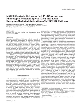
  • 4. MMP-9 CONTROLS SCHWANN MITOSIS AND TROPHIC SIGNALING 1319 This data suggests that Schwann cells produce MMP-9 after proinflammatory but not trophic stimulation. MMP-9 Gene Deletion Promotes Schwann Cell Mitosis In Vivo Sciatic nerve axotomy was used to spatially separate events of Wallerian degeneration (distal stump) and regeneration (proximal stump). Nerves were analyzed at 4 days post-injury, when Schwann cell proliferation occurs in wild-type nerve injury models (Cheng and Zochodne, 2002; Clemence et al., 1989) and when contri- bution of infiltrating immune cells in MMP-92/2 mice is minimal (Shubayev et al., 2006). A statistically signifi- cant increase in DAPI-positive profiles was observed in Fig. 1. MMP-9 expression in primary Schwann cells. Taqman qPCR the proximal but not distal nerve stumps of MMP-92/2 for MMP-9 in primary Schwann cells after 24 h of stimulation with compared with wild-type mice (Fig. 2A). In fact, the dis- BSA, TNF-a, NGF, LPS, or NRG-1. Expressed as mRNA fold increase compared with low serum media, using GAPDH as normalizer. Data tal stump demonstrated a 17% decline in cell number in represents the mean 6 SEM of N 5 4/group, by one-way ANOVA and knockout versus wild-type mice that was not quite stat- Tukey–Kramer post-hoc test (*P 0.05; **P 0.01). istically significant (P 5 0.0594). In vivo BrdU incorporation studies in axotomized into OCT compound in liquid N2, and cut into 10-lm- MMP-92/2 mice were performed to assess the role of thick transverse sections. Cells were fixed with 4% PFA MMP-9 in cell mitosis. A 2.1-fold increase in cell prolif- in TBS for 10 min, washed with TBS, and permeabilized eration was found in proximal nerve stumps of MMP- with 0.1% Triton X in TBS. Nonspecific binding was 92/2 compared with wild-type mice (Fig. 2B), whereas blocked with 10% goat serum. In tissue sections, endoge- no difference was observed in the distal stump. BrdU- nous aldehydes were blocked with 0.5% sodium borohy- positive cells colocalized with S100, a phenotypic marker dride in 1% dibasic sodium phosphate for 5 min and for Schwann cells, suggesting that MMP-9 suppresses Dako antigen retrieval (Carpinteria, CA) was applied for Schwann cell proliferation in vivo. 5 min at 95°C, then for 20 min at RT. Primary antibod- ies were diluted in TBS containing 1% FBS and applied sequentially, the first primary antibody for 1 h at RT, MMP-9 Activates ERK1/2 and Reduces Growth then rinsed, followed by Alexa 564 conjugated (red) first of Primary Schwann Cells secondary goat antibody for 1 h at RT and second pri- mary antibody application overnight at 4°C. Slides were Because ERK MAPK signals cell cycle arrest in rinsed in PBS containing 0.1% Tween 20 and incubated Schwann cells (Harrisingh et al., 2004; Lloyd et al., with second Alexa 488 conjugated (green) secondary 1997), we studied the effect of rhMMP-9 on ERK1/2 acti- goat antibody for 1 h at 22°C. DAPI (1:20,000) was vation, and its correlation to Schwann cell growth. applied for 5 min. Replacement of primary antibody rhMMP-9 produced a dose-dependent activation of with the respective normal IgG was done to control sig- ERK1/2 (Fig. 3A). Based on this data, rhMMP-9 of 100 nal specificity. Imaging was performed using a Leica nM was selected for subsequent experiments. Temporal DMR bright-light and fluorescence microscope using analyses of rhMMP-9 effect in Schwann cells demon- Openlab 4.0 software (Improvision). All micrographs strated biphasic pERK1/2 activation with acute (15 min) represent at least three independent in vitro experi- and sustained (3212 h) phases (Fig. 3B). Immunofluo- ments and N 5 3 animals per group. rescence for pERK1/2 (Fig. 3C) demonstrates its cyto- solic distribution at 15 min and 6 h after rhMMP-9 stimulation, consistent with its phosphorylated state in RESULTS Schwann cell lysates seen by western blot. MMP-9 Is Induced in Schwann Cells by The effect of rhMMP-9 on ERK1/2 activation was cor- Proinflammatory but Not Trophic Factors related to Schwann cell growth kinetics. A significantly reduced Schwann cell growth was observed after daily To identify the stimuli for MMP-9 mRNA expression, rhMMP-9 treatment over a course of 72 h (Fig. 3D). primary Schwann cells were treated with TNF-a, LPS, To test whether MMP-9-induced ERK1/2 activation NGF, or NRG-1. MMP-9 mRNA expression was signifi- depends on the state of Schwann cell differentiation, the cantly induced 100-fold by LPS and 160-fold by TNF-a, latter was stimulated with dibutyryl cyclic AMP whereas the changes in NGF and NRG-1 were not sig- (dbcAMP) for 48 h, as suggested (Harrisingh et al., nificant (see Fig. 1). BSA, used as a control TNF-a car- 2004), followed by treatment with rhMMP-9 (Fig. 4A). rier, increased MMP-9 mRNA by 14-fold, consistent with Successful differentiation was confirmed by the expres- our observations in nerve (Chattopadhyay et al., 2007). sion of myelin protein zero (P0). Biphasic activation of GLIA
  • 5. 1320 CHATTOPADHYAY AND SHUBAYEV Fig. 2. Schwann cell proliferation in axotomized nerves of MMP- tal sciatic nerve stumps 4 days after axotomy. Mean 6 SEM 1,000 lm2 92/2 knockout mice. A, DAPI profiles (blue) in proximal and distal sci- endoneurial area, N 5 5/group, two sections/N, three areas/section at atic nerve stumps 4 days after axotomy. Mean 6 SEM per 100 lm2 objective magnification 340 (scale bar 5 50 lm), by unpaired Student’s endoneurial area, N 5 4/group, two sections/N, three areas/section, by t-test (*P 0.05). Dual-immunofluorescence for BrdU (green) and S100 unpaired Student’s t-test (*P 0.05). Objective magnification 340 (Schwann cell marker, red) in MMP-92/2 nerves (scale bar 5 20 lm). (scale bar 5 50 lm). B, BrdU incorporation (green) in proximal and dis- pERK, peaking at 15 min and 6 h of rhMMP-9 stimula- from 30 min to 8 days of axotomy in both stumps (Sheu tion in myelinating Schwann cells (Fig. 4B), was consist- et al., 2000), we established its initiation within 15 min ent with the findings in undifferentiated cells. of axotomy in both distal and proximal stumps (Fig. 5C). Early changes in MMP-9 mRNA expression after sci- atic nerve axotomy have not been reported. Using Taq- man qPCR (Fig. 5D), we found no significant change in Correlation of MMP-9 and ERK1/2 MMP-9 mRNA at 10 min (i.e., preceding ERK activa- Expression in Injured Nerve tion), a 3.5-fold induction at 1 h in the distal stump, and a 70-fold increase in distal and a 146-fold increase in Exogenous MMP-9-PEX can activate ERK1/2 in proximal stumps at 1 day postaxotomy, representing injured sciatic nerve (Mantuano et al., 2008). Here, we about two-fold higher MMP-9 expression level in proxi- correlated the patterns of endogenous MMP-9 expression mal versus distal stumps. and ERK1/2 activation after axotomy, with the focus on immediate changes. At 24 h after nerve injury, both MMP-9 and pERK are coordinately expressed in distal and proximal stumps of axotomized sciatic nerves (Fig. MMP-9 Activates MEK/ERK1/2 Pathway via ErbB, 5A), colocalizing in Schwann cells, as determined by a IGF-1, and PDGF Tyrosine Kinase Receptors characteristic crescent morphology (Fig. 5B). There was no visible difference between the stumps observed and, MMPs activate trophic signaling in various cells (Page- thus, only one representative micrograph is shown (Fig. McCaw et al., 2007). We analyzed whether MMP-9 5B). While ERK1/2 activation has been shown to sustain induced ERK1/2 signaling by activation of trophic tyro- GLIA
  • 6. MMP-9 CONTROLS SCHWANN MITOSIS AND TROPHIC SIGNALING 1321 Fig. 3. MMP-9 activates ERK1/2 and suppresses Schwann cell objective magnification 3100 (representative micrographs of 3 inde- growth. A, MMP-9 activates ERK1/2 in a dose-dependent manner. 100 pendent experiments). D, Schwann cell growth curve studies using nM rhMMP-9 was selected for use in the subsequent experiments. B, A crystal violet, with and without daily rhMMP-9 stimulation for 72 h. time-course of rhMMP-9 treatment demonstrating a biphasic activation Data represents the mean 6 SD of N 5 3/group, analyzed by one-way of ERK1/2. C, Immunofluorescence for pERK (green) and DAPI (blue) ANOVA (*P 0.05). confirms the biphasic reactivity of cytosolic pERK at 15 min and 6 h, Fig. 4. rhMMP-9 stimulates biphasic ERK1/2 activation in myelinat- firmed by myelin protein zero (P0) expression; b-actin was used as load- ing Schwann cells. Schwann cell differentiation was induced with ing control. B, An extended time-course of rhMMP-9 stimulation dis- dbcAMP (500 lM) for 48 h, followed by treatment with rhMMP-9 (100 played biphasic activation of ERK1/2, as seen in undifferentiated cells nM) for 15 min. A, rhMMP-9 stimulates transient ERK1/2 activation in Fig. 3. over a 1 h period. Successful Schwann cell differentiation was con- sine kinase receptors involved in regulation of Schwann PDK/MEK pathway (see Fig. 8). GM6001 (10 lM), a cell mitosis, including ErbB (Corfas et al., 2004), PDGF broad-spectrum MMP inhibitor, reduced MMP-9 effect. and IGF-1 receptors (Delaney et al., 1999; Meier et al., To evaluate selectivity of rhMMP-9-induced ERK1/2 1999), and/or the MEK/ERK pathway (Fig. 6A). The re- activation, we analyzed the changes in JNK or p38 at 6 sults are summarized in a schematic diagram (see Fig. 8). h of rhMMP-9 stimulation (Fig. 6B). No change in phos- Focus on sustained and not transient ERK1/2 activation pho-JNK or phospho-p38 activation was observed. UV (i.e., 6 h after MMP-9 stimulation) is based on its role in (250 J/m2) stimulation for 15 min was used as positive suppression of cell mitosis (Lloyd et al., 1997; Marshall, controls for p38 and JNK activation, and NRG-1 (10 ng/ 1995). mL) as positive controls for ERK1/2 activation. Note MMP-9-stimulated pERK1/2 levels declined after that pretreatment with GM6001 (a broad-spectrum treatment with general ErbB1/2/4 inhibitor by 49%, but MMP inhibitor) stimulated JNK or p38 MAPK activa- not the specific ErbB2 inhibitor. PDGF receptor inhibitor tion above MMP-9 or basal levels. (PDGFR-I) blocked MMP-9-stimulated pERK activation by 36%, while IGF-1 receptor inhibitor (IGF1R-I) virtu- ally ablated it. MEK (PD98059) and MEK1/2 (U0126) ErbB Inhibition Reversed MMP-9-Induced inhibitors reduced MMP-9-stimulated pERK increase by Suppression of Schwann Cell Mitosis 79 and 93%, respectively, while control PI3K inhibitor (LY294002) produced little effect. The latter also indi- Using an in vitro BrdU incorporation assay, we cates that MMP-9 does not activate ERK via the PI3K/ assessed rhMMP-9 effect on Schwann cell mitosis and GLIA
  • 7. 1322 CHATTOPADHYAY AND SHUBAYEV Fig. 5. Endogenous ERK1/2 and MMP-9 in axotomized rat sciatic activation in distal and proximal stumps at 15 min, 1 h, and 1 day after nerve. A, Western blot for ERK1/2 and MMP-9 in the proximal (P) and axotomy, relative to sham (sh) and contralateral (c) nerves. Duplicate distal (D) stumps 1 day after rat sciatic nerve axotomy relative to contra- representative of N 5 4/group. D, Real-time Taqman qPCR for MMP-9 in lateral (C) nerves; representative of N 5 4/group. B, MMP-9 (red) and axomotized rat nerves normalized to GAPDH and calibrated to sham. pERK (green) colocalize in Schwann cells of a proximal stump, a repre- Data represents the mean 6 SEM of N 5 4/group, by one-way ANOVA sentative micrograph of N 5 3 (scale bar 5 20 lm). C, Sustained ERK1/2 and Tukey–Kramer post-hoc test (*P 0.05; **P 0.01). Fig. 6. rhMMP9 activates MEK–ERK pathway via IGF-1, ErbB, (GM6001, 10 lM). MMP-9-induced ERK activation was inhibited by and PDGF receptors. A, Western blot for ERK1/2 in primary Schwann MEK, ErbB1/2/4, IGF-1R, and PDGFR, but not ErbB2 or PI3K inhibi- cells 6 h after 100 nM rhMMP-9 stimulation, with or without 15 min of tors. B, Western blot for MAPKs of Schwann cells lysates 6 h after pretreatment with the inhibitors to ErbB1/2/4 receptor (ErbB-I, 10 lM), treatment with 100 nM rhMMP-9. MMP-9 produced no effect on activa- ErbB2 receptor (ErbB2-I, 10–50 lM), PDGF receptor (PDGFR-I, 10 tion of p38 and JNK, whereas pretreatment with GM6001 induced it. lM), IGF1 receptor (IGFR-I, 10 lM), MEK (PD98059, 10 lM), MEK1/2 b-actin was used as loading control. Data is representative of three in- (U0126, 10 lM), PI3K (LY294002, 50 lM), and MMP inhibitor dependent experiments. its relationship to an MMP-9-dependent ErbB trophic relative to low-serum media. Both ErbB-I and control pathway (see Fig. 7) that participates in suppression MMPi, GM6001, reversed antimitogenic action of MMP- of Schwann cell proliferation (Chen et al., 2003). 9. GM6001 treatment without MMP-9 stimulation fur- rhMMP-9 stimulation inhibited Schwann cell mitosis ther promoted mitosis. PI3K inhibitor (LY294002) pro- GLIA
  • 8. MMP-9 CONTROLS SCHWANN MITOSIS AND TROPHIC SIGNALING 1323 Fig. 7. MMP-9 inhibits Schwann cell proliferation in vitro. BrdU incorporation is measured 8 h after treatment with 100 nM rhMMP-9. The GM6001 (50 lM), ErbB-I (10 lM), or LY294002 (50 lM) were applied 15 min before stimulation with rhMMP-9. Complete media con- taining 10% FBS and promitotic bovine pituitary extract were used as a positive control. Media containing 1% FBS was used for all experi- mental treatments. Data shown represents the mean 6 SEM of three independent experiments performed in quadruplicate, analyzed by one- way ANOVA and Tukey–Kramer post-hoc test (**P 0.05, *P 0.01). duced no effect on rhMMP-9-stimulated reduction in mitosis. Fig. 8. MMP-9 activation of trophic signaling in Schwann cells (a schematic diagram). MMP-9 expression is induced by proinflammatory stimuli (see Fig. 1). MMP-9 stimulates MAPK p44/42 (or ERK1/2) sig- naling in Schwann cells via activation of IGF-1, ErbB4, and PDGF tyro- sine kinase receptor and Ras/Raf/MEK pathway (shown in black), but DISCUSSION not PI3K/PDK/MEK pathway (shown in grey), as determined using the specified pharmacologic inhibitors (see Fig. 6). These data are the first to implicate MMP-9 in regula- tion of Schwann cell proliferation or trophic signaling. We find that MMP-9 can activate the Ras/Raf/MEK– Supporting data for MMP-mediated signaling by ERK1/2 signal transduction pathway via ErbB, IGF-1, direct receptor binding is only surfacing. A recent study and PDGF tyrosine kinase receptors, as summarized in suggests that hemopexin (substrate-binding) MMP-9 Fig. 8. The exact mechanisms need to be clarified, as domain fused with a GST protein (GST-MMP-9-PEX) MMPs control cell signaling via regulatory proteolysis of activates ERK1/2 in Schwann cells via low-density lipo- latent signaling factors localized in the extracellular protein receptor-related protein 1 (LRP-1) (Mantuano matrix or by (nonproteolytic) direct receptor binding. et al., 2008), a hybrid scavenger, and signaling receptor Evidence for proteolytic MMP function in activation of (Herz and Strickland, 2001). Although the study pro- trophic systems is sound. For example, release of trophic vides no evidence for its direct LRP-1 binding, GST- factors from their regulatory proteins depends on cata- MMP-9-PEX contains a binding site for LRP-1, among lytic activity of MMPs (Page-McCaw et al., 2007), such other surface receptors and substrates (Burg-Roderfeld as IGF-1 release from IGF binding protein, IGFBP-6 in et al., 2007; Roeb et al., 2002). Consistently, GST-MMP- CNS (Larsen et al., 2006). IGF-1 can stimulate MMP- 9-PEX was sufficient to activate ERK in other cells mediated release of the EGF ligand from its heparin- (Dufour et al., 2008). Thus, MMP-9 utilizes several bound form (HB-EGF), leading to cumulative transacti- Schwann cell receptor systems to activate ERK signal- vation of its own and EGF receptors and Ras/Raf/MEK ing, including trophic tyrosine kinase and other signal- signaling (El-Shewy et al., 2004; Roudabush et al., ing receptors, such as LRP-1. Because proteolytic and 2000). In other cells, MMP-9 controls ERK1/2 via activa- receptor agonist MMP actions are not mutually exclu- tion via the EGF receptor (Roelle et al., 2003). Of the sive, either or both potentially relate to the trophic EGF receptor family, Schwann cells express ErbB2, 3, systems. But MMPs intrinsically lacking the hemopexin and 4 (Corfas et al., 2004) and ErbB2 is dispensable in domain, such as MMP-7, are potent inducers of trophic Schwann cell survival after nerve injury (Atanasoski signaling, including that of IGF, EGF, and ErbB (Ii et al., 2006). Considering that ErbB3 has no functional et al., 2006; Sanderson et al., 2006), presumably through kinase domain (Pearson and Carroll, 2004) and ErbB2-I a proteolytic mechanism. was ineffective in blocking MMP-9-induced ERK1/2 acti- MMP-9-induced activation of trophic signaling and sup- vation, the effects of the general ErbB-I seen here is pression of Schwann cell mitosis are independent find- likely to result from ErbB4 block. Proteolytic processing ings. Their relationship was evidenced by ErbB receptor of ErbB4 and its subsequent nuclear translocation block of MMP-9-induced Schwann cell mitosis. ErbB42/2 depend on metalloproteases (Vecchi and Carpenter, in Schwann cells can lead to excessive mitosis (Chen 1997), identified in various cells as ADAM-17, MMP-3, et al., 2003). Because state of Schwann cell differentiation MMP-7, and MMP-9 (Dempsey et al., 2002; Ii et al., and microenvironment influence the functional outcome 2006; Rio et al., 2000; Sanderson et al., 2006). of NRG-1/ErbB system action (Corfas et al., 2004; Jessen GLIA
  • 9. 1324 CHATTOPADHYAY AND SHUBAYEV and Mirsky, 2005) and MMP-9 stimulates ERK1/2 in un- they induce MMP-9 expression in denervated Schwann differentiated and differentiated (myelinating) Schwann cells within 1 h after axotomy. We have already demon- cells, it is important to establish how propensity to strated an over 200-fold increase in MMP-9 mRNA by myelinate and axonal contact affect the outcomes of 6 h of sciatic nerve damage (Shubayev et al., 2006). MMP-9-induced ERK1/2 activation. In our study, it corre- Cytokines induce MMP-9 mRNA in nerves to promote lates with reduced Schwann cell growth and suppressed neuroinflammatory remodeling (Chattopadhyay et al., mitosis, consistent with its role in prodifferentiating func- 2007; Shubayev et al., 2006). Thus, induction of MMP-9 tions of migration (Mantuano et al., 2008) and myelin pro- mRNA in response to proinflammatory (LPS and TNF- tein maintenance (Kobayashi et al., 2008). a), but not trophic (NGF and NRG-1) stimuli in cultured MMP-9 selectively activates ERK but not p38 or JNK Schwann cells is consistent with this earlier developed signaling in Schwann cells, as seen in other cells (Roelle paradigm. rhMMP-9-induced ERK activation seen et al., 2003). Because growth arrest and suppression of in vitro correlates with MMP-9 ability to activate ERK mitosis is signaled through sustained and not transient in injured sciatic nerve (Mantuano et al., 2008). But Ras/Raf/ERK activation (Lloyd et al., 1997; Marshall, because ERK1/2 activation precedes endogenous MMP-9 1995), sustained ERK activation (i.e., 6 h after MMP-9 expression, we suggest that MMP-9 is not the initial stimulation) was the focus of this study. It will be impor- stimulus to ERK1/2 activation after axotomy. This is not tant to determine the mechanisms of MMP-9 induced surprising given that ERK1/2 signals for a plethora of transient (15 min) ERK1/2 activation or ability of MMP- cytokines and trophic factors after nerve damage (Ji and 9 to induce p38 or JNK pathways in Schwann cells at Woolf, 2001). Moreover, our results do not rule out the other time-points in future studies. Interestingly, possibility that ERK signaling is utilized (e.g. by cyto- GM6001 (specific and broad-spectrum MMP inhibitor) kines) to induce MMP-9, as found in cortical astrocytes activated JNK and p38 and stimulated in vitro BrdU (Arai et al., 2003). incorporation above basal levels, implicating endogenous In conclusion, MMP-9 emerges as a potent modulator MMPs in suppressing these signaling pathways and of Schwann cell signaling and phenotypic remodeling Schwann cell mitosis. For example, MMP-3 can generate after nerve injury. It suppresses Schwann cell mitosis antimitogenic fibronectin fragments in Schwann cells and supports functions of differentiation, such as migra- (Muir and Manthorpe, 1992). tion and myelin protein maintenance (Kobayashi et al., Increased Schwann cell mitosis in axotomized MMP- 2008). 92/2 nerves was consistent with antimitogenic proper- ties of MMP-9 in primary Schwann cells (both were determined by BrdU incorporation). While other cells ACKNOWLEDGMENTS types might have contributed to the increased number of mitotic cells, neuronal cell bodies were excluded from We thank Jennifer Dolkas and Julie Janes for techni- analyses and MMP-92/2 nerves were deficient in mac- cal assistance and Amber Millen for help in editing the rophages (Shubayev et al., 2006). An interesting finding manuscript. is that excessive mitosis was uncompensated only in the proximal (regenerating) but not distal (degenerating) stump of axotomized MMP-92/2 nerves. The failure REFERENCES of distal MMP-92/2 nerves to recruit macrophages Arai K, Lee SR, Lo EH. 2003. Essential role for ERK mitogen-activated (Shubayev et al., 2006) is consistent with the diminished protein kinase in matrix metalloproteinase-9 regulation in rat cortical contribution of macrophage-released factors to promote astrocytes. Glia 43:254–264. Schwann cell mitosis (Baichwal et al., 1988). Besides, Atanasoski S, Scherer SS, Sirkowski E, Leone D, Garratt AN, Birchme- ier C, Suter U. 2006. ErbB2 signaling in Schwann cells is mostly dis- the patterns of Schwann cell proliferation are intrinsi- pensable for maintenance of myelinated peripheral nerves and prolif- cally different between the stumps. It spans from 48 h eration of adult Schwann cells after injury. J Neurosci 26:2124–2131. Baichwal RR, Bigbee JW, DeVries GH. 1988. Macrophage-mediated to 14 days after axotomy in the proximal stump, accom- myelin-related mitogenic factor for cultured Schwann cells. Proc Natl panied by dedifferentiation of myelinating Schwann cells Acad Sci USA 85:1701–1705. (Cheng and Zochodne, 2002), also regulated by the Ras/ Brockes JP, Fields KL, Raff MC. 1979. Studies on cultured rat Schwann cells. I. Establishment of purified populations from cultures of periph- Raf/MEK pathway (Harrisingh et al., 2004). In the dis- eral nerve. Brain Res 165:105–118. tal stump, myelinating Schwann cells remain mitotic Burg-Roderfeld M, Roderfeld M, Wagner S, Henkel C, Grotzinger J, longer than nonmyelinating phenotype (Clemence et al., Roeb E. 2007. MMP-9-hemopexin domain hampers adhesion and migration of colorectal cancer cells. Int J Oncol 30:985–992. 1989), owing to promitogenic action of myelin degrada- Chattopadhyay S, Machado-Pinilla R, Manguan-Garcia C, Belda-Iniesta tion products (Clemence et al., 1989; Murinson et al., C, Moratilla C, Cejas P, Fresno-Vara JA, de Castro-Carpeno J, 2005). Thus, reduced degradation of myelin basic protein Casado E, Nistal M, Gonzalez-Barn M, Perona R. 2006. MKP1/ o CL100 controls tumor growth and sensitivity to cisplatin in non- in distal MMP-92/2 nerves (Kobayashi et al., 2008) can small-cell lung cancer. Oncogene 25:3335–3345. also explain their reduced mitogenic activity. Overall, Chattopadhyay S, Myers RR, Janes J, Shubayev V. 2007. Cytokine reg- ulation of MMP-9 in peripheral glia: Implications for pathological these differential responses of axotomized stumps to processes and pain in injured nerve. Brain Behav Immun 21:561– MMP-9 gene deletion support a model of distinct MMP-9 568. actions in nerve degeneration and regeneration. Chen S, Rio C, Ji RR, Dikkes P, Coggeshall RE, Woolf CJ, Corfas G. 2003. Disruption of ErbB receptor signaling in adult non-myelinating Although Schwann cells express low levels of MMP-9 Schwann cells causes progressive sensory loss. Nat Neurosci 6:1186– in normal nerve (Shubayev and Myers, 2000, 2002), 1193. GLIA
  • 10. MMP-9 CONTROLS SCHWANN MITOSIS AND TROPHIC SIGNALING 1325 Cheng C, Zochodne DW. 2002. In vivo proliferation, migration and phe- binding to low-density lipoprotein receptor-related protein. J Neurosci notypic changes of Schwann cells in the presence of myelinated 28:11571–11582. fibers. Neuroscience 115:321–329. Marshall CJ. 1995. Specificity of receptor tyrosine kinase signaling: Clemence A, Mirsky R, Jessen KR. 1989. Non-myelin-forming Schwann Transient versus sustained extracellular signal-regulated kinase acti- cells proliferate rapidly during Wallerian degeneration in the rat sci- vation. Cell 80:179–185. atic nerve. J Neurocytol 18:185–192. Meier C, Parmantier E, Brennan A, Mirsky R, Jessen KR. 1999. Devel- Corfas G, Velardez MO, Ko CP, Ratner N, Peles E. 2004. Mechanisms and oping Schwann cells acquire the ability to survive without axons by roles of axon–Schwann cell interactions. J Neurosci 24:9250–9260. establishing an autocrine circuit involving insulin-like growth factor, Delaney CL, Cheng HL, Feldman EL. 1999. Insulin-like growth factor-I neurotrophin-3, and platelet-derived growth factor-BB. J Neurosci 19: prevents caspase-mediated apoptosis in Schwann cells. J Neurobiol 3847–3859. 41:540–548. Muir D, Manthorpe M. 1992. Stromelysin generates a fibronectin frag- Demestre M, Wells GM, Miller KM, Smith KJ, Hughes RA, Gearing ment that inhibits Schwann cell proliferation. J Cell Biol 116:177– AJ, Gregson NA. 2004. Characterisation of matrix metalloproteinases 185. and the effects of a broad-spectrum inhibitor (BB-1101) in peripheral Murinson BB, Archer DR, Li Y, Griffin JW. 2005. Degeneration of my- nerve regeneration. Neuroscience 124:767–779. elinated efferent fibers prompts mitosis in Remak Schwann cells of Dempsey PJ, Garton K, Raines EW. 2002. Emerging roles of TACE as a uninjured C-fiber afferents. J Neurosci 25:1179–1187. key protease in ErbB ligand shedding. Mol Interv 2:136–141. Page-McCaw A, Ewald AJ, Werb Z. 2007. Matrix metalloproteinases Dufour A, Sampson NS, Zucker S, Cao J. 2008. Role of the hemopexin and the regulation of tissue remodelling. Nat Rev Mol Cell Biol 8: domain of matrix metalloproteinases in cell migration. J Cell Physiol 221–233. 217:643–651. Pearson RJ Jr, Carroll SL. 2004. ErbB transmembrane tyrosine kinase El-Shewy HM, Kelly FL, Barki-Harrington L, Luttrell LM. 2004. Ecto- receptors are expressed by sensory and motor neurons projecting into domain shedding-dependent transactivation of epidermal growth fac- sciatic nerve. J Histochem Cytochem 52:1299–1311. tor receptors in response to insulin-like growth factor type I. Mol Pfaffl MW. 2001. A new mathematical model for relative quantification Endocrinol 18:2727–2739. in real-time RT-PCR. Nucleic Acids Res 29:e45. Harrisingh MC, Perez-Nadales E, Parkinson DB, Malcolm DS, Mudge Platt CI, Krekoski CA, Ward RV, Edwards DR, Gavrilovic J. 2003. AW, Lloyd AC. 2004. The Ras/Raf/ERK signalling pathway drives Extracellular matrix and matrix metalloproteinases in sciatic nerve. Schwann cell dedifferentiation. EMBO J 23:3061–3071. J Neurosci Res 74:417–429. Herz J, Strickland DK. 2001. LRP: A multifunctional scavenger and Rio C, Buxbaum JD, Peschon JJ, Corfas G. 2000. Tumor necrosis fac- signaling receptor. J Clin Invest 108:779–784. tor-alpha-converting enzyme is required for cleavage of erbB4/HER4. Ii M, Yamamoto H, Adachi Y, Maruyama Y, Shinomura Y. 2006. Role of J Biol Chem 275:10379–10387. matrix metalloproteinase-7 (matrilysin) in human cancer invasion, Roeb E, Schleinkofer K, Kernebeck T, Potsch S, Jansen B, Behrmann I, apoptosis, growth, and angiogenesis. Exp Biol Med (Maywood) 231: Matern S, Grotzinger J. 2002. The matrix metalloproteinase 9 (mmp- 20–27. 9) hemopexin domain is a novel gelatin binding domain and acts as Jessen KR, Mirsky R. 2005. The origin and development of glial cells in an antagonist. J Biol Chem 277:50326–50332. peripheral nerves. Nat Rev Neurosci 6:671–682. Roelle S, Grosse R, Aigner A, Krell HW, Czubayko F, Gudermann T. Ji RR, Woolf CJ. 2001. Neuronal plasticity and signal transduction in 2003. Matrix metalloproteinases 2 and 9 mediate epidermal growth nociceptive neurons: Implications for the initiation and maintenance factor receptor transactivation by gonadotropin-releasing hormone. of pathological pain. Neurobiol Dis 8:1–10. J Biol Chem 278:47307–47318. Kobayashi H, Chattopadhyay S, Kato K, Dolkas J, Kikuchi S, Myers Roudabush FL, Pierce KL, Maudsley S, Khan KD, Luttrell LM. 2000. RR, Shubayev VI. 2008. MMPs initiate Schwann cell-mediated MBP Transactivation of the EGF receptor mediates IGF-1-stimulated shc degradation and mechanical nociception after nerve damage. Mol phosphorylation and ERK1/2 activation in COS-7 cells. J Biol Chem Cell Neurosci 39:619–627. 275:22583–22589. La Fleur M, Underwood JL, Rappolee DA, Werb Z. 1996. Basement Sanderson MP, Dempsey PJ, Dunbar AJ. 2006. Control of ErbB signal- membrane and repair of injury to peripheral nerve: Defining a poten- ing through metalloprotease mediated ectodomain shedding of EGF- tial role for macrophages, matrix metalloproteinases, and tissue in- like factors. Growth Factors 24:121–136. hibitor of metalloproteinases-1. J Exp Med 184:2311–2326. Sheu JY, Kulhanek DJ, Eckenstein FP. 2000. Differential patterns of Larsen PH, DaSilva AG, Conant K, Yong VW. 2006. Myelin formation ERK, STAT3 phosphorylation after sciatic nerve transection in the during development of the CNS is delayed in matrix metalloprotei- rat. Exp Neurol 166:392–402. nase-9 and -12 null mice. J Neurosci 26:2207–2214. Shubayev VI, Angert M, Dolkas J, Campana WM, Palenscar K, Myers Leppert D, Hughes P, Huber S, Erne B, Grygar C, Said G, Miller KM, RR. 2006. TNFalpha-induced MMP-9 promotes macrophage recruit- Steck AJ, Probst A, Fuhr P. 1999. Matrix metalloproteinase upregula- ment into injured peripheral nerve. Mol Cell Neurosci 31:407–415. tion in chronic inflammatory demyelinating polyneuropathy and non- Shubayev VI, Myers RR. 2000. Upregulation and interaction of TNFal- systemic vasculitic neuropathy. Neurology 53:62–70. pha and gelatinases A, B in painful peripheral nerve injury. Brain Livak KJ, Schmittgen TD. 2001. Analysis of relative gene expression Res 855:83–89. data using real-time quantitative PCR, the 2(-Delta Delta C(T)) Shubayev VI, Myers RR. 2002. Endoneurial remodeling by TNFalpha- method. Methods 25:402–408. and TNFalpha-releasing proteases. A spatial and temporal co-local- Lloyd AC, Obermuller F, Staddon S, Barth CF, McMahon M, Land H. ization study in painful neuropathy. J Peripher Nerv Syst 7:28–36. 1997. Cooperating oncogenes converge to regulate cyclin/cdk com- Vecchi M, Carpenter G. 1997. Constitutive proteolysis of the ErbB-4 re- plexes. Genes Dev 11:663–677. ceptor tyrosine kinase by a unique, sequential mechanism. J Cell Mantuano E, Inoue G, Li X, Takahashi K, Gaultier A, Gonias SL, Cam- Biol 139:995–1003. pana WM. 2008. The hemopexin domain of matrix metalloproteinase- Werb Z. 1997. ECM and cell surface proteolysis: Regulating cellular 9 activates cell signaling and promotes migration of Schwann cells by ecology. Cell 91:439–442. GLIA