Osteogenesis imperfecta
•Download as PPTX, PDF•
9 likes•1,428 views

Lecture on Osteogenesis Imperfecta for MBBS students.

Recommended
More Related Content
What's hot
What's hot (20)
Similar to Osteogenesis imperfecta
Similar to Osteogenesis imperfecta (20)
Recently uploaded
Recently uploaded (20)
Osteogenesis imperfecta
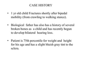
- 1. CASE HISTORY • 1 yr old child Fractures shortly after bipedal mobility (from crawling to walking stance). • Biological father has also has a history of several broken bones as a child and has recently begun to develop bilateral hearing loss. • Patient is 75th percentile for weight and height for his age and has a slight bluish gray tint to the sclera.
- 2. Osteogenesis Imperfacta Hereditary condition resulting from a decrease in the amount of normal Type I collagen Type I collagen ( important for ) Bone Ligaments Teeth White Sclera Skin
- 3. Type I collagen deficiency can result from decreased collagen secretion production of abnormal collagen Both Autosomal dominant and Autosomal recessive forms Can be severe or mild (Tarda) Osteogenesis Imperfecta
- 4. Epidemiology Incidence : 1 casefor every 20,000livebirths Equallycommon in males andfemales Nopredilection fora particular race
- 5. • Earliest known case of osteogenesis imperfecta in a partially mummified infant’s skeleton from ancient Egypt now housed in the British Museum in London. • OTHER NAMES • LOBSTEIN DISEASE • BRITTLE- BONE DISEASE • BLUE-SCLERA SYNDROME • FRAGILE-BONE DISEASE
- 6. GENETICS Quantitativedefectsof type1collagen: mutations on COL1A gene Qualitativedefectsof type1collagen: autosomal dominant mutations on either the COL1A or the COL1Bgene
- 9. Pathologic changes seen in all tissues in which type 1 collagen is an important constituent -bone, ligament, dentin, and sclera
- 10. Orthopaedic manifestations Bone fragility and fractures fractures heal in normal fashion initially but the bone is does not remodel- lead to progressive bowing Ligamentous laxity Short stature Basilar invagination-the tip of the odontoid process projects above the foramen magnum Olecranon apophyseal avulsionfx
- 11. Scoliosis Codfish vertebrae(compressionfx) Wormian skullbones (puzzle pieceintrasutural skullbones)
- 12. Non-Orthopaedic manifestations Blue sclera Hearing loss Dentinogenesis imperfecta brownishopalescentteeth
- 13. Symptoms Mild cases multiple fractures duringchildhood Severe cases present with fractures at birth and can be fatal Number of fractures typically decreases as patient ages and usually stops after puberty But deformity persist. Basilar invagination Brain Stem dysfunction apnea, altered consciousness, ataxia, or myelopathy usually in third or fourth decade of life, but can be as early as teenage years
- 14. Physical exam Multiple fractures leads to Saber shin appearance of tibia Bowing of long bones Scoliosis
- 15. Physical Examination Sillence classification : 4 types on basis of clinical and radiologicfeatures Dentinogenesis imperfecta denoted as subtype B,whereas OI without dentinogenesis imperfecta isdenoted as subtypeA
- 16. Sillence Classification of Osteogenes Imperfecta (simplified) Type I Mildest form. Presents at preschool age (Tarda). Autosomal dominant Blue sclera Hearing deficit in 50%. Divided into type A and B based on tooth involvement Type II Autosomal recessive / AD Blue sclera Lethal in perinatal period
- 17. Type III Autosomal recessive Normal sclerea Fractures at birth. Progressively short stature. Most severe survivable form Type IV Autosomal dominant normal Moderate severity. Bowing bones and vertebral fractures are common. Hearing normal. Divided into type Aand B based on tooth involvement
- 18. Type5 • Autosomal dominent • Samefeatures astype 4 • Different histologically ( bone appears mesh like) • Calcification of radio ulnarinterosseous membrane
- 21. Diagnosis Diagnosis is based on family history associated with typical radiographic and clinical features No commercially available diagnostic test Laboratory values are typically within normal range Fibroblast culturing to analyze type I collagen (positive in 80% of Type IV) can be used for confirmation of diagnosis in equivocal cases
- 22. Skin biopsy: collagen can be isolated from cultured fibroblasts and assessed for defects, with an accuracy of 85-87% Bone biopsy : show changes in concentrations of noncollagenous bone proteins, such as osteonectin, sialoprotein, and decorin
- 23. DNA DNA blood testing for gene defects has an accuracy of 60- 94%. Samples are obtained during chorionic villus sampling performed under ultrasonographic guidance when a mutation in another member of the family is already known
- 24. Prenatalultrasonography : Useful in evaluating OItypesII andIII Detects limb-length abnormalities at 15-18weeks Supervisualization of intracranial contents causedby decreased mineralization of calvaria(also calvarial compressibility), bowing ofthe long bones,decreasedbone length (especially of the femur), and multiple rib fractures
- 25. Radiographs Thin cortices Generalized osteopenia Long bone thin and bowed-shepherd’s crook deformities of the femurs Excessive callus formation and popcorn bones Pelvis may show acetabular protrusion Fractures that are at different stages of healing The vertebra maybe biconcave. Skull changes - Wormian bones
- 27. TREATMENT NOCURE Orthotics: limited role, to stabilize lax joints (eg,ankle and subtalar joints withankle-foot orthoses) and to prevent progressive deformities andfractures. Provide walking aids, specialized wheelchairs, and home adaptation devices to help improve patient’s mobility andfunction
- 29. PatientEducation Patients with OI: well motivated and keen to achieve as much as possible despite their physical limitations Education of parents and families :to know how to position child in crib and how to hold child so as to minimize risk of fractures while maintaining bonding and physical stimulation
- 30. DIETANDACTIVITY Nutritional evaluation and intervention paramount to ensure appropriate intakeof calcium, phosphorus, and vitamin D Caloric management important, particularly in adolescents and adults with severeforms ofOI Physicaltherapy, in form of comprehensive rehabilitation programs, directed toward improving joint mobility and developingmuscle strength
- 31. Treatment of Fractures Fracture prevention Early bracing Decrease deformity. Stabilize lax joints. Decrease fractures incidence. Bisphosphonates
- 32. BISPHOSPHONATES Synthetic analogues of pyrophosphate that inhibit osteoclast- mediated bone resorption on the endosteal surface of bone by binding to hydroxyapatite. Unopposed osteoblastic new bone formation on the periosteal surface results in an increase in cortical thickness.
- 33. Pamidronate Injectable bisphosphonate (Cyclic Intravenous ) Increases cortical bone thickness Increase bone mass and density. Decreases the incidence of fractures. Relieves chronic bonepain. Increases activity levels. Decreases the reliance on mobility aids. Increases the height of the collapsed vertebral bodies. BUT Notdecreasetheincidenceofscoliosis. Zebralines Radiographically Pamidronate therapy creates growth lines in the bone
- 34. Growth hormone: act on growth plate, stimulate osteoblast function, possibly via IGF-1 ,IGFBP-3
- 35. Bone marrowtransplantation Used with somesuccess Introduces normal marrow stem cells that could potentially differentiate into normal osteoblasts, Problems of graft rejection and graft versus host reactions limit thisapproach. FUTURE
- 36. Fracture treatment Nonoperative child is less than 2 years treataschildwithoutOI Operative Fixation with INTRAMEDULLARY RODS (TELESCOPING) patients > 2 years allow continued growth
- 37. conservative Treatment in OIFractures Children <24 months
- 39. Treatment of Long Bone Deformities Realignment Osteotomy with rod fixation (Sofield-Miller procedure)- KEBAB OSTEOTOMY Indicated in severe deformity to Correct the deformity Reduce fracture rates Techniques include Nontelescopic devices Telescopic devices
- 40. Treatment of Scoliosis Observation Curve less than 45° Bracing is ineffective Operative posterior spinal fusion Indications for curves > 45 °in mild formsand > 35 °in severe forms Technique Challenging due to fragility of bones Use allograft instead of iliac crest autograft Large blood loss
- 41. . Acute / Stress Fracture Deformity with Chronic StressFracture
- 42. FRACTURE --------OSTEOPOROSIS---------REFRACTURE Early Surgery and PreparatoryPamidronate
- 43. Never useplate in OI
- 44. Plating MUST BEAVOIDEDAS HIGH RISK OFSTRESS FRACTURE.
- 45. 7year old child withOI Plating done forfemur fracture Deformity appears above and belowthe plate due to stress shielding of bone
- 46. Revised to F– D TelescopingRod
- 47. 20 month old girl, repeated femur fractures, unable to stand ?delayed milesones
- 49. Nine Months post-op Standing withsupport
- 50. • 10 year old child • OI?TypeIII, progressive deformity • 5 cycles of Pamidronate received • No surgery done
- 51. How early to NAIL? Rodding :before orafter bisphosphonatetreatment Choice ofRod
- 52. ClassicalShishKabab Surgery Very Narrow IM Canal:2mm Rodinserted Spicafor 6 weeks and followed by splints
- 55. ThankYou