Call Girls Bareilly Just Call 9907093804 Top Class Call Girl Service Available
Rheuumatic heart disese
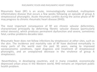
1. RHEUMATIC FEVER AND RHEUMATIC HEART DISEASE
Rheumatic fever (RF) is an acute, immunologically mediated, multisystem
inflammatory disease that occurs a few weeks following an episode of group A
streptococcal pharyngitis. Acute rheumatic carditis during the active phase of RF
may progress to chronic rheumatic heart disease (RHD).
The most important consequence of RF are chronic valvular deformities,
characterized principally by deforming fibrotic valvular disease (particularly
mitral stenosis), which produces permanent dysfunction and severe, sometimes
fatal, cardiac problems decades later.
Rheumatic fever does not follow infections by streptococci at other sites, such as
the skin. The incidence and mortality rate of RF have declined remarkably in
many parts of the world over the past 30 years, owing to improved
socioeconomic conditions, rapid diagnosis and treatment of streptococcal
pharyngitis, and an unexplained decrease in the virulence of group A
streptococci.
Nevertheless, in developing countries, and in many crowded, economically
depressed urban areas in the Western world, RHD remains an important public
health problem.
2. Morphology
During acute RF,
• focal inflammatory lesions are found in various tissues.
They are most distinctive within the heart, where they
are called Aschoff bodies. They consist of foci of
swollen eosinophilic collagen surrounded by
lymphocytes (primarily T cells), occasional plasma cells,
and plump macrophages called Anitschkow cells
(pathognomonic for RF). These distinctive cells have
abundant cytoplasm and central round-to-ovoid nuclei
in which the chromatin is disposed in a central, slender,
wavy ribbon (hence the designation "caterpillar cells").
Some of the larger macrophages become
multinucleated to form Aschoff giant cells.
3. Acute and chronic rheumatic heart disease. A, Acute rheumatic mitral valvulitis superimposed on
chronic rheumatic heart disease. Small vegetations (verrucae) are visible along the line of closure of the mitral
valve leaflet (arrows). Previous episodes of rheumatic valvulitis have caused fibrous thickening and fusion of
the chordae tendineae. B, Microscopic appearance of Aschoff body in a patient with acute rheumatic carditis.
The myocardial interstitium has a circumscribed collection of mononuclear inflammatory cells, including some
large histiocytes with prominent nucleoli and a prominent binuclear histiocyte, and central necrosis.
4. • During acute RF, diffuse inflammation and Aschoff bodies
may be found in any of the three layers of the heart—
pericardium, myocardium, or endocardium—hence the
lesion is called a pancarditis. In the pericardium, the
inflammation is accompanied by a fibrinous or
serofibrinous pericardial exudate, described as a "bread-
and-butter" pericarditis, which generally resolves without
sequelae.
• The myocardial involvement—myocarditis—takes the form
of scattered Aschoff bodies within the interstitial
connective tissue, often perivascular.
• Subendocardial lesions, perhaps exacerbated by regurgitant
jets, may induce irregular thickenings called MacCallum
plaques, usually in the left atrium.
5. C and D, Mitral stenosis with diffuse fibrous thickening and distortion of the
valve leaflets, commissural fusion (arrows), and thickening and shortening of
the chordae tendineae. Marked dilation of the left atrium is noted in the left
atrial view (C). D, Opened valve. Note neovascularization of anterior mitral
leaflet (arrow).
6. • Chronic RHD is characterized by organization of the
acute inflammation and subsequent fibrosis. In
particular, the valvular leaflets become thickened and
retracted, causing permanent deformity.
• The cardinal anatomic changes of the mitral (or
tricuspid) valve are leaflet thickening, commissural
fusion and shortening, and thickening and fusion of
the tendinous cords .
• In chronic disease, the mitral valve is virtually always
abnormal, but involvement of another valve, such as
the aortic, may be the most clinically important in
some cases. Microscopically there is diffuse
7. E, Surgically removed specimen of rheumatic aortic stenosis,
demonstrating thickening and distortion of the cusps with
commissural fusion
8. Pathogenesis.
• It is strongly suspected that acute rheumatic fever is a
hypersensitivity reaction induced by group A
streptococci, but the exact pathogenesis remains
uncertain despite many years of investigation.
• It is thought that antibodies directed against the M
proteins of certain strains of streptococci cross-react
with glycoprotein antigens in the heart, joints, and
other tissues. The onset of symptoms 2 to 3 weeks
after infection and the absence of streptococci from
the lesions support the concept that RF results from an
immune response against the offending bacteria.
Because the nature of cross-reacting antigens has been
difficult to define, it has also been suggested that the
streptococcal infection evokes an autoimmune
response against self-antigens. Only a minority of
infected patients develop RF, suggesting that genetic
susceptibility influences the hypersensitivity reaction.
9.
10. Clinical Features.
• RF is characterized by a constellation of findings that
includes as major manifestations (1) migratory polyarthritis
of the large joints, (2) carditis, (3) subcutaneous nodules,
(4) erythema marginatum of the skin, and (5) Sydenham
chorea, a neurologic disorder with involuntary purposeless,
rapid movements.
• The diagnosis is established by the so-called Jones criteria:
evidence of a preceding group A streptococcal infection,
with the presence of two of the major manifestations listed
above or one major and two minor manifestations
(nonspecific signs and symptoms that include fever,
arthralgia, or elevated blood levels of acute phase
reactants).
11. • Acute rheumatic fever typically occurs 10 days to 6 weeks after an episode
of pharyngitis caused by group A streptococci in about 3% of patients.
Acute RF appears most often in children between ages 5 and 15, but about
20% of first attacks occur in middle to later life. Although pharyngeal
cultures for streptococci are negative by the time the illness begins,
antibodies to one or more streptococcal enzymes, such as streptolysin O
and DNAse B, are present and can be detected in the sera of most
patients.
• The predominant clinical manifestations are those of arthritis and carditis.
Arthritis is far more common in adults than in children. It typically begins
with migratory polyarthritis accompanied by fever in which one large joint
after another becomes painful and swollen for a period of days and then
subsides spontaneously, leaving no residual disability. Clinical features
related to acute carditis include pericardial friction rubs, weak heart
sounds, tachycardia, and arrhythmias. The myocarditis may cause cardiac
dilation that may evolve to functional mitral valve insufficiency or even
heart failure. Overall the prognosis for the primary attack is generally
good, and only 1% of patients die from fulminant RF.
12. Congenital Heart Disease
Congenital heart disease is a general term used to
describe abnormalities of the heart or great vessels
that are present from birth. Most such disorders
arise from faulty embryogenesis during gestational
weeks 3 through 8, when major cardiovascular
structures develop. The most severe anomalies may
be incompatible with intrauterine survival.
• Congenital heart defects compatible with
embryologic maturation and birth are generally
morphogenetic defects of individual chambers or
regions of the heart, with the remainder of the heart
developing relatively normally.
13. Table 12-2 -- Frequencies of Congenital Cardiac Malformations *
Malformation Incidence per Million Live Births %
Ventricular septal defect 4482 42
Atrial septal defect 1043 10
Pulmonary stenosis 836 8
Patent ductus arteriosus 781 7
Tetralogy of Fallot 577 5
Coarctation of aorta 492 5
Atrioventricular septal defect 396 4
Aortic stenosis 388 4
Transposition of great arteries 388 4
Truncus arteriosus 136 1
Total anomalous pulmonary venous
connection 120 1
Tricuspid atresia 118 1
TOTAL 9757
Source: Hoffman JIE, Kaplan S: The incidence of congenital heart
disease. J Am Coll Cardiol
39:1890, 2002.