2. 376 The Amygdala – A Discrete Multitasking Manager
addition to these symptoms, AD patients frequently show personality changes that affect
their activities of daily living and the interaction with their caregivers. Personality changes
may appear in any phase of dementia but often precede other early clinical manifestations of
the disease, such as cognitive impairment and mood changes. These changes may therefore
help in the clinical diagnosis of AD at early stages (Robins Wahlin and Byrne, 2011).
Interestingly, personality changes and some of the neuropsychiatric symptoms (agitation,
dysphoria and apathy) are better correlated with the severity of cognitive, functional and
behavioral signs than with the patient’s age, gender, education or disease duration (Mega et
al., 1996; Talassi et al., 2007). Thus, personality changes and neuropsychiatric symptoms may
reflect the impact of progressive brain damage in AD (Robins Wahlin and Byrne, 2011).
3. Emotional memory in AD
Emotional memory is a form of episodic memory defined as memory of arousing emotional
events. These memories are sometimes referred to as "flashbulb" memories (Hamann et al.,
2000). Results from studies in animals and humans have strongly implicated the amygdala
in this memory type (LaBar, 2003; Brierley et al., 2004; Richter-Levin, 2004). While it is
recognized that normal people better remember events associated with an emotional
component, there is a controversy regarding the strength of emotional memory in AD
patients (Satler et al., 2007; Schultz et al., 2009; Huijbers et al., 2011; Nashiro and Mather,
2011; Sundstrom, 2011). Since the amygdala is one of the structures damaged in early stages
of the AD pathology, it has been hypothesized that emotional memory should be impaired
in AD patients. Indeed, data have shown that unlike healthy individuals, AD patients do not
show memory enhancement for emotional events (enhanced memory for emotional
compared to neutral stimuli) in spite of normal emotional reactions (Hamann et al., 2000).
Notably, the degree of emotional memory impairment has positively been correlated with
the extent of the amygdaloid atrophy (Mori et al., 1999a, b; Fleming et al., 2003).
4. Pathology of the amygdala in AD patients
While normal aging primarily affects the prefrontal cortex but relatively spares limbic
regions, AD mainly affects limbic regions. The amygdala of AD patients shows a
considerable shrinkage, distortion and loss of neurons, and widespread gliosis (Vereecken et
al.; Herzog and Kemper, 1980; Cuenod et al., 1993). The amygdaloid atrophy in AD is the
result of neuronal death (especially in the magnocellular basolateral amygdalar nuclei
group) and loss of dendrites and axons. The accumulation of intraneuronal neurofibrillary
tangles, Lewy bodies and extracellular Amyloid peptide (A deposits in plaques also
contribute significantly to the atrophy. Detailed pathological examination of the amygdala
of AD patients reveals that many neurofibrillary tangles and A plaques are located in the
accessory basal and cortical nuclei and in the cortical transition area, whereas the
mediobasal nucleus is less affected (Kromer Vogt et al., 1990). The medial, lateral,
laterobasal and central nuclei are relatively free of neurofibrillary tangles and A plaques
(Kromer Vogt et al., 1990). Interestingly, it has been observed that the morphological
3. Amygdala in Alzheimer's Disease 377
deformation of the amygdala in AD patients is associated with intrinsic damage to its
subnuclei and their reciprocal connectivity with other brain areas. Specifically, it has been
reported that amygdaloid nuclei receiving input from and giving rise to hippocampal
projections are consistently affected by neuropathological alterations in AD. In contrast,
amygdaloid nuclei which receive strong cholinergic input from nucleus basalis of Meynert
(e.g. laterobasal nucleus) are less affected (Kromer Vogt et al., 1990). To conclude,
histological analysis of the amygdala of AD patients allows a thorough examination of this
region thus rendering possible to detect nucleus-specific pathologies. Nevertheless, the
major limitation of post-mortem analysis is that it is typically performed on brains taken
from patients at late stages of the disease. Thus, information is lacking regarding
neuropathological alterations in early stages of AD.
5. Imaging of the amygdala in vivo
Whereas histological procedures are used to investigate the anatomical complexity of the
amygdala in brains from AD patients, a standard magnetic resonance imaging (MRI)
technique can only detect few internal details and similar resolution cannot be obtained. The
discovery that neuronal loss is a cause of amygdaloid atrophy provided the basis for later
studies correlating amygdaloid volumetry, as measured with MRI, with the cognitive status
of individual AD patients. Indeed, MRI-based volumetry is now regularly used as a research
tool to explore the relationship between amygdaloid volume and the onset and progression
of AD (de Leon et al., 1996; Mori et al., 1999a; Vasconcelos et al., 2011). While in the past the
use of MRI was limited to clinical studies, the recent rise in MRI accessibility allowed its
utilization for non-clinical studies aimed at investigating the involvement of the amygdala
in emotion, memory processes and personality (Mori et al., 1999a). The main disadvantage
of MRI-based amygdaloid volumetry consists in the difficulty to precisely and reliably
delineate the contours of the amygdala in vivo. This difficulty arises from the similarity
in MRI signal intensities between the amygdala and other temporal lobe structures
surrounding it (hippocampus proper, subiculum, entorhinal cortex, claustrum and tail of the
caudate)(Convit et al., 1999). Nevertheless, new imaging techniques such as ultrahigh field
structural MRI enable clear in vivo detection and even segmentation of the amygdala
(Solano-Castiella et al., 2011), and might be used to investigate the anatomical features of
different amygdaloid nuclei in AD patients.
Numerous studies measuring the amygdaloid volume (normalized to intracranial volume)
in AD patients at different clinical stages and in healthy age-matched controls showed a
correlation of this factor with the neuropsychological performance of each patient. These
studies have consistently demonstrated a decrease in amygdaloid volume in AD patients
when compared to healthy controls (Horinek et al., 2007; Beacher et al., 2009; Cherubini et
al., 2010; Lehmann et al., 2010; Vasconcelos Lde et al., 2011). Importantly, atrophy of the
amygdala was found even in preclinical stages of the disease (Fox et al., 1996; Heun et al.,
1997; Golebiowski et al., 1999). In fact, in the very early stages of AD, amygdaloid volume
reductions were at least as large as hippocampal volume reductions although at this stage
4. 378 The Amygdala – A Discrete Multitasking Manager
some overlap does exist between patients and healthy controls. Still, the volume of the
amygdala has been suggested to be an independent variable in predicting conversion from
mild cognitive impairment to AD (Liu et al., 2010).
Functional MRI. Increasing number of neuroimaging studies using functional MRI (fMRI)
are used to examine the neuronal activity of the amygdala by detecting changes in local
blood perfusion, blood volume or blood oxygenation. Injecting contrast agents are often
used in this technique. In some studies, voxel-based morphometry (VBM) on MRI was
combined with Positron Emission Tomography (PET) to compare activity in specific brain
areas in AD patients and healthy controls (Kawachi et al., 2006). Functional MRI studies
showed that the amygdala is excessively responsive to human faces (both novel emotional
and familiar neutral expressions) in mild AD patients relative to elderly controls (Wright et
al., 2007). On the other hand, AD patients presented deficits in the recognition of some facial
expressions of emotion (happy, sad, fearful, and neutral expressions)(Kohler et al., 2005).
These alterations in the normal activity of the amygdala probably contribute to the
significant social and behavioral defects observed in AD patients.
Positron Emission Tomography (PET). PET is almost exclusively used to image the brain, and
may be used to detect functional abnormalities early in the course of AD, way before
anatomical changes occur. For example, PET was used to examine acetylcholine esterase
activity in vivo in the amygdala and cerebral cortex (Shinotoh et al., 2003). To note, levels of
acetylcholine are significantly decreased in AD due to degeneration of the cholinergic
magnocellular neurons of the nucleus basalis of Meynert (nbM) that send cholinergic
projection mainly to the amygdala (Mesulam, 2004). In fact, the degree of the cholinergic
loss is positively correlated with the severity of dementia in AD (Perry et al., 1981), probably
due to the importance of nbM in emotional memory consolidation. PET measurements of C-
11-labeled N-methyl-4-piperidyl-acetate (MP4A, a specific substrate of AChE) have shown
that AChE activity is significantly reduced in patients with AD in both the amygdala and
cerebral cortex (Shinotoh et al., 2003). Importantly, these deficits are present in mild to
moderate AD, supporting the notion that cortical and amygdaloid functional changes of the
cholinergic system occur early in AD (Herholz et al., 2004). These functional alterations are
therefore suggested to serve as a physiologic and noninvasive marker for certain
neuropsychiatric manifestations of mild AD. In addition, these finding suggest that the
amygdala should receive an important attention in studies of the mild or even prodromal
stages of AD (Basso et al., 2006) even though considerable evidences continue to support the
focus on the hippocampus in MRI studies of AD.
6. The use of AD mice model to study the A-dependent changes in the
amygdala
In modern AD research, transgenic mice bearing infrequent mutations leading to familial
forms of AD are being used to characterize in details the physiological, morphological and
behavioral consequences of AD neuropathology in order to understand the anatomical and
synaptic basis of dementia (Selkoe, 1996). These mutations include mutations in amyloid
5. Amygdala in Alzheimer's Disease 379
precursor protein (APP), the precursor of the Aβ peptide, or in presenilin (PS) 1 or 2, the
catalytic subunit of the gamma secretase complex, which cleaves APP to form Aβ.
Transgenic AD mice model represents an important tool to examine the consequences of in
vivo Aβ accumulation and were proved to mimic many of the pathological features of AD
(Spires and Hyman, 2005; Spires-Jones and Knafo, 2012). APP and APP/PS1 mice present
abundant extracellular Aβ plaques, synaptic dysfunction and loss, astrocytosis, activation of
microglia and cognitive deficits (Games et al., 1995). The fact that Aβ plaques occupy a
minor fraction (less than 5%) of the neuropil (see Fig. 1) in cognitively impaired transgenic
mice (Knafo et al., 2009; Merino-Serrais et al., 2011) and the lack of correlation between the
plaque load and the degree of cognitive impairment in AD patients (Terry et al., 1991; Terry,
2000), support the notion that fibrillar Aβ in plaques does not contribute significantly to
dementia in AD patients. Instead, soluble Aβ assemblies (i.e. oligomeric or protofibrillary
Aβ species that linger in aqueous solution after high-speed centrifugation) seem to be the
main factors responsible for the structural, synaptic and cognitive deficits in these mice and
probably also in initiating disease in AD patients (Selkoe, 2002).
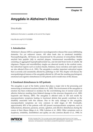
A section stained with the anti-Aβ antibody and Nissl.
BLA = basolateral nucleus of the amygdala; EC = external capsule; LA = lateral nucleus of the amygdala; PIR = piriform
cortex. Scale bar, 350 m (Knafo et al., 2009)
Figure 1. Coronal sections through the amygdala and adjacent regions showing the pattern of
distribution of amyloid plaques.
In a recent study, transgenic mice expressing a chimeric mouse/human amyloid precursor
protein (Mo/HuAPP695swe) and a mutant human presenilin 1 (PS1-dE9) (APP/PS1, Borchelt
6. 380 The Amygdala – A Discrete Multitasking Manager
et al., 1997) were used to study the morphological basis for amygdala-dependent cognitive
impairment (Knafo et al., 2009). In this study, the authors first showed a clear impairment of
auditory fear conditioning in APP/PS1 mice, a learning task that depends on the lateral
nucleus of the amygdala (LA) (Knafo et al., 2009). Importantly, this cognitive deficit did not
result from changes in anxiety or sensitivity to shock. Then, the authors used intracellular
injection of Alexa594 into projection neurons in the LA, combined with thioflavin-S plaque
staining (Fig. 2) and three-dimensional reconstructions of the dendritic trees and spines. The
results of this study show that in APP/PS1 mice the morphology of projection neurons in the
amygdala is modified, as reflected by changes in dendritic complexity, and that there is a
(a) Panoramic confocal (10×) views of the lateral amygdala showing Alexa594-injected neurons and thioflavin-s-
positive plaques in a Tg- mouse (left) and an APP/PS1 mouse (right).
(b) Representative images of projection neurons from a Tg- mouse (left) and an APP/PS1 mouse (right).
(c) The method used to distinguish dendrites and spines within and outside plaques. Left: a plaque suspected of
containing a dendrite due to the rotation of its three-dimensional image. Center: the plaque surface is marked with the
aid of the IsoSurface tool of Imaris software. Right: the voxels outside the surface are set to zero, leaving only the
dendritic segment within the plaque (Knafo et al., 2009).
Figure 2. Intracellular injections
7. Amygdala in Alzheimer's Disease 381
significant decrease in number of large spines on these neurons (Knafo et al., 2009). The
authors emphasized the finding that the morphological alteration in dendrites and spines
occur mainly in plaque-free areas that occupy most of the neuropil. Thus, as spines are main
postsynaptic elements of excitatory synapses in the brain (Gray, 1959) and are fundamental
in memory, learning and cognition (Lamprecht and LeDoux, 2004) the authors suggested
that these changes, rather than changes detected within plaques contribute to the cognitive
impairment seen in APP/PS1 mice.
To summarize, amygdala is significantly and consistently affected by A both in patients
with AD and in mouse models of this disease. Therefore, this region is a central participant
in the pathology of AD (Unger et al., 1991) and its damage may be the structural substrate to
the frequent emotional, psychological, and memory disturbances seen in this devastating
disorder.
Author details
Shira Knafo
Severo Ochoa Center for Molecular Biology, Spanish National Research Council
(CSIC)/Autonomous University of Madrid, Madrid, Spain
7. References
Basso M, Yang J, Warren L, MacAvoy MG, Varma P, Bronen RA, van Dyck CH (2006)
Volumetry of amygdala and hippocampus and memory performance in Alzheimer's
disease. Psychiatry Research: Neuroimaging 146:251-261.
Beacher F, Daly E, Simmons A, Prasher V, Morris R, Robinson C, Lovestone S, Murphy K,
Murphy DG (2009) Alzheimer's disease and Down's syndrome: an in vivo MRI study.
Psychological medicine 39:675-684.
Borchelt DR, Ratovitski T, van Lare J, Lee MK, Gonzales V, Jenkins NA, Copeland NG, Price
DL, Sisodia SS (1997) Accelerated amyloid deposition in the brains of transgenic mice
coexpressing mutant presenilin 1 and amyloid precursor proteins. Neuron 19:939-945.
Brierley B, Medford N, Shaw P, David AS (2004) Emotional memory and perception in
temporal lobectomy patients with amygdala damage. Journal of neurology,
neurosurgery, and psychiatry 75:593-599.
Cherubini A, Peran P, Spoletini I, Di Paola M, Di Iulio F, Hagberg GE, Sancesario G, Gianni
W, Bossu P, Caltagirone C, Sabatini U, Spalletta G (2010) Combined volumetry and DTI
in subcortical structures of mild cognitive impairment and Alzheimer's disease patients.
J Alzheimers Dis 19:1273-1282.
Convit A, McHugh P, Wolf OT, de Leon MJ, Bobinski M, De Santi S, Roche A, Tsui W (1999)
MRI volume of the amygdala: a reliable method allowing separation from the
hippocampal formation. Psychiatry Res 90:113-123.
Cuenod C-A, Denys A, Michot J-L, Jehenson P, Forette F, Kaplan D, Syrota A, Boller F (1993)
Amygdala Atrophy in Alzheimer's Disease: An In Vivo Magnetic Resonance Imaging
Study. In, pp 941-945.
8. 382 The Amygdala – A Discrete Multitasking Manager
de Leon MJ, Convit A, George AE, Golomb J, de Santi S, Tarshish C, Rusinek H, Bobinski M,
Ince C, Miller D, Wisniewski H (1996) In vivo structural studies of the hippocampus in
normal aging and in incipient Alzheimer's disease. Annals of the New York Academy
of Sciences 777:1-13.
Fleming K, Kim SH, Doo M, Maguire G, Potkin SG (2003) Memory for emotional stimuli in
patients with Alzheimer's disease. Am J Alzheimers Dis Other Demen 18:340-342.
Fox NC, Warrington EK, Freeborough PA, Hartikainen P, Kennedy AM, Stevens JM, Rossor
MN (1996) Presymptomatic hippocampal atrophy in Alzheimer's disease. A
longitudinal MRI study. Brain 119 ( Pt 6):2001-2007.
Games D et al. (1995) Alzheimer-type neuropathology in transgenic mice overexpressing
V717F [beta]-amyloid precursor protein. Nature 373:523-527.
Golebiowski M, Barcikowska M, Pfeffer A (1999) Magnetic resonance imaging-based
hippocampal volumetry in patients with dementia of the Alzheimer type. Dement
Geriatr Cogn Disord 10:284-288.
Gray EG (1959) Electron microscopy of synaptic contacts on dendrite spines of the cerebral
cortex. Nature 183:1592-1593.
Hamann SB, Monarch ES, Goldstein FC (2000) Memory enhancement for emotional stimuli
is impaired in early Alzheimer's disease. Neuropsychology 14:82-92.
Herholz K, Weisenbach S, Z√ºndorf G, Lenz O, Schröder H, Bauer B, Kalbe E, Heiss WD
(2004) In vivo study of acetylcholine esterase in basal forebrain, amygdala, and cortex in
mild to moderate Alzheimer disease. Neuroimage 21:136-143.
Herzog AG, Kemper TL (1980) Amygdaloid Changes in Aging and Dementia. In, pp 625-629.
Heun R, Mazanek M, Atzor KR, Tintera J, Gawehn J, Burkart M, Gansicke M, Falkai P,
Stoeter P (1997) Amygdala-hippocampal atrophy and memory performance in
dementia of Alzheimer type. Dement Geriatr Cogn Disord 8:329-336.
Horinek D, Varjassyova A, Hort J (2007) Magnetic resonance analysis of amygdalar volume
in Alzheimer's disease. Current opinion in psychiatry 20:273-277.
Huijbers MJ, Bergmann HC, Olde Rikkert MG, Kessels RP (2011) Memory for emotional
pictures in patients with Alzheimer's dementia: comparing picture-location binding and
subsequent recognition. Journal of aging research 2011:409364.
Izquierdo A, Murray EA (2007) Selective Bilateral Amygdala Lesions in Rhesus Monkeys
Fail to Disrupt Object Reversal Learning. The Journal of Neuroscience 27:1054-1062.
Izquierdo A, Suda RK, Murray EA (2005) Comparison of the Effects of Bilateral Orbital
Prefrontal Cortex Lesions and Amygdala Lesions on Emotional Responses in Rhesus
Monkeys. The Journal of Neuroscience 25:8534-8542.
Kaufer DI, Cummings JL, Christine D, Bray T, Castellon S, Masterman D, MacMillan A,
Ketchel P, DeKosky ST (1998) Assessing the impact of neuropsychiatric symptoms in
Alzheimer's disease: the Neuropsychiatric Inventory Caregiver Distress Scale. Journal
of the American Geriatrics Society 46:210-215.
Kawachi T, Ishii K, Sakamoto S, Sasaki M, Mori T, Yamashita F, Matsuda H, Mori E (2006)
Comparison of the diagnostic performance of FDG-PET and VBM-MRI in very mild
Alzheimer’s disease. European journal of nuclear medicine and molecular imaging
33:801-809.
Knafo S, Venero C, Merino-Serrais P, Fernaud-Espinosa I, Gonzalez-Soriano J, Ferrer I,
Santpere G, DeFelipe J (2009) Morphological alterations to neurons of the amygdala and
9. Amygdala in Alzheimer's Disease 383
impaired fear conditioning in a transgenic mouse model of Alzheimer's disease. The
Journal of pathology 219:41-51.
Kohler CG, Anselmo-Gallagher G, Bilker W, Karlawish J, Gur RE, Clark CM (2005) Emotion-
Discrimination Deficits in Mild Alzheimer Disease. American Journal of Geriatric Psych
13:926-933.
Kromer Vogt LJ, Hyman BT, Van Hoesen GW, Damasio AR (1990) Pathological alterations
in the amygdala in Alzheimer's disease. Neuroscience 37:377-385.
LaBar KS (2003) Emotional memory functions of the human amygdala. Current neurology
and neuroscience reports 3:363-364.
Lamprecht R, LeDoux J (2004) Structural plasticity and memory. Nature reviews 5:45-54.
Lehmann M, Douiri A, Kim LG, Modat M, Chan D, Ourselin S, Barnes J, Fox NC (2010)
Atrophy patterns in Alzheimer's disease and semantic dementia: a comparison of
FreeSurfer and manual volumetric measurements. Neuroimage 49:2264-2274.
Liu Y, Paajanen T, Zhang Y, Westman E, Wahlund LO, Simmons A, Tunnard C, Sobow T,
Mecocci P, Tsolaki M, Vellas B, Muehlboeck S, Evans A, Spenger C, Lovestone S, Soininen H
(2010) Analysis of regional MRI volumes and thicknesses as predictors of conversion from
mild cognitive impairment to Alzheimer's disease. Neurobiology of aging 31:1375-1385.
Lyketsos CG, Lopez O, Jones B, Fitzpatrick AL, Breitner J, DeKosky S (2002) Prevalence of
neuropsychiatric symptoms in dementia and mild cognitive impairment: results from
the cardiovascular health study. Jama 288:1475-1483.
Mega MS, Cummings JL, Fiorello T, Gornbein J (1996) The spectrum of behavioral changes
in Alzheimer's disease. Neurology 46:130-135.
Merino-Serrais P, Knafo S, Alonso-Nanclares L, Fernaud-Espinosa I, DeFelipe J (2011) Layer-
specific alterations to CA1 dendritic spines in a mouse model of Alzheimer's disease.
Hippocampus 21:1037-1044.
Mesulam M (2004) The Cholinergic Lesion of Alzheimer's Disease: Pivotal Factor or Side
Show? Learning & Memory 11:43-49.
Mori E, Ikeda M, Hirono N, Kitagaki H, Imamura T, Shimomura T (1999a) Amygdalar
volume and emotional memory in Alzheimer's disease. Am J Psychiatry 156:216-222.
Mori E, Ikeda M, Hirono N, Kitagaki H, Imamura T, Shimomura T (1999b) Amygdalar
volume and emotional memory in Alzheimer's disease. The American journal of
psychiatry 156:216-222.
Nashiro K, Mather M (2011) Effects of emotional arousal on memory binding in normal
aging and Alzheimer's disease. The American journal of psychology 124:301-312.
Perry EK, Blessed G, Tomlinson BE, Perry RH, Crow TJ, Cross AJ, Dockray GJ, Dimaline R,
Arregui A (1981) Neurochemical activities in human temporal lobe related to aging and
Alzheimer-type changes. Neurobiology of aging 2:251-256.
Price JL, Davis PB, Morris JC, White DL (1991) The distribution of tangles, plaques and
related immunohistochemical markers in healthy aging and Alzheimer's disease.
Neurobiology of aging 12:295-312.
Richter-Levin G (2004) The amygdala, the hippocampus, and emotional modulation of
memory. The Neuroscientist : a review journal bringing neurobiology, neurology and
psychiatry 10:31-39.
Robins Wahlin TB, Byrne GJ (2011) Personality changes in Alzheimer's disease: a systematic
review. Int J Geriatr Psychiatry 26:1019-1029.
10. 384 The Amygdala – A Discrete Multitasking Manager
Satler C, Garrido LM, Sarmiento EP, Leme S, Conde C, Tomaz C (2007) Emotional arousal
enhances declarative memory in patients with Alzheimer's disease. Acta neurologica
Scandinavica 116:355-360.
Schafe GE, Doyere V, LeDoux JE (2005) Tracking the fear engram: the lateral amygdala is an
essential locus of fear memory storage. J Neurosci 25:10010-10014.
Schultz RR, de Castro CC, Bertolucci PH (2009) Memory with emotional content, brain
amygdala and Alzheimer's disease. Acta neurologica Scandinavica 120:101-110.
Selkoe DJ (1996) Amyloid beta-protein and the genetics of Alzheimer's disease. The Journal
of biological chemistry 271:18295-18298.
Selkoe DJ (2002) Alzheimer's disease is a synaptic failure. Science (New York, NY 298:789-791.
Shinotoh H, Fukushi K, Nagatsuka S, Tanaka N, Aotsuka A, Ota T, Namba H, Tanada S, Irie
T (2003) The Amygdala and Alzheimer's Disease. Annals of the New York Academy of
Sciences 985:411-419.
Solano-Castiella E, Schäfer A, Reimer E, Türke E, Pröger T, Lohmann G, Trampel R, Turner
R (2011) Parcellation of human amygdala in vivo using ultra high field structural MRI.
Neuroimage 58:741-748.
Spires TL, Hyman BT (2005) Transgenic models of Alzheimer's disease: Learning from
animals. NeuroRx 2:423-437.
Spires-Jones T, Knafo S (2012) Spines, Plasticity, and Cognition in Alzheimer's Model Mice.
Neural Plasticity 2012.
Sundstrom M (2011) Modeling recall memory for emotional objects in Alzheimer's disease.
Neuropsychology, development, and cognition Section B, Aging, neuropsychology and
cognition 18:396-413.
Talassi E, Cipriani G, Bianchetti A, Trabucchi M (2007) Personality changes in Alzheimer's
disease. Aging Ment Health 11:526-531.
Terry RD (2000) Cell Death or Synaptic Loss in Alzheimer Disease. Journal of
Neuropathology & Experimental Neurology 59:1118-1119.
Terry RD, Masliah E, Salmon DP, Butters N, DeTeresa R, Hill R, Hansen LA, Katzman R
(1991) Physical basis of cognitive alterations in alzheimer's disease: Synapse loss is the
major correlate of cognitive impairment. Annals of neurology 30:572-580.
Unger JW, Lapham LW, McNeill TH, Eskin TA, Hamill RW (1991) The amygdala in
Alzheimer's disease: neuropathology and Alz 50 immunoreactivity. Neurobiology of
aging 12:389-399.
Vasconcelos Lde G, Jackowski AP, Oliveira MO, Flor YM, Bueno OF, Brucki SM (2011)
Voxel-based morphometry findings in Alzheimer's disease: neuropsychiatric symptoms
and disability correlations - preliminary results. Clinics (Sao Paulo) 66:1045-1050.
Vasconcelos LdG, Jackowski AP, Oliveira MOd, Flor YMR, Bueno OFA, Brucki SMD (2011)
Voxel-based morphometry findings in Alzheimer's disease: neuropsychiatric symptoms
and disability correlations - preliminary results. Clinics 66:1045-1050.
Vereecken THLG, Vogels OJM, Nieuwenhuys R Neuron loss and shrinkage in the amygdala
in Alzheimer's disease. Neurobiology of aging 15:45-54.
Wright CI, Dickerson BC, Feczko E, Negeira A, Williams D (2007) A Functional Magnetic
Resonance Imaging Study of Amygdala Responses to Human Faces in Aging and Mild
Alzheimer’s Disease. Biological Psychiatry 62:1388-1395.