
Recommended
More Related Content
What's hot
What's hot (20)
Similar to Liver trauma
Similar to Liver trauma (20)
More from syed ubaid
Recently uploaded
Recently uploaded (20)
Liver trauma
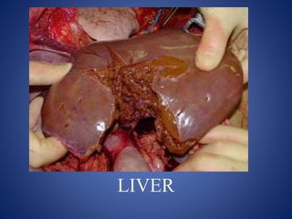
- 1. LIVER
- 2. DR.SYED UBAID M.S(KEM MUMBAI) FMAS LAPAROSCOPIC AND GENERAL SURGEON ASSOCIATE PROFESSOR OF SURGERY IIMSR,JALNA
- 4. Surface anatomy • In RUQ • 5th ICS in midclavicular line to the Rt costal margin. • Weighs 1400 g n women and 1800g n men . • Span 10 cm +/-2
- 5. Surface anatomy • Superior, anterior, and right lateral surfaces – fit diaphragm. – Falciform ligament • Posterior surface – Rt lobe: colon, right kidney, and duodenum – Lt lobe: stomach
- 8. • The liver covered by fibrous capsule that reflects on the diaphragm and post abdominal wall • Leaving a bear area that connects the liver to the retroperitoneum directly
- 9. Ligaments Liver supported by: • Coronary lig • Rt & Lt Triangular lig • Falciform lig
- 10. Fissures
- 11. 1. The liver is divided into right and left lobes of almost equal size by a major fissure (Cantlie’s line) running from the gallbladder fossa in front to the IVC fossa behind. 2. This division is based on the right and left branches of the hepatic artery and the portal vein , with tributaries of bile (hepatic) ducts following. 3.The middle hepatic vein (MHV) lies in Cantlie's line.
- 12. Liver segments •Couinaud divided the liver into 8 segments. • Divided vertically by • the 3 main hepatic veins and transversely by the right and left portal branches.
- 15. Blood Supply • Portal vein • Hepatic artery • Hepatic vein
- 17. Blood Supply – Portal Vein • Superior Mesentric and Splenic veins • Posterior to hepatic artery and bile duct at the hepatodudenal junction. • Valveless • 75% of total blood supply the liver • Pressure 3-5 mmHg
- 18. Blood supply – Hepatic artery • Intrahepatic anatomy; part of portal tried follows segmental anatomy. • Extrahepatic anatomy; highly variable: – Commonest ( in 60%) anatomy: abdominal aorta celiac trunk CHA proper hepatic art Rt and Lt hepatic artery – LHA seg 1,2,3 and middle hepatic artery seg 4. – RHA cystic art , Rt liver
- 20. Blood supply – Hepatic vein • Rt hepatic vein Drain seg 5,6,7,8 vena cava. • Middle hepatic vein Drain seg 4,5,8 • Lt hepatic vein Drain seg 2,3 [ seg 1 drain by short hepatic vena cava]
- 22. LIVER INJURY
- 23. Introduction • Most commonly injured organ in Blunt abdominal trauma • 2nd most commonly injured organ in Penetrating abdominal trauma after Bowel. • Motor vehicle collision is the most common injury mechanism • The posterior portion of the right lobe is the most common site of hepatic injury in blunt trauma
- 24. Why the liver… • Large organ • Friable parenchyma, thin capsule, fixed position in relation to spine prone to blunt injury • Wide bore, thin walled blood vessels with high blood flow Excessive blood loss • Right lobe larger, closer to ribs more injury
- 25. • Isolated liver injury occurs in less than 50% of patients. • Blunt trauma 45% with spleen • Rib fracture 33% with Liver injury Associations:
- 26. Injuries • Parenchymal damage • Subcapsular hematoma • Laceration • Contusion • Hepatic vascular disruption • Bile duct injury
- 28. • Blood Loss • Peritonism • Symptoms – Abdominal Pain – Radiation to shoulder – Altered Sensoium • Signs – Hypotension – RUQ tenderness, and guarding – Generalized Peritonism • Hemoperitoneum • Biliary Peritonitis – Delayed – Intra-abdominal abscess
- 29. Management • Initial resuscitation as per ATLS protocol • It is important to note the mechanism of injury • Clinical picture may vary from mild RUQ pain through to peritonism to haemorrhagic shock • Stable patients undergo CT imaging • Unstable patients require resuscitation and laparotomy
- 31. INVESTIGATIONS
- 32. Labs & Radiology • Hematologic • Elevated LFTs • DPL -- high sensitivity • CT scan is the diagnostic procedure of choice. • USG • MRI ?? • Diagnostic Laparoscopy
- 34. Angiography • Active bleeding • Transcatheter embolization • Embolization & stenting for fistulas.
- 36. Grade VI Hepatic Avulsion
- 37. CT Scans • Accurate in localizing the site of liver injury and any associated injuries • Used to monitor healing • CT criteria for staging liver trauma uses AAST liver injury scale • Grades 1-6
- 39. II-Parenchymal laceration 1-3cm deep, subcapsular hematoma1-3 cm thick.
- 40. III-Parenchymal laceration> 3cm deep and subcapsular hematoma> 3cm diameter.
- 41. IV-Parenchymal/supcapsular hematoma> 10cm in diameter, lobar destruction,
- 42. V- Global destruction or devascularization of the liver.
- 44. Gallbladder injuries… • Rare • Predisposing factors. • contusions, avulsions, lacerations or perforations.
- 45. MANAGEMENT
- 46. • Remember associated injuries – Spleen – Pancreas – Bowel • Resuscitate • Consider Cryoprecipitate, FFP • Assessment of injury – Spiral CT – Laparotomy ♦ Treatment ♦ OM ♦ NOM Management
- 47. Management • Initial management is done according to ATLS protocol. Criteria for non Operative Management: (1) haemodynamic stability, or stability achieved with minimal resuscitation(1-2 litres of crystalloid) (2) absence of other abdominal injuries requiring laparotomy (3) preserved consciousness allowing serial examination of abdomen (4) absence of peritonism (5) absence of ongoing bleeding on CT scan
- 48. • Non-operative management (NOM) consists of – close observation of the patient complemented with angio-embolization, if necessary. – Observational management involves • admission to a unit and the monitoring of vital signs, • strict bed rest, • frequent monitoring of hemoglobin concentration • serial abdominal examinations
- 49. Criteria for Operative Management 1) any patient who is haemodynamically unstable with suspected liver trauma (2) multiple transfusions required to maintain haemodynamic stability (3) signs of peritonism, or development of peritonism on serial abdominal examinations (4) active arterial blush on CT for which interventional techniques have failed and/or ongoing bleeding on CT scan with focal pooling of contrast (5) penetrating trauma
- 50. Operative management • In hemodynamically unstable patient • Grade IV, V and VI injuries • Goal is to arrest Hemorrhage • Initial control of hemorrhage is attained by – Perihepatic packing – Mannual compression
- 51. 4 Ps of operative management • Operative management can be summarized as – PUSH – PRINGLE – PLUG – PACK
- 52. Perihepatic packing • Lobes of the liver must be compressed back to normal position • Packs should never be inserted into the hepatic wound – tear the vessels and will increase the bleeding • To reduce the risk of abdominal compartment syndrome due to aggresive packing, – some advocate closing the upper part of the wound to enhance the tamponade effect but leaving the lower two-thirds open covered by bagota.
- 53. • The right costal margin is elevated, and the pads are strategically placed over and around the bleeding site • Additional pads should be placed between the liver, diaphragm, and anterior chest wall until the bleeding has been controlled. • Sometimes 10 to 15 pads may be required to control the hemorrhage from an extensive right lobar injury
- 56. Perihepatic packing • Packing is not as effective for the injuries to the left hemiliver, • With the abdomen open, there is insufficient abdominal and thoracic wall anterior to the left hemiliver to provide adequate compression. • Fortunately, haemorrhage from the left hemiliver can be controlled by – dividing the left triangular and left coronary ligaments and compressing the left hemiliver between the hands.
- 57. • Packs are removed after 36 to 48 hours provided the patient is stable • Should NOT be removed before 24 hours as there are chances of rebleed • Perihepatic packing will control profuse haemorrhage in up to 80% of patients
- 58. • A Pringle maneuver can help delineate the source of hemorrhage. • In fact, hemorrhage from hepatic artery and portal vein injuries will halt with the application of a vascular clamp across the portal triad; • whereas, bleeding from the hepatic veins and retrohepatic vena cava will continue.
- 59. The Pringle maneuver, performed with a vascular clamp, occludes the hepatic pedicle containing the portal vein, hepatic artery, and common bile duct
- 61. Hemorrhage control • Ligation upto common hepatic artery is tolerated due to collaterols • Common hepatic artery should be repaired • If right hepatic artery is ligated the cholecystectomy should be performed
- 62. Hemorhage control “Hepatorraphy” • A running suture is used to approximate the edges of shallow lacerations, • Deeper lacerations are approximated using interrupted horizontal mattress sutures placed parallel to the edge of the laceration. • When the suture is tied, tension is adequate when – visible hemorrhage ceases or – liver blanches around the suture
- 64. Mesh Wrapping • Highly selective tight compression without increased intraabdominal pressure • Key points – Apply mesh under enough tension to create a tamponade effect – mesh should be attached into two anchoring stable points • diaphragmatic crus and • the falciform ligament
- 65. Hepatotomy and selective vascular ligation: • Pachter et al. recommend a rapid and extensive finger fracture, often through normal parenchyma, to reach the site of injury • Hepatotomy is done under Pringle manoeuvre – finger fracture method is used to divide the parenchyma to ligate the bleeding vessels • Pringle clamp is released intermittently to identify bleeding vessels. Finger fracture technique (digitoclasy), liver parenchyma is crushed between the thumb and one finger isolating vessels and bile ducts, which can then be ligated and divided.
- 66. Non-anatomical resection of liver • Removal of devitalised parenchyma using the line of injury as the boundary of the resection rather than standard anatomical planes • Used in conjunction with inflow control and hepatotomy
- 67. Anatomical resection of liver • Mortality exceeds 50 percent so performed rarely
- 68. Hepatic segments Resections • Right hemihepatectomy (segments 5 to 8); AKA as Right hepatectomy or right hepatic lobectomy • Right trisectionectomy (segments 4 to 8); AKA as Right lobectomy or Rrisegmentectomy of Starzl • Left hemihepatectomy (segments 1 to 4); AKA as Left hepatectomy or Left hepatic lobectomy • Left lateral sectionectomy (segments 1 to 3); AKA as Left lobectomy or Left lateral segmentectomy
- 69. Intrahepatic balloon tamponade • Useful for transhepatic penetrating injury. • Foley catheter and Penrose drain or a Sengstaken-Blakemore tube can be used • Passed into the length of the tract and then inflated. • Radio-opaque contrast fluid is used so integrity and position can be later confirmed radiologically. • Once patient is stabilized it is removed through re laprotomy
- 71. Total vascular exclusion • Last resort limited to specialist centres • Used for extensive retrohepatic venous injuries, • Involves clamping of the portal triad and infra- and supra-hepatic IVC. • Used to manage grade V penetrating injuries
- 72. Post Op Complications • Post operative hemorrhage – Surgical haemorrhage (ie discrete bleeding) – disseminated intravascular coagulation account for the majority of causes – Chances increased if packs removed <36 hours • Coagulapathy is corrected first if hemorrhage is persistant then angiography with embolisation or re laprotomy is consisdered.
- 73. Sepsis and abscess • 12-32% of patients • CT with intravenous and oral contrast should be performed to diagnose the cause of sepsis • Most intraabdominal abscess can be drained percutaneously under USG or Ct guidance • If not possible then operative drainage is done
- 74. Summary • Non operative management with interventional tehniques is preferred • Grade IV and V injuries in stable patients can also be managed conservatively • In surgical management damage control surgery is preferred than definite procedures