

Recomendados
Más contenido relacionado
La actualidad más candente
La actualidad más candente (20)
Destacado
Similar a Lecture 1 cell
Similar a Lecture 1 cell (20)
Más de Taw Alzu
Último
Último (20)
Lecture 1 cell
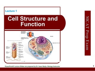
- 1. Lecture 1 PowerPoint® Lecture Slides are prepared by Dr. Isaac Barjis, Biology Instructor MCAT Prep Exam Cell Structure and Function 1
- 2. Outline Cellular Level of Organization Prokaryotic Cells Eukaryotic Cells Cell theory Cell size Organelles Nucleus and Ribosome Endomembrane System Other Vesicles and Vacuoles Energy related organelles Cytoskeleton Centrioles, Cilia, and Flagella 2
- 3. Cell Theory Cell was not discovered untill the development of Microscope Detailed study of the cell began in the 1830s A unifying concept in biology States that: All organisms are composed of cells All cells come only from preexisting cells Cells are the smallest structural and functional unit of organisms Cells carry genetic information in the form of DNA 3
- 4. Sizes of Living Things 4
- 5. Cell Size Cells range in size from one millimeter down to one micrometer Cells need a large surface area of plasma membrane to adequately exchange materials. The surface‑ area‑ to‑ volume ratio requires that cells be small 5
- 6. Surface to Volume Ratio 6
- 7. Microscopy Today: Compound Light Microscope Light passed through specimen Focused Max by glass lenses magnification about 1000X objects separated by 0.2 µm, 500X better than human eye Resolves Resolution is limited by the wavelength of light (nanometer) 7
- 8. Compound Light Microscope Diaphragm – controls amount of light – important for image contrast Coarse Adjustment Knob – focuses the image Fine Adjustment Knob – finely focuses the image 8
- 9. Microscopy Today: Transmission Electron Microscope Abbreviated T.E.M. Uses a beam of electrons to allow 100 fold higher magnification Because it uses beam of electrons, its resolution is at the atomic level (picometer) Tissue Can must be fixed and sectioned living specimen be examined by T.E.M? 9
- 11. Microscopy Today: Immunofluorescence Light Microscope Antibodies developed against a specific protein Fluorescent dye molecule attached to antibody molecules Specimen exposed to fluorescent antibodies Ultra-violet specimen light (black light) passed through Fluorescent dye glows in color where antigen is located Emitted light is focused by glass lenses onto human retina Allows in cell mapping distribution of a specific protein 11
- 12. Microscopy and Amoeba proteus 12
- 13. Cells Under the Microscope phase-contrast light microscope - look at unstained living animal cells. electron microscope - look at organelles e.g. ribosomes. fluorescence microscope - look at a living cell expressing green fluorescent protein or to do confocal microscopy.
- 14. Autoradiography Radioactive compounds decay or transform into other compounds or elements. An autoradiograph is an image on an xray film or nuclear emulsion produced by the pattern of decay emissions (e.g., beta particles or gamma rays) from a distribution of a radioactive substance Autoradiography can also uses radioactive molecule to study biochemical activity, 14
- 15. Cell Fractionation and Differential Centrifugation Cell fractionation is the breaking apart of cellular components Differential Allows separation of cell parts Separated Works centrifugation: out by size & density like spin cycle of washer The faster the machine spins, the smaller the parts that are settled out 15
- 16. Cell Fractionation and Differential Centrifugation 16
- 17. Eukaryotes Vs Prokaryotes Eukaryotic Cells Prokaryotic Cells The cells of “complex” organisms, including all plants, Protists, fungi and animals Contain a nucleus and membrane bound organelles “Simple” organisms, including bacteria and cyanobacteria (blue-green algae) Lack a nucleus and other membrane-encased organelles. Can specialize for certain functions, such as absorbing nutrients from food or transmitting nerve impulses; multicellular organs and organisms Usually exist as single, virtually identical cells Cell Wall present in Plants and Fungi only Ribosome: 40s, 60S Cell Wall Ribosome: 30S, 50S 17
- 18. The Structure of Bacteria Occur in three basic shapes: Spherical coccus, Rod-shaped bacillus, Spiral spirillum (if rigid) or spirochete (if flexible). Cell Envelope includes: Plasma membrane - lipid bilayer with imbedded and peripheral protein Cell wall - maintains the shape of the cell 18
- 19. The Structure of Bacteria 19
- 20. The Structure of Bacteria 20
- 21. The Structure of Bacteria Cytoplasm & Appendages Cytoplasm Semifluid solution Bounded by plasma membrane Contains water, inorganic and organic molecules, and enzymes. Nucleoid is a region that contains the single, circular DNA molecule. Plasmids are small accessory (extrachromosomal) rings of DNA Appendages Flagella – Provide motility Fimbriae – small, bristle-like fibers that sprout from the cell surface Sex pili – rigid tubular structures used to pass DNA from cell to cell 21
- 22. Eukaryotic Cells Domain Eukarya includes: Fungi Plants Protists Animals Cells contain: Membrane-bound nucleus that houses DNA Specialized organelles Plasma membrane Much larger than prokaryotic cells Some cells (e.g., plant cells) have a cell wall 22
- 23. Hypothesized Origin of Eukaryotic Cells 23
- 24. Eukaryotic Cells: Organelles Eukaryotic cells are compartmentalized They contain small structures called organelles Perform specific functions Isolates reactions from others Two classes of organelles: Endomembrane system: Organelles that communicate with one another Via membrane channels Via small vesicles Energy related organelles Mitochondria & chloroplasts Basically independent & self-sufficient 24
- 28. Cytosole Cytosol, contains many long, fine filaments of protein that are responsible for cell shape and structure and thereby form the cell’s cytoskeleton 28
- 29. Nucleus Command center of cell, usually near center Separated from cytoplasm by nuclear envelope Consists of double layer of membrane Nuclear pores permit exchange between nucleoplasm & cytoplasm Contains chromatin in semifluid nucleoplasm Chromatin contains DNA of genes, and proteins (Histones) Condenses to form chromosomes Chromosomes are formed during cell division Nucleolus is a dense structure in the nucleus Synthesize ribosome RNA (rRNA) 29
- 30. Anatomy of the Nucleus 30
- 31. Ribosomes Are the site of protein synthesis in the cell Composed of rRNA and protein Consists of a large subunit and a small subunit Subunits made in nucleolus May be located: On the endoplasmic reticulum (thereby making it “rough”), or Free in the cytoplasm 31
- 32. Nucleus, Ribosomes, & ER 32
- 33. Endomembrane System Series of intracellular membranes that compartmentalize the cell Restrict enzymatic reactions to specific compartments within cell Consists of: Nuclear envelope Membranes of endoplasmic reticulum Golgi apparatus Vesicles Several types Transport materials between organelles of system 33
- 34. Endomembrane System: The Endoplasmic Reticulum A system of membrane channels and saccules (flattened vesicles) continuous with the outer membrane of the nuclear envelope Rough ER Studded with ribosomes on cytoplasmic side Protein anabolism Synthesizes proteins Modifies and processes proteins Adds sugar to protein Results in glycoproteins Smooth ER No ribosomes Synthesis of lipids Site of various synthetic processes, detoxification, and storage Forms transport vesicles 34
- 36. Endomembrane System: The Golgi Apparatus Golgi Apparatus Consists of flattened, curved saccules Resembles Modifies stack of hollow pancakes proteins and lipids Receives vesicles from ER on cis (or inner face) Modifies them and repackages them in vesicles Release the vesicles from trans (or outer face) Within cell Export from cell (secretion, exocytosis) 36
- 38. Endomembrane System: Lysosomes Membrane-bound vesicles (not in plants) Produced by the Golgi apparatus Contain powerful digestive enzymes and are highly acidic Digestion of large molecules Recycling of cellular debris and resources Autolysis may occur in injured or dying cell to cause apoptosis (programmed cell death, like tadpole losing tail) 38
- 39. Lysosomes 39
- 40. Endomembrane System: Summary Proteins produced in rough ER and lipids from smooth ER are carried in vesicles to the Golgi apparatus. The Golgi apparatus modifies these products and then sorts and packages them into vesicles that go to various cell destinations. Secretory vesicles carry products to the membrane where exocytosis produces secretions. Lysosomes fuse with incoming vesicles and digest macromolecules. 40
- 41. Endomembrane System: A Visual Summary 41
- 42. Peroxisomes Similar to lysosomes Membrane-bounded vesicles Enclose enzymes that rid the cell of toxic peroxides Participate in the metabolism of fatty acids and many other metabolites 42
- 43. Peroxisomes 43
- 44. Vacuoles Membranous sacs that are larger than vesicles Store materials that occur in excess Others very specialized (contractile vacuole) Plants cells typically have a central vacuole Up to 90% volume of some cells Functions in: Storage of water, nutrients, pigments, and waste products Development of turgor pressure Some functions performed by lysosomes in other eukaryotes 44
- 45. Vacuoles 45
- 46. Energy-Related Organelles: Chloroplast Structure Bounded Inner by double membrane membrane infolded Forms disc-like thylakoids, which are stacked to form grana Suspended in semi-fluid stroma Chlorophyll Green photosynthetic pigment Chlorophyll capture solar energy 46
- 47. Energy-Related Organelles: Chloroplasts Serve as the site of photosynthesis Captures light energy to drive cellular machinery Photosynthesis Synthesizes carbohydrates from CO2 & H2O Makes own food using CO2 as only carbon source Inorganic molecules (Energy-poor compounds) are converted to organic molecules (energy-rich compounds) Only plants, algae, and certain bacteria are capable of conducting photosynthesis 47
- 49. Energy-Related Organelles: Mitochondria Smaller than chloroplast Contain ribosomes and their own DNA Surrounded by a double membrane Inner membrane surrounds the matrix and is convoluted (folds) to form cristae. Matrix – Inner semifluid containing respiratory enzymes Break down carbohydrates Involved in cellular respiration Produce most of ATP utilized by the cell Contain their own DNA and ribosome i.e. they are semiautonomous Inherited from Oocyte 49
- 52. The Cytoskeleton Maintains Assists Aids cell shape in movement of cell and organelles movement of materials in and out of cells Three types of macromolecular fibers Microfilament Intermediate Filaments Microtubules Assemble and disassemble as needed 52
- 53. The Cytoskeleton: Actin Filaments Microfilament are rods of actin Extremely thin filaments like twisted pearl necklace Support for microvilli in intestinal cells Intracellular traffic control For moving stuff around within cell Cytoplasmic streaming Function in pseudopods of amoeboid cells Important component in muscle contraction 53
- 54. The Cytoskeleton: Actin Filament Operation 54
- 55. The Cytoskeleton: Intermediate Filaments Intermediate microtubules Rope-like in size between actin filaments and assembly of fibrous polypeptides Functions: Support nuclear envelope Cell-cell junctions, like those holding skin cells tightly together 55
- 56. The Cytoskeleton: Microtubules Hollow cylinders made of two globular proteins called α and β tubulin Spontaneous pairing of α and β tubulin molecules form structures called dimers Dimers then arrange themselves into tubular spirals of 13 dimers around Assembly: Under control of Microtubule Organizing Center (MTOC) Most important MTOC is centrosome Function: Provide framework for movement of organelle within cell Direct separation of chromosomes during cell division (e.g. Centrioles are composed of microtubules) Provide locomotion and movement (e.g. flagella and cilia) 56
- 57. The Cytoskeleton: Microtubule Operation 57
- 59. Microtubular Arrays: Centrioles Short, One hollow cylinders pair per animal cell Located in centrosome of animal cells Oriented at right angles to each other Separate during mitosis to determine plane of division 59
- 61. Microtubular Arrays: Cilia and Flagella Hair-like projections from cell surface that aid in cell movement In eukaryotes, cilia are much shorter than flagella Cilia move in coordinated waves like oars Flagella move like a propeller or cork screw 61
- 62. Structure of a Flagellum 62
- 63. Comparison of Prokaryotic and Eukaryotic Cells 63
- 64. Lecture 1 PowerPoint® Lecture Slides are prepared by Dr. Isaac Barjis, Biology Instructor MCAT Exam Prep Membrane Structure and Function 64
- 65. Outline Membrane Models Fluid-Mosaic Plasma Membrane Structure and Function Phospholipids Proteins Plasma Membrane Permeability Diffusion Osmosis Transport Via Carrier Proteins Cell Surface Modifications 65
- 66. Structure and Function: The Phospholipid Bilayer The plasma membrane is common to all cells Separates: Internal living cytoplasmic from External environment of cell Phospholipid bilayer: External surface lined with hydrophilic polar heads Cytoplasmic surface lined with hydrophilic polar heads Nonpolar, hydrophobic, fatty-acid tails sandwiched in between 66
- 67. Unit Membrane 67
- 68. Membrane Models Fluid-Mosaic Model Three components: Basic membrane referred to as phospholipid bilayer Protein molecules Float around like icebergs on a sea Membrane proteins may be peripheral or integral Peripheral proteins are found on the inner membrane surface Integral proteins are partially or wholly embedded (transmembrane) in the membrane Some have carbohydrate chains attached Cholesterol 68
- 69. The Fluid Mosaic Model 69
- 71. Functions of Membrane Proteins Channel Proteins: Carrier Proteins: Provides unique chemical ID for cells Help body recognize foreign substances Receptor Proteins: Combine with substance to be transported Assist passage of molecules through membrane Cell Recognition Proteins: Tubular Allow passage of molecules through membrane Binds with messenger molecule Causes cell to respond to message Enzymatic Proteins: Carry out metabolic reactions directly 71
- 73. Science Focus: Cell Signaling 73
- 74. Types of Transport: Active vs. Passive Plasma membrane is differentially (selectively) permeable Allows some material to pass Inhibits passage of other materials Passive Transport: No ATP requirement Molecules follow concentration gradient Active Transport Requires carrier protein Requires energy in form of ATP 74
- 75. Passage of Molecules Across the Membrane 75
- 76. Types of Membrane Transport: Overview 76
- 77. Types of Transport: Diffusion A solution consists of: A solvent (liquid), and A solute (dissolved solid) Diffusion Net movement of solute molecules down a concentration gradient Molecules move both ways along gradient Molecules move from high to low Equilibrium: When NET change stops Solute concentration uniform – no gradient 77
- 78. Gas Exchange in Lungs: Diffusion Across Lung 78
- 79. Types of Transport: Osmosis Osmosis: Special case of diffusion Focuses on solvent (water) movement rather than solute Diffusion of water across a differentially (selectively) permeable membrane Solute concentration on one side high, but water concentration low Solute concentration on other side low, but water concentration high Water diffuses both ways across membrane but solute can’t Net movement of water is toward low water (high solute) concentration Osmotic pressure is the pressure that develops due to osmosis 79
- 80. Types of Transport: Carrier Proteins Facilitated Transport Small molecules Can’t get through membrane lipids Combine Follow Active with carrier proteins concentration gradient Transport Small molecules Move against concentration gradient Combining Requires with carrier proteins energy 80
- 81. Types of Transport: Carrier Proteins Facilitated Transport Small molecules Can’t get through membrane lipids Combine Follow Active with carrier proteins concentration gradient Transport Small molecules Move against concentration gradient Combining Requires with carrier proteins energy 81
- 82. Types of Transport: Membrane-Assisted Transport Macromolecules transported into or out of the cell inside vesicles Exocytosis – Vesicles fuse with plasma membrane and secrete contents Endocytosis – Cells engulf substances into pouch which becomes a vesicle Phagocytosis Pinocytosis vesicle – Large, solid material into vesicle – Liquid or small, solid particles go into Receptor-Mediated using a coated pit – Specific form of pinocytosis 82
- 83. Cell Surface Modifications: Junctions Cell Surfaces in Animals Junctions Between Cells Adhesion Intercellular filaments between cells Tight Junctions Form impermeable barriers Gap Junctions Junctions Plasma membrane channels are joined (allows communication) 83
- 84. Cell Surface Modifications Extracellular Matrix External meshwork of polysaccharides and proteins Found in close association with the cell that produced them Plant Cell Walls Plants have freely permeable cell wall, with cellulose as the main component Plasmodesmata penetrate cell wall Each contains a strand of cytoplasm Allow passage of material between cells 84