
Recomendados
Más contenido relacionado
La actualidad más candente
La actualidad más candente (20)
Similar a Adrenal glands
Similar a Adrenal glands (20)
Más de vanajayarrlagadda
Más de vanajayarrlagadda (20)
Último
Último (20)
Adrenal glands
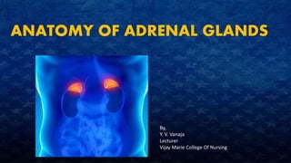
- 1. ANATOMY OF ADRENAL GLANDS By, Y. V. Vanaja Lecturer Vijay Marie College Of Nursing
- 2. The paired adrenal glands, one of which lies superior to each kidney in the retroperitoneal space. Flattened pyramidal shape. Each adrenal gland is 3-5 cm in height, 2-3 cm inn width, and a little less than 1 cm thick, with a mass of 3.5 -5g,only half its size at birth. During embryonic development, the adrenal glands differentiate into two structurally and functionally distinct regions • Peripherally located adrenal cortex – comprising 80 – 90% of the gland • Small , centrally located adrenal medulla. Connective tissue capsule covers the gland. The adrenal glands are highly vascularized The adrenal cortex produces steroid hormones that are essential for life The adrenal medulla produces three catecholamine hormones – norepinephrine, epinephrine, and a small amount of dopamine
- 3. Adrenal Cortex The adrenal cortex is subdivided into 3 zones Zona glomerulosa Zona fasciculate Zona reticularis • Outer zone – Zonaglomerulosa • Its cells are closely packed and arranged in spherical clusters and arched columns • It secrete hormones called mineralocorticoids – affect mineral homeostatsis. • Middle zone or Zona fasciculata • It is the widest of the three zones and consists of cells arranged in long, straight columns. • The cells of the zona fasciculata secrete mainly glucocorticoids, primarily cortisol, so named because they affect glucose homeostatsis • Inner zone – zona reticularis • Arranged in branching cords • They synthesize small amounts of weak androgens, steroid hormones that have masculinizing effects
- 5. Mineralocorticoids: • Aldosterone is the major mineralocorticoid • It regulates homeostasis of 2 mineral ions- namely, sodium ions, and potassium ions- and helps adjust blood pressure and the blood volume. • aldosterone also promotes excretion of hydrogen in the urine • This removal of acids from the body can help prevent acidosis. Glucocorticoids: • The glucocorticoids, which regulate metabolism and resistance to stress, include • cortisol also called as hydrocortisone. • Corticosterone • Cortisone • These 3 hormones secreted by the zona faciculata • Cortisol is the most abundant, accounting for the 95% of glucocorticoid activity
- 6. Androgens • In both males and females, the adrenal cortex secretes small amounts of weak androgens. • The major androgen secreted by the adrenal gland is dehydroepiandrosterone (DHEA) • After puberty in males, the androgen testosterone is also released in much greater quantity by the testes. • In females, however, adrenal androgens play important roles. • They promote libido and are converted into estrogens by other body tissues. • Adrenal androgens also stimulate growth of axillary and pubic hair in boys and girls and contribute to the prepubertal growth spurt. • The hormone that stimulates its secretion is ACTH
- 7. Adrenal medulla • The inner region of the adrenal gland, the adrenal medulla, is a modified sympathetic ganglion of the autonomic nervous system. • It develops from the same embryonic tissue as all other sympathetic ganglia, but its cells, which lacks axons, form clusters around large blood vessels. • Rather than releasing a neurotransmitter, the cells of the adrenal medulla secrete hormones. • The hormone producing cells called chromaffin cells.. • Because the ANS exerts direct control over the chromaffin cells, hormone release can occur very quickly. • Two major hormones released by adrenal medulla are • Epinephrine or adrenaline • Norepinephrine or noradrenaline • The chromaffin cells of the adrenal medulla secrete an unequal amount of these hormones – about 80% epinephrine and 20% norepinephrine • The hormones of the adrenal medulla intensify sympathetic responses that occur in other parts of the body.
- 8. In stressful situations and during exercise , impulses from the hypothalamus stimulate sympathetic preganglionic neurons, which in turn stimulate the chromaffin cells to secrete epinephrine and norepinephrine. These two hormones greatly augment the fight –or –flight response • By increasing the HR and force of contraction, epinephrine and norepinephrine increase the output of the heart, which increases B.P • These also increases blood flow to the heart, liver, skeletal muscles, and adipose tissue; • Dilate airways of lungs • Increase blood levels of glucose and fatty acids
- 9. PHYSIOLOGY OF ADRENAL GLANDS
- 10. Renin – angiotensin- aldosterone or RAA pathway Stimuli that initiate the renin-angiotensin-aldosterone pathway include dehydration, sodium deficiency or hemorrhage These conditions cause a decrease in blood volume Decreased blood volume leads to decrease d blood pressure Lowered blood pressure stimulates certain cells of the kidneys, called juxtaglomerular cells, to secrete the enzyme renin. The level of renin in the blood increases Renin converts angiotensinogen a plasma protein produced by the liver, into angiotensin I Blood containing increased levels of angiotensin I circulates in the body As blood flows through the capillaries, particularly those of the lungs, the enzyme angiotensin- converting enzyme converts angiotensin1 into the hormone angiotensin II
- 11. Blood level of angiotensin II increases Angiotensin II stimulates the adrenal cortex to secrete aldosterone Blood containing increased levels of aldosterone circulates to the kidneys In the kidneys, aldosterone increases reabsorption of sodium, which in turn causes reabsorption of water by osmosis • As a result water is lost in the urine • Aldosterone also stimulates the kidneys to increase secretion of potassium and hydrogen into the urine With increase reabsorption by the kidneys, blood volume increases As blood volume increases, blood pressure increases to normal
- 12. Angiotensin II also stimulates contraction of smooth muscle in the walls of arterioles The resulting vasoconstriction of the arterioles increases blood pressure and thus helps raise blood pressure to normal Besides angiotensin II, a second stimulator of aldosterone secretion is an increase in the potassium concentration of blood. a decrease in the blood potassium level has the opposite effect .
- 14. Regulation of glucocorticoids • Control of glucocorticoid secretion occurs via a typical negative feedback system. • Low blood levels of glucocorticoids, mainly cortisol • Stimulates neurosecretory cells in the hypothalamus to secrete corticotropin releasing hormone(CRH) • Promotes the release of ACTH from the anterior pituitary • ACTH flows in the blood to the adrenal cortex, stimulates glucocorticoid secretion
- 16. Effects of Glucocorticoids Protein Break down: • Glucocorticoids increase the rate of protein breakdown, mainly in , muscle fibers, and thus liberation of amino acids into the blood stream • The amino acids may be used by body cells for synthesis of new proteins or for ATP production Glucose Formation: • On stimulation by glucocorticoids liver cells may convert certain amino acids or lactic acid to glucose, which neurons and other cells can use for ATP production. Lipolysis; • Breakdown of triglycerides and release of fatty acids from adipose tissue into the blood Resistance to stress; • The additional glucose supplied by the liver cells provides tissues with a ready source of ATP to combat a range of stresses • Glucocorticoids makes blood vessels more sensitive to other hormones that cause vasoconstriction, they raise B.P
- 17. Anti-inflammatory Effect; • Glucocorticoids inhibit WBC that participate in inflammatory responses. • Unfortunately, glucocorticoids also retard tissue repair, as a result, they slow wound healing • Glucocorticoids are very useful in the treatment of chronic inflammatory disorders. Depression of Immune responses: • High doses of glucocorticoids depress immune responses • Glucocorticoids are prescribed for organ transplant recipients to retard tissue rejection by the immune system.
- 18. Cushing syndrome Introduction; Cushing syndrome occurs when body is exposed to high levels of the hormone cortisol for a long time. Cushing syndrome, sometimes called hypercortisolism, may be caused by the use of oral corticosteroid medication. The condition can also occur when body makes too much cortisol on its own. Definition; Cushing syndrome is a spectrum of clinical abnormalities caused by excess corticosteroids, particularly glucocorticoids
- 19. Causes of cushing syndrome • Prolonged administration of high doses of corticosteroids • ACTH secreting pituitary tumor • Cortisol – secreting neoplasm within the adrenal cortex that can be either carcinoma or adenoma • Excess secretion of ACTH from carcinoma of the lung or other malignant growth outside the pituitary or adrenal glands. • body may produce high levels of cortisol for a variety of reasons, including: • high stress levels, including stress related to an acute illness, surgery, injury, or pregnancy, especially in the final trimester • malnutrition • alcoholism • depression, panic disorders, or high levels of emotional stress
- 20. •High blood sugar •increased thirst •increased urination •osteoporosis •high blood pressure (hypertension) • headache •mood swings •anxiety •irritability •depression •an increased incidence of infections Signs and symptoms
- 21. In addition to the symptoms above, children with Cushing’s syndrome may also have: •obesity •slower rate of growth •high blood pressure (hypertension)
- 22. MANAGEMENT • The primary goal of treatment for cushing’s disease is to normalize hormone secretion. • If the underlying cause is pituitary adenoma, the standard treatment is surgical removal of the pituitary tumor • Radiation to the pituitary adenoma may be necessary if the surgical outcomes are not optimal.
- 23. Addisons syndrome Definition; Addison's disease, also known as primary adrenal insufficiency and hypocortisolism, is a long-term endocrine disorder in which the adrenal glands do not produce enough steroid hormones. Causes; • Autoimmune response • T.B • Infarction • Fungal disease • AIDS • Metastatic cancer
- 25. Management Treatment of adrenocortical insufficiency is replacement therapy Hydrocortisone is the most commonly used form of replacement therapy, has both glucocorticoid and mineralocorticoid properties Large volumes of 0.9% saline solutions and 5% dextrose are administered to reverse hypotension and electrolyte imbalance until B.p returns to normal.
- 33. ADENAL GLANDS