Most Beautiful Call Girl in Bangalore Contact on Whatsapp
pathophysiology of myocardial infraction
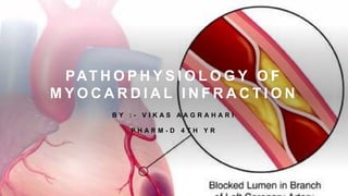
1. PAT H O P H Y S I O L O G Y O F
M Y O C A R D I A L I N F R A C T I O N
B Y : - V I K A S A A G R A H A R I
P H A R M - D 4 T H Y R
2. I N T R O D U C T I O N :
• Myocardial infarction is an irreversible injury to a part of the
heart or myocardial tissue that results from ischemia and
hypoxia finally necrosis of particular cells.
• Usually this is because one of the coronary arteries that
supplies blood to the heart develops a blockage due to an
unstable buildup of atherosclerotic plaques and other blood
cells.
• Myocardial infarction is one of the leading killer in the
United States.
• This is called Acute MI if it is sudden and serious.
• 64% of cases does not have chest pain or other
symptoms.
• This is called Silent myocardial infarction.
3.
4. T Y P E S O F
M I :
According to
PATHOLOGY:
Transmural AMI: ST
section higher than the
baseline is called an ST
elevation MI (STEMI)
which usually requires
more aggressive
treatment.
Subendocardial
AMI: Non-ST elevation
myocardial infarction
(NSTEMI), managed with
medication, although
angioplasty may be
required if the person is
considered to be at high
risk.
5. T Y P E S O F M I
2007 consensus document classifies MI into five main types:
• Type 1 – spontaneous MI related to ischemia due to a primary coronary
event such as plaque erosion and/or rupture, fissuring, or dissection
• Type 2 – MI secondary to ischemia due to either increased oxygen
demand or decreased supply, e.g. coronary artery spasm, coronary
embolism, anemia, arrhythmias, hypertension, or hypotension
• Type 3 – sudden unexpected cardiac death, including cardiac arrest,
often with symptoms suggestive of myocardial ischemia, accompanied
by new ST elevation,
• Type 4 – associated with coronary angioplasty or stents:
Type 4a – MI associated with Percutaneous coronary intervention
(PCI)
Type 4b – MI associated with stent thrombosis as documented by
angiography or at autopsy
• Type 5 – MI associated with CABG (Coronary Artery Bypass Graft)
6.
7. S Y M P T O M S :
Sudden chest pain that is felt behind the sternum and sometimes travels to the left arm or
the left side of the neck.
Shortness of breath
Sweating
Nausea
Vomiting
Abnormal heartbeats
Anxiety
Weakness
Feeling of indigestion
Fatigue
8. R I S K
F A C T O R S :
Cardiovascular disease
Old age
Tobacco smoking
High blood levels of LDL, low levels
of HDL,
Diabetes
High blood pressure
Lack of physical activity
Obesity
Chronic kidney disease
Excessive alcohol consumption
Use of Cocaine and Amphetamines
9.
10. P A T H O P H Y S I O L O G Y
:
The pathophysiology of acute myocardial infarction is
complex. Loss of viable myocardium impairs cardiac function,
which can lead to reduced cardiac output, if damage is
severe, cause cardiogenic shock. Infarction is tissue death
caused by ischemia. AMI occurs when localized myocardial
ischemia causes the development of a defined region of
necrosis. A collagen scar forms in the necrosis place.
Apoptosis also plays a role in the process of tissue damage
subsequent to MI. As a result, the patient's heart will be
permanently damaged. Ischemia can cause arrhythmias and
conduction blocks that can further impair function and
become life-threatening in some cases. Reduced cardiac
output and arterial pressure can elicit baroreceptors, that lead
to activation of sympathetic nerves and the RAAS.
11. M E C H A N I S M S
A N D
C O N S E Q U E N C E S
O F P L A Q U E
R U P T U R E :
• Coronary plaques which are prone to rupture are typically
small and nonobstructive, with a large lipid-rich core
covered by a thin fibrous cap. Activated macrophages and
T-lymphocytes localized at the site of plaque rupture are
thought to release metalloproteases and cytokines which
weaken the fibrous cap, rendering it liable to tear due to the
stress exerted by the blood flow. Plaque rupture reveals
subendothelial collagen, which serves as a site of platelet
adhesion, activation and aggregation.
12. 1 The release of substances such as TXA2
(thromboxane A2) , fibrinogen, 5-HT, PAF
and ADP, which further promote platelet
aggregation.
2 Activation of the clotting cascade,
leading to fibrin formation
and propagation and stabilization of
the occlusive thrombus.
13. • The resulting deficit of antithrombotic factors such as Thrombomodulin and Prostacyclin
enhances thrombus formation.
• Platelet-derived factors (e.g. TXA2, 5-HT) to cause vasoconstriction. This may promote the
development of local vasospasm, which worsens coronary occlusion.
• Sudden death and acute coronary syndrome is peaking at 9 a.m. and 11 p.m.
• Increased levels of Catecholamines at this time resulting in increased platelet aggregability,
vascular tone, heart rate and blood pressure, which may trigger plaque rupture and
thrombosis.
• Increased physical and mental stress can also cause MI and sudden death, supporting a
role for increases in catecholamines in MI pathophysiology.
14. • The degree of coronary occlusion and myocardial damage caused by plaque rupture
probably depends on systemic catecholamine levels, as well as local factors such as
plaque location and morphology, the depth of plaque rupture, and the extent to which
coronary vasoconstriction occurs.
• Severe and prolonged ischemia produces a region of necrosis spanning the entire
thickness of the myocardial wall (STEMI).
• Less severe and protracted ischemia (NSTEMI)can arise when:
1. Coronary occlusion is followed by spontaneous reperfusion.
2. The infarct-related artery is not completely occluded.
3. The oxygen demand in the affected zone of myocardium is smaller.
15. Most patients who sustain an MI have coronary atherosclerosis.
The thrombus formation occurs most often at the site of an atherosclerotic
lesion, thus obstructing blood flow to the myocardial tissues.
Plaque rupture is believed to be the triggering mechanism for the
development of the thrombus in most patients with an MI.
When the plaques rupture, a thrombus is formed at the site that can occlude
blood flow, thus resulting in an MI.
16. D I A G N O S I S :
• ECG - differentiate between
two types of myocardial
infarctions.
• Blood tests are Troponin
and Creatine kinase (CK-
MB).
17. T R E A T M E N T:
• Aspirin - which prevents further blood from
clotting, chest pain.
• Nitroglycerin – Vasodilator.
• STEMI is treated by reperfusion therapy,
angioplasty (arteries are pushed open)
Thrombolytics.
• People who have multiple blockages of their
coronary arteries, particularly if they also
have diabetes, treated with CABG).