Short case publication Version 3.17
•
2 likes•201 views

Short case...Neuromyelitis optica http://yassermetwally.com http://yassermetwally.net

Recommended
Recommended
More Related Content
Viewers also liked
Viewers also liked (20)
Similar to Short case publication Version 3.17
Similar to Short case publication Version 3.17 (20)
More from Professor Yasser Metwally
More from Professor Yasser Metwally (20)
Short case publication Version 3.17
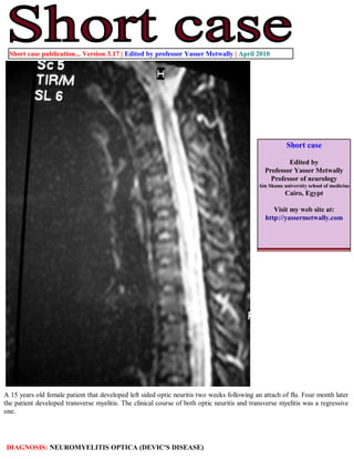
- 1. Short case publication... Version 3.17 | Edited by professor Yasser Metwally | April 2010 Short case Edited by Professor Yasser Metwally Professor of neurology Ain Shams university school of medicine Cairo, Egypt Visit my web site at: http://yassermetwally.com A 15 years old female patient that developed left sided optic neuritis two weeks following an attach of flu. Four month later the patient developed transverse myelitis. The clinical course of both optic neuritis and transverse myelitis was a regressive one. DIAGNOSIS: NEUROMYELITIS OPTICA (DEVIC'S DISEASE)
- 2. Figure 1. MRI FLAIR images showing periventricular white matter changes that are not typical for multiple sclerosis. Figure 2. MRI postcontrast T1 image (A) and MRI T2 image (B) showing a longitudinally extensive central hypointensity (A) and Hyperintensity (B) extending from C1 to D5. Notice the mild peripheral enhancement (A). Also notice the mild cord enlargement
- 3. Figure 3. MRI T2 images showing cord enlargement, central hyperintensities involving more that 2/3 of the spinal cord in cross section and the central dot sign. References 1. Metwally, MYM: Textbook of neurimaging, A CD-ROM publication, (Metwally, MYM editor) WEB-CD agency for electronic publishing, version 11.1a December 2010 Addendum A new version of short case is uploaded in my web site every week (every Saturday and remains available till Friday.) To download the current version follow the link "http://pdf.yassermetwally.com/short.pdf". You can download the long case version of this short case during the same week from: http://pdf.yassermetwally.com/case.pdf or visit web site: http://pdf.yassermetwally.com To download the software version of the publication (crow.exe) follow the link: http://neurology.yassermetwally.com/crow.zip At the end of each year, all the publications are compiled on a single CD-ROM, please contact the author to know more details. Also to view a list of the previously published case records follow the following link (http://wordpress.com/tag/case- record/) or click on it if it appears as a link in your PDF reader To inspect the patient's full radiological study, click on the attachment icon (the paper clip icon in the left pane) of the acrobat reader then double click on the attached file Click here to download the long case version of this short case in PDF format