Expression and inhibition of il 23 by colon cancer cells a promising approach in prevention of ibd.
•
1 recomendación•278 vistas

International journals call for papers, http://www.iiste.org/

Recomendados
Recomendados
Más contenido relacionado
La actualidad más candente
La actualidad más candente (20)
Similar a Expression and inhibition of il 23 by colon cancer cells a promising approach in prevention of ibd.
Similar a Expression and inhibition of il 23 by colon cancer cells a promising approach in prevention of ibd. (20)
Más de Alexander Decker
Más de Alexander Decker (20)
Último
Último (20)
Expression and inhibition of il 23 by colon cancer cells a promising approach in prevention of ibd.
- 1. Journal of Biology, Agriculture and Healthcare ISSN 2224-3208 (Paper) ISSN 2225 Vol.3, No.4, 2013 Expression and Inhibition Promising Approach Dr. Vishal Bhargava 1. Department of Biochemistry, G.R. Medical College 2. Department of Biotechnology DRDO Gwalior * E-mail of the corresponding author: The research is financed by G.R. Medical College Autonomous Society Abstract Inflammatory bowel disease is chronic uncontrolled inflamma activated leukocytes. Here we have developed an epithelial cell line HT-29 expressed elevated IL strains. In this in-vitro model of IBD, IL anti-inflammatory drug Sulfasalizine was studied and was found to block the up regulation of IL Keywords: IBD (Inflammatory Bowel Disease), HT (Lipopolysaccharide). 1. Introduction Inflammation can be defined as a series of non mediators respond to tissue injury (Elenkov IJ 2005). Inflammation, the response of tissue to injury, is characterized in the acute phase by increased blood flow and vascular permeability along with the accumulation of fluid, leukocytes, and inflammatory mediators such as cy development of specific humoral and cellular immune responses to the pathogen(s) present at the site of tissue injury (Carol A. 1997). Most cytokines involved in the inflammation processes their effects locally or systemically in an autocrine or paracrine manner (Carol A. 1997). Cytokines are involved in extensive networks that involve synergistic (proinflammatory cytokines) as (anti-inflammatory cytokines) and exhibit both negative and positive regulatory effects on various target cells (Adams G 2003). Sometimes immune cells fail to distinguish among the foreign and its native cells, as a result inflammation is targeted towards its own cells, the condition is termed as Auto inflammation. One such condition is Inflammatory Bowel Disease interacted with its own underlying immun there by leading to the destruction of tissue as such Inflammatory bowel disease (IBD) Ulcerative colitis (UC) and Crohn’s disease (CD). uncontrolled inflammation characterized by intense mucosal recruitment of activated leukocytes (Hanauer. S 2006). Although the diseases have some features in common, there ar Over the past 15 years wide variety of candidate genes have been studied for IBD. Significant linkages have been reported on chromosomes 1,3,6,7,12,14,16 and 19. Detailed mapping of chromosome 16 resulted in identification of the gene responsible, at least in part for this linkage (Podolsky D.K. 2002).This gene encodes a cytoplasmic protein designated nucleotide-binding oligomerization domain domain CARD15 (Hanauer B 2006) immune system. It is the first gene to be clearly associated with IBD, and >60 mutations have been recognized, 3 of which have been linked to development of CD. The mechanism where development of IBD remains unclear. The NOD2 gene is expressed mainly in monocyte/macrophage cell lines, where it has a role in host signaling pathway. Reasonable data suggest th Crohn’s disease is dominated by CD4+ lymphocytes with a type 1 helper Journal of Biology, Agriculture and Healthcare 3208 (Paper) ISSN 2225-093X (Online) 25 nd Inhibition of Il-23 by Colon Cancer Cells: A Promising Approach in Prevention of Ibd. Dr. Vishal Bhargava1* Dr. Neelima Singh1 , Dr.Richa Sijoria Department of Biochemistry, G.R. Medical College, Gwalior (M.P.) India. (474009) Department of Biotechnology DRDO Gwalior (M.P) India mail of the corresponding author: bhargavavishal6@gmail.com The research is financed by G.R. Medical College Autonomous Society Inflammatory bowel disease is chronic uncontrolled inflammation characterized by intense mucosal recruitment of activated leukocytes. Here we have developed an in vitro model for IBD using colon cancer cell line HT 29 expressed elevated IL-23 levels when induced with LPS isolated model of IBD, IL-23 was playing a key role in inflammation cascade. Action of inflammatory drug Sulfasalizine was studied and was found to block the up regulation of IL IBD (Inflammatory Bowel Disease), HT-29, IL-23 (Interleukin- Inflammation can be defined as a series of non-specific defense mechanism of body, in which cells and different o tissue injury (Elenkov IJ 2005). Inflammation, the response of tissue to injury, is characterized in the acute phase by increased blood flow and vascular permeability along with the accumulation of fluid, leukocytes, and inflammatory mediators such as cytokines. In the sub acute/chronic phase it is characterized by the development of specific humoral and cellular immune responses to the pathogen(s) present at the site of tissue injury Most cytokines involved in the inflammation processes are multifunctional. They are pleiotropic molecules that elicit their effects locally or systemically in an autocrine or paracrine manner (Carol A. 1997). Cytokines are involved in extensive networks that involve synergistic (proinflammatory cytokines) as well as antagonistic interactions inflammatory cytokines) and exhibit both negative and positive regulatory effects on various target cells Sometimes immune cells fail to distinguish among the foreign and its native cells, as a result inflammation is targeted towards its own cells, the condition is termed as Auto inflammation. Inflammatory Bowel Disease in which the gut micro biota if through any pathway gets interacted with its own underlying immune cells; the process of inflammation towards these microorganisms begins there by leading to the destruction of tissue as such Inflammatory bowel disease (IBD) refers to two chronic diseases that cause inflammation of the intestines: Crohn’s disease (CD). The hallmark of inflammatory bowel disease is chronic uncontrolled inflammation characterized by intense mucosal recruitment of activated leukocytes (Hanauer. S 2006). Although the diseases have some features in common, there are some important differences. Over the past 15 years wide variety of candidate genes have been studied for IBD. Significant linkages have been reported on chromosomes 1,3,6,7,12,14,16 and 19. Detailed mapping of chromosome 16 resulted in identification of the gene responsible, at least in part for this linkage (Podolsky D.K. 2002).This gene encodes a cytoplasmic protein binding oligomerization domain 2 NOD2 also known as caspase activation and recruitment domain CARD15 (Hanauer B 2006). This is a polymorphic gene the product of which is involved in the innate immune system. It is the first gene to be clearly associated with IBD, and >60 mutations have been recognized, 3 of which have been linked to development of CD. The mechanism whereby defects in the NOD2 gene lead to the development of IBD remains unclear. The NOD2 gene is expressed mainly in monocyte/macrophage cell lines, where it has a role in host signaling pathway. Reasonable data suggest that mucosa of the patients with establi Crohn’s disease is dominated by CD4+ lymphocytes with a type 1 helper-T-cell (Th1) phenotype, characterized by www.iiste.org y Colon Cancer Cells: A f Ibd. , Dr.Richa Sijoria2 Gwalior (M.P.) India. (474009) India bhargavavishal6@gmail.com tion characterized by intense mucosal recruitment of model for IBD using colon cancer cell line HT-29. Colon 23 levels when induced with LPS isolated from various bacterial 23 was playing a key role in inflammation cascade. Action of inflammatory drug Sulfasalizine was studied and was found to block the up regulation of IL-23. -23), Sulfasalizine, LPS specific defense mechanism of body, in which cells and different o tissue injury (Elenkov IJ 2005). Inflammation, the response of tissue to injury, is characterized in the acute phase by increased blood flow and vascular permeability along with the accumulation of fluid, tokines. In the sub acute/chronic phase it is characterized by the development of specific humoral and cellular immune responses to the pathogen(s) present at the site of tissue injury are multifunctional. They are pleiotropic molecules that elicit their effects locally or systemically in an autocrine or paracrine manner (Carol A. 1997). Cytokines are involved in well as antagonistic interactions inflammatory cytokines) and exhibit both negative and positive regulatory effects on various target cells Sometimes immune cells fail to distinguish among the foreign and its native cells, as a result the process of inflammation is targeted towards its own cells, the condition is termed as Auto inflammation. in which the gut micro biota if through any pathway gets e cells; the process of inflammation towards these microorganisms begins refers to two chronic diseases that cause inflammation of the intestines: The hallmark of inflammatory bowel disease is chronic uncontrolled inflammation characterized by intense mucosal recruitment of activated leukocytes (Hanauer. S 2006). e some important differences. Over the past 15 years wide variety of candidate genes have been studied for IBD. Significant linkages have been reported on chromosomes 1,3,6,7,12,14,16 and 19. Detailed mapping of chromosome 16 resulted in identification of the gene responsible, at least in part for this linkage (Podolsky D.K. 2002).This gene encodes a cytoplasmic protein also known as caspase activation and recruitment . This is a polymorphic gene the product of which is involved in the innate immune system. It is the first gene to be clearly associated with IBD, and >60 mutations have been recognized, 3 of by defects in the NOD2 gene lead to the development of IBD remains unclear. The NOD2 gene is expressed mainly in monocyte/macrophage cell lines, at mucosa of the patients with established cell (Th1) phenotype, characterized by
- 2. Journal of Biology, Agriculture and Healthcare ISSN 2224-3208 (Paper) ISSN 2225 Vol.3, No.4, 2013 production of IFN-γ, IL-2 IL-8 and IL by CD4+ lymphocytes with an atypical type 2 helper transforming growth factor β (TGF- 2. Materials and Methods 2.1 HT-29 These cells are also adherent colonic epithelial cells obtaine with different cellular products mainly carcinoembryonic antigen (CEA); transforming growth factor beta binding protein; mucin. The cell line used in the present study was purchased from American Type Cu (ATCC), Manassas VA 20108, USA. 2.2 Reagents DMEM Powder, Bovine serum albumin (BSA), DMSO, Sodium bi carbonate, Trizma, EDTA and others were obtained from Sigma (Sigma Aldrich co. St. Louis MO USA). 2.3 ELISA kit Kit for both IL-23 was quantitated from the cell supernatants using DuoSet ELISA Development KITS. ELISAs are sensitive enzyme immunoassays that measure soluble levels of proteins in biological samples. R&D SYSTEM provides Complete ELISA kits that offer accurate and DuoSet kits contain the basic components required to develop an immunoassay to measure natural or recombinant proteins. All these were obtained from 3. Methodology In our study HT-29 cells were cultured with a medium consisting of DMEM, 10%FETAL BOVINE SERUM (FBS) and 1% penicillin –streptomycin antibiotic solution .The cells were incubated at 37 of 5% CO2 in air. 3.1 Seeding Around 80% confluency stage, the cells were sub cultured. Medium contained in the flask was completely discarded using a sterile disposable pipette. Cells were washed with 1X PBS so as to remove the dead cells and the cell debris. Cells were treated with trypsin-EDTA solution (0.25%W/V) and kept at37 cells were detached from the substratum, as monitored under the phase contrast microscope. Once the cells were completely detached, immediately excess amount of prolonged exposure of cells with the trypsin the complete medium acts as an inhibitor of trypsin. Flushing gently with a pipette t From this cell suspension, a sample was subjected to counting using a hemocytometer. The desired no. of cells was selected by diluting it with complete medium. Seed the cell suspension diluted with the medium in the resp plate. (12, 24, or 96 well plate) according to the requirement of the experiment. Incubate the respective plates over night at 37°C in the CO2 incubator prior to any treatment, for ensuring proper growth and spreading of cells. 3.2Treatment with LPS and the drug addition Specific cell line was selected based on the experiment and the cells were allowed to get confluent. Post confluent cells were seeded in 96 well plate as described above, with a concentration of 30,000 then left at CO2 incubator for over night incubation. As designed for the experiment 1) if the cells are to be pre-incubated with drug followed by LPS treatment, the cells in the plate were incubated with drug in different concentrations. Based on the solubility of the drug if soluble in water was directly diluted with medium and applied to the cells or if insoluble was the diluted in DMSO and the applied. 3 hours later of drug incubation cells were treated with LPS to get inflamed. LPS treatment was optimiz efficient strain to cause inflammation was chosen for the experiment. Based on the volume of the cell suspension in the well LPS was introduced respectively. Followed this plate containing cells were incub overnight. Next day supernatant from each well was collected by centrifuging plate at 1500 rpm for 15 min. Supernatant from the plate was collected in sterile eppendorfs and was stored as samples for ELISA at plate with the adherent cells was then undertaken for the MTT assay for measuring the cell viability. To assess the maximum tolerance of cells for the given concentration of drug MTT assay was performed. ELISA per the user manual provided with the R Observations of ELISA are depicted graphically as follows with interpretations. Journal of Biology, Agriculture and Healthcare 3208 (Paper) ISSN 2225-093X (Online) 26 8 and IL-23. In contrast the mucosa in patients with ulcerative colitis may be dominated th an atypical type 2 helper-T-cell (Th2) phenotype, characterized by the production of -β) and IL-5 (Podolsky. D.K 2002). are also adherent colonic epithelial cells obtained from the patients of colorectal adenocarcinoma but with different cellular products mainly carcinoembryonic antigen (CEA); transforming growth factor beta binding protein; mucin. The cell line used in the present study was purchased from American Type Cu (ATCC), Manassas VA 20108, USA. DMEM Powder, Bovine serum albumin (BSA), DMSO, Sodium bi carbonate, Trizma, EDTA and others were (Sigma Aldrich co. St. Louis MO USA). quantitated from the cell supernatants using DuoSet ELISA Development KITS. ELISAs are sensitive enzyme immunoassays that measure soluble levels of proteins in biological samples. R&D SYSTEM provides Complete ELISA kits that offer accurate and reproducible results with no development time DuoSet kits contain the basic components required to develop an immunoassay to measure natural or recombinant proteins. All these were obtained from R&D Systems, Inc 614 McKinley Place NE, Minneapolis, USA. 29 cells were cultured with a medium consisting of DMEM, 10%FETAL BOVINE SERUM (FBS) streptomycin antibiotic solution .The cells were incubated at 37°C under a humidified atmosphere Around 80% confluency stage, the cells were sub cultured. Medium contained in the flask was completely discarded using a sterile disposable pipette. Cells were washed with 1X PBS so as to remove the dead cells and the cell debris. EDTA solution (0.25%W/V) and kept at37°C in the CO2 incubator for 2 cells were detached from the substratum, as monitored under the phase contrast microscope. Once the cells were completely detached, immediately excess amount of complete medium was added to the cells. This is because a prolonged exposure of cells with the trypsin -EDTA solution is known to cause cell death. The serum contained in the complete medium acts as an inhibitor of trypsin. Flushing gently with a pipette tip mixed the contents of the flask. From this cell suspension, a sample was subjected to counting using a hemocytometer. The desired no. of cells was selected by diluting it with complete medium. Seed the cell suspension diluted with the medium in the resp plate. (12, 24, or 96 well plate) according to the requirement of the experiment. Incubate the respective plates over C in the CO2 incubator prior to any treatment, for ensuring proper growth and spreading of cells. and the drug addition Specific cell line was selected based on the experiment and the cells were allowed to get confluent. Post confluent cells were seeded in 96 well plate as described above, with a concentration of 30,000-40,000 cells/well. Plate was hen left at CO2 incubator for over night incubation. As designed for the experiment 1) if the cells are to be incubated with drug followed by LPS treatment, the cells in the plate were incubated with drug in different ility of the drug if soluble in water was directly diluted with medium and applied to the cells or if insoluble was the diluted in DMSO and the applied. 3 hours later of drug incubation cells were treated with LPS to get inflamed. LPS treatment was optimized using different strains of bacteria and maximum efficient strain to cause inflammation was chosen for the experiment. Based on the volume of the cell suspension in the well LPS was introduced respectively. Followed this plate containing cells were incub overnight. Next day supernatant from each well was collected by centrifuging plate at 1500 rpm for 15 min. Supernatant from the plate was collected in sterile eppendorfs and was stored as samples for ELISA at the adherent cells was then undertaken for the MTT assay for measuring the cell viability. To assess the maximum tolerance of cells for the given concentration of drug MTT assay was performed. ELISA per the user manual provided with the R&D Duo kit system. Observations of ELISA are depicted graphically as follows with interpretations. www.iiste.org 23. In contrast the mucosa in patients with ulcerative colitis may be dominated cell (Th2) phenotype, characterized by the production of d from the patients of colorectal adenocarcinoma but with different cellular products mainly carcinoembryonic antigen (CEA); transforming growth factor beta binding protein; mucin. The cell line used in the present study was purchased from American Type Culture collection DMEM Powder, Bovine serum albumin (BSA), DMSO, Sodium bi carbonate, Trizma, EDTA and others were quantitated from the cell supernatants using DuoSet ELISA Development KITS. Sandwich ELISAs are sensitive enzyme immunoassays that measure soluble levels of proteins in biological samples. R&D reproducible results with no development time DuoSet kits contain the basic components required to develop an immunoassay to measure natural or recombinant , Inc 614 McKinley Place NE, Minneapolis, USA. 29 cells were cultured with a medium consisting of DMEM, 10%FETAL BOVINE SERUM (FBS) C under a humidified atmosphere Around 80% confluency stage, the cells were sub cultured. Medium contained in the flask was completely discarded using a sterile disposable pipette. Cells were washed with 1X PBS so as to remove the dead cells and the cell debris. C in the CO2 incubator for 2-5 mins. The cells were detached from the substratum, as monitored under the phase contrast microscope. Once the cells were complete medium was added to the cells. This is because a EDTA solution is known to cause cell death. The serum contained in ip mixed the contents of the flask. From this cell suspension, a sample was subjected to counting using a hemocytometer. The desired no. of cells was selected by diluting it with complete medium. Seed the cell suspension diluted with the medium in the respective plate. (12, 24, or 96 well plate) according to the requirement of the experiment. Incubate the respective plates over C in the CO2 incubator prior to any treatment, for ensuring proper growth and spreading of cells. Specific cell line was selected based on the experiment and the cells were allowed to get confluent. Post confluent 40,000 cells/well. Plate was hen left at CO2 incubator for over night incubation. As designed for the experiment 1) if the cells are to be incubated with drug followed by LPS treatment, the cells in the plate were incubated with drug in different ility of the drug if soluble in water was directly diluted with medium and applied to the cells or if insoluble was the diluted in DMSO and the applied. 3 hours later of drug incubation cells were ed using different strains of bacteria and maximum efficient strain to cause inflammation was chosen for the experiment. Based on the volume of the cell suspension in the well LPS was introduced respectively. Followed this plate containing cells were incubated in CO2 incubator for overnight. Next day supernatant from each well was collected by centrifuging plate at 1500 rpm for 15 min. Supernatant from the plate was collected in sterile eppendorfs and was stored as samples for ELISA at -80°C. The the adherent cells was then undertaken for the MTT assay for measuring the cell viability. To assess the maximum tolerance of cells for the given concentration of drug MTT assay was performed. ELISA was performed as
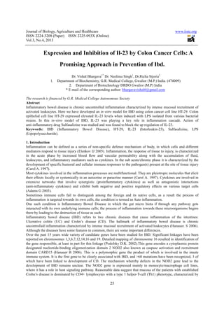
- 3. Journal of Biology, Agriculture and Healthcare ISSN 2224-3208 (Paper) ISSN 2225 Vol.3, No.4, 2013 4. Observations and Results. The Inflammatory Bowel Disease (IBD) is a chronic autoinflammatory disease where intestinal epithelial cells (IEC) gets differentiated as cancer cells when induced by some inflammatory agents. Normally in remain separated from underlying immune cells by a membrane lining. In most of the cases of IBD the underlying immune cells comes in direct contact (Hendrickson BA 2002), thereby leading to uncontrolled tissue destruction (Weber CR 2007). The process of inflammation could be studied with the help of certain tumour markers. In prepa model for IBD several cytokines were used as prominent markers whose upregulation determined the stages of inflammation (Carol A 1997). In-vitro model for IBD could be developed by using colon cancer cell line like HT which show some inflammatory responses. In our experiment we have used HT proteins indicating inflammatory responses (Haller D 2004). Development of inflammatory conditions in HT studies the bacterial DNA or bacteria as a whole were confirmed to express inflammatory responses (Haller D 2000 & Mahmood A 2003). The idea to introduce Lipopolysaccharide (LPS), a product of gram negative bacterial cell wal as an inflammatory agent is a new approach of our research laboratory for this study since no study in this regard has been made so far. In normal intestinal flora (in-vivo) numbers of bacterial strains exist but few of them have been found to be pathogenic in nature. LPS was isolated from different bacterial strains and were introduced in HT selection of LPS we used strains of E.coli B E.coli B-4 did not induce much inflammatory conditions in HT of bacteria like Salmonella Minnesota (S.Minn.), Salmonella Enteritidis (S.Ent.) and Pseudomonas to study the inflammatory reactions. It was observed that S.Minn and After developing inflammatory conditions in colon cancer cell line HT S.Minn and S.Ent we then checked inflammatory responses with cytokines as a marker (Neurath MF 2003). Cytokines are categorized under pro Interleukin-23 (IL-23) cytokine for observing the effect of LPS. LPS induction expressed different levels of IL with respect to each bacterial strain (Puleston J 2005). Upregu the inflammatory conditions in given cell line. Further in our study to check the inflammation process developed, the effect of certain anti studied in the same (Botoman VA 1991). concentration was introduced to inhibit the upregulated cytokine IL inhibition of upregulated IL-23 with minimum concentration of Sulfasalizine was mimic the in-vivo model of IBD (Mulder J 1988) (Fig.1). Dose response curve (DRC) was set up for each given concentration of drug with respect to bacterial strain and cell line to study the cytotoxic effect and most effect 4.1Figure.1 4.2 Figure.2 5. Conclusion HT-29 cells expressed different levels of inflammatory processes for in Lipopolysaccharide (LPS) isolated from strains of gram-negative bacteria induced inflammation in the monolayer and co were highly expressed in the process of inflammation. Sulfasalizine at different concentration was found effective in treatment of inflammatory bowel disease. 6. References Araki, Y. Sugihara, H. & Hattori T. (2006) In vitro effects of dextran sulfate sodium on a Caco plausible mechanisms for dextran sulfate sodium Bisping, G. Lügering, N. Lütke-Brintrup, S. Pauels, H.G. Schürmann, G, Domschke, W. & Kucharzik, T. (2001) Patients with inflammatory bowel disease (IBD) reveal increased induction capacity of intracellular interferon-gamma (IFN-gamma) in peripheral CD8+ lymphocytes co Journal of Biology, Agriculture and Healthcare 3208 (Paper) ISSN 2225-093X (Online) 27 The Inflammatory Bowel Disease (IBD) is a chronic autoinflammatory disease where intestinal epithelial cells (IEC) rentiated as cancer cells when induced by some inflammatory agents. Normally in remain separated from underlying immune cells by a membrane lining. In most of the cases of IBD the underlying immune cells comes in direct contact with micro-biota of intestine resulting in the process of inflammation (Hendrickson BA 2002), thereby leading to uncontrolled tissue destruction (Weber CR 2007). The process of inflammation could be studied with the help of certain tumour markers. In prepa model for IBD several cytokines were used as prominent markers whose upregulation determined the stages of vitro model for IBD could be developed by using colon cancer cell line like HT-29, CaCo which show some inflammatory responses. In our experiment we have used HT-29 cell which expressed various proteins indicating inflammatory responses (Haller D 2004). Development of inflammatory conditions in HT-29 cell line could be achieved through various ways. In several studies the bacterial DNA or bacteria as a whole were confirmed to express inflammatory responses (Haller D 2000 & Mahmood A 2003). The idea to introduce Lipopolysaccharide (LPS), a product of gram negative bacterial cell wal as an inflammatory agent is a new approach of our research laboratory for this study since no study in this regard has vivo) numbers of bacterial strains exist but few of them have been found to be nic in nature. LPS was isolated from different bacterial strains and were introduced in HT selection of LPS we used strains of E.coli B-4 (present in abundance in intestine) to elicit infection. It was found that much inflammatory conditions in HT-29 cells. Then LPS was isolated from different strains of bacteria like Salmonella Minnesota (S.Minn.), Salmonella Enteritidis (S.Ent.) and Pseudomonas to study the inflammatory reactions. It was observed that S.Minn and S.Ent were most potent inflammatory agents. After developing inflammatory conditions in colon cancer cell line HT-29 with LPS isolated from bacterial strains of S.Minn and S.Ent we then checked inflammatory responses with cytokines as a marker (Neurath MF 2003). Cytokines are categorized under pro-inflammatory and anti-inflammatory in nature. In our present study we selected 23) cytokine for observing the effect of LPS. LPS induction expressed different levels of IL with respect to each bacterial strain (Puleston J 2005). Upregulation of IL-23 cytokine (proinflammatory) confirmed the inflammatory conditions in given cell line. Further in our study to check the inflammation process developed, the effect of certain anti studied in the same (Botoman VA 1991). The popularly used drug for IBD like Sulfasalizine at different concentration was introduced to inhibit the upregulated cytokine IL-23 (Bonner GB 1996). Complete or above 70% 23 with minimum concentration of Sulfasalizine was regarded as efficiency of drug to vivo model of IBD (Mulder J 1988) (Fig.1). Dose response curve (DRC) was set up for each given concentration of drug with respect to bacterial strain and cell line to study the cytotoxic effect and most effective concentration with least cytotoxic effect was analyzed (Fig 2). 29 cells expressed different levels of inflammatory processes for in-vitro model of inflammatory bowel disease. ed from strains of Salmonella Enteritidis and Salmonella Minnesota negative bacteria induced inflammation in the monolayer and co-culture system of inflammation. IL were highly expressed in the process of inflammation. Sulfasalizine at different concentration was found effective in tory bowel disease. Araki, Y. Sugihara, H. & Hattori T. (2006) In vitro effects of dextran sulfate sodium on a Caco plausible mechanisms for dextran sulfate sodium-induced colitis. Oncol Rep, 16,1357-62. Brintrup, S. Pauels, H.G. Schürmann, G, Domschke, W. & Kucharzik, T. (2001) Patients with inflammatory bowel disease (IBD) reveal increased induction capacity of intracellular gamma) in peripheral CD8+ lymphocytes co-cultured with intestinal epithelial cells. www.iiste.org The Inflammatory Bowel Disease (IBD) is a chronic autoinflammatory disease where intestinal epithelial cells (IEC) rentiated as cancer cells when induced by some inflammatory agents. Normally in-vivo these epithelial cells remain separated from underlying immune cells by a membrane lining. In most of the cases of IBD the underlying biota of intestine resulting in the process of inflammation (Hendrickson BA 2002), thereby leading to uncontrolled tissue destruction (Weber CR 2007). The process of inflammation could be studied with the help of certain tumour markers. In preparing the in-vitro model for IBD several cytokines were used as prominent markers whose upregulation determined the stages of 29, CaCo-2, SW-620 and others 29 cell which expressed various ough various ways. In several studies the bacterial DNA or bacteria as a whole were confirmed to express inflammatory responses (Haller D 2000 & Mahmood A 2003). The idea to introduce Lipopolysaccharide (LPS), a product of gram negative bacterial cell wall as an inflammatory agent is a new approach of our research laboratory for this study since no study in this regard has vivo) numbers of bacterial strains exist but few of them have been found to be nic in nature. LPS was isolated from different bacterial strains and were introduced in HT-29 cells. In the 4 (present in abundance in intestine) to elicit infection. It was found that 29 cells. Then LPS was isolated from different strains of bacteria like Salmonella Minnesota (S.Minn.), Salmonella Enteritidis (S.Ent.) and Pseudomonas to study the S.Ent were most potent inflammatory agents. 29 with LPS isolated from bacterial strains of S.Minn and S.Ent we then checked inflammatory responses with cytokines as a marker (Neurath MF 2003). inflammatory in nature. In our present study we selected 23) cytokine for observing the effect of LPS. LPS induction expressed different levels of IL-23 23 cytokine (proinflammatory) confirmed Further in our study to check the inflammation process developed, the effect of certain anti-inflammatory drugs was The popularly used drug for IBD like Sulfasalizine at different 23 (Bonner GB 1996). Complete or above 70% regarded as efficiency of drug to Dose response curve (DRC) was set up for each given concentration of drug with respect to bacterial strain and cell ive concentration with least cytotoxic effect was analyzed (Fig 2). vitro model of inflammatory bowel disease. Salmonella Minnesota of culture system of inflammation. IL-23 levels were highly expressed in the process of inflammation. Sulfasalizine at different concentration was found effective in Araki, Y. Sugihara, H. & Hattori T. (2006) In vitro effects of dextran sulfate sodium on a Caco-2 cell line and . Brintrup, S. Pauels, H.G. Schürmann, G, Domschke, W. & Kucharzik, T. (2001) Patients with inflammatory bowel disease (IBD) reveal increased induction capacity of intracellular ed with intestinal epithelial cells. Clin
- 4. Journal of Biology, Agriculture and Healthcare ISSN 2224-3208 (Paper) ISSN 2225 Vol.3, No.4, 2013 Exp Immunol.123, 15-22. Bonner, G.B. & Ruderman,W.B., (1993) 5 disease. In: Inflammopharmacology. Norwell, Mass.: Kluwer Academic, 247 Bonner, G.F., (1996) Current medical therapy for inflammatory bowel disease. Botoman, V.A, Kozarek, R.A. & Taylor RB, (1991). Inflammatory bowel disease difficult medical management. Philadelphia: Saunders, 374-86 Carol, A. Feghali & Timothy M. Wright. (1997) Cytokines in acute and chronic inflammation Frontiers in Bioscience 2, d12-26, 12 Farmer, R.G, Whelan G & Fazio, V.W, (1985) Long between the clinical pattern and prognosis. Griffiths, A.M., Ohlsson, A. Sherman, P.M., & Sutherland LR. (1995) Meta primary treatment of active Crohn's disease. Haller, D. Bode, C. Hammes, W.P., Pfeifer, A.M., Schiffrin, E.J &Blum, S. (2000) differential cytokine response by intestinal epithelial cell/leucocyte co Haller D, Holt L, Parlesak A, Zanga J, Bäuerlein A, Sartor RB & on non-pathogenic Gram-negative bacteria expression in intestinal epithelial cells. Helzer JE, Chammas S, Norland CC, Stillings WA, & Alpers DH. (1984) A study of the association between Crohn's disease and psychiatric illness. Gastroenterology Hendrickson BA, Gokhale R & Cho JH. (2002) Clinical aspects and pathophysiology of inflammatory bowel disease Clin Microbiol Rev.; 15.79-94. Kaplan MA, Korelitz BI. (1988) Narcotic dependence in inflammatory bowel disease. 10:275-8. M. I. Torres, M. Le Discorde, P. Lorite, A. Ríos, M. A. Gassull, A. Gil, J. Maldonado, J. Dausset (2004) Expression of HLA-G in inflammatory ulcerative colitis and Crohn’s disease. Mahmood Akhtar, James L. Watson, Aisha Nazli, and Derek M. McK production by a MAPK-dependent, NFκΒ 10.1096/fj.02-0950fje. Markus F Neurath (2003) Inflammatory bowel disease: Cytokines and cytokine therapies 4075–4082. Mulder CJ, Tytgat GN, Weterman IT, Dekker W, Blok P. & Schrijver M. (1988) Double slow-release 5-aminosalicylate and sulfasalazine in remission maintenance in ulcerative colitis. 95,1449-53. Nugent FW & Roy MA. (1989) Duodenal Crohn's disease: an analysis of 89 cases. Owen D. Bayless TM (1989) Endoscopic biopsy Current management of inflammatory bowel disease. Philadelphia: Decker, 1989,13-6. Podolsky DK. (2002) Inflammatory bowel disease Puleston J, Cooper M, Murch S, Bid K, Makh S, Ashwood P, Bingham AH, Green H, Moss P, Dhillon A, Morris R, Strobel S, Gelinas R, Pounder RE, Platt A. (2005) Dist Aliment Pharmacol Ther. 21, 109-120 Rampton D.S & Phil D. (1998) New treatments for inflammatory bowel disease. Roberts SE, Williams JG, Yeates D & Goldacre MJ (2007) Mortality in patients with and without colectomy admitted to hospital for ulcerative colitis and Crohn's disease: record linkage studies.BMJ. 17; Satsu, H., Ishimoto Y, Nakano T, Mochizuki T, Iwanaga T & Shimizu M. (2006) Induction by activated macrophage-like THP-1 cells of apoptotic and necro tumor necrosis factor-alpha. Exp Cell Res. 15; Weber CR & Turner JR. (2007) Inflammatory bowel disease: is it really just another break in the wall? Gut; Journal of Biology, Agriculture and Healthcare 3208 (Paper) ISSN 2225-093X (Online) 28 Bonner, G.B. & Ruderman,W.B., (1993) 5-Aminosalicylic acid preparations in the treatment of inflammatory bowel disease. In: Inflammopharmacology. Norwell, Mass.: Kluwer Academic, 247-62. onner, G.F., (1996) Current medical therapy for inflammatory bowel disease. South Med J Botoman, V.A, Kozarek, R.A. & Taylor RB, (1991). Inflammatory bowel disease difficult medical management. & Timothy M. Wright. (1997) Cytokines in acute and chronic inflammation Frontiers in Bioscience Farmer, R.G, Whelan G & Fazio, V.W, (1985) Long-term follow-up of patients with Crohn's disease. Relationship ognosis. Gastroenterology; 88, 1818-25. Griffiths, A.M., Ohlsson, A. Sherman, P.M., & Sutherland LR. (1995) Meta-analysis of enteral nutrition as a primary treatment of active Crohn's disease. Gastroenterology 108:1056-67. ., Pfeifer, A.M., Schiffrin, E.J &Blum, S. (2000) Non- differential cytokine response by intestinal epithelial cell/leucocyte co-cultures. Gut. ; 47 Haller D, Holt L, Parlesak A, Zanga J, Bäuerlein A, Sartor RB & Jobin C. (2004) Differential effect of immune cells negative bacteria-induced nuclear factor-kappaB activation and pro expression in intestinal epithelial cells. Immunology.,112,310-20. nd CC, Stillings WA, & Alpers DH. (1984) A study of the association between Crohn's Gastroenterology; 86:324-30. Hendrickson BA, Gokhale R & Cho JH. (2002) Clinical aspects and pathophysiology of inflammatory bowel disease Kaplan MA, Korelitz BI. (1988) Narcotic dependence in inflammatory bowel disease. M. I. Torres, M. Le Discorde, P. Lorite, A. Ríos, M. A. Gassull, A. Gil, J. Maldonado, J. Dausset inflammatory bowel disease provides a potential way to distinguish between disease. International Immunology, 16, 579-583. Mahmood Akhtar, James L. Watson, Aisha Nazli, and Derek M. McKay (2003) Bacterial DNA evokes epithelial IL dependent, NFκΒ-independent pathway The FASEB Journal Inflammatory bowel disease: Cytokines and cytokine therapies Mulder CJ, Tytgat GN, Weterman IT, Dekker W, Blok P. & Schrijver M. (1988) Double aminosalicylate and sulfasalazine in remission maintenance in ulcerative colitis. Nugent FW & Roy MA. (1989) Duodenal Crohn's disease: an analysis of 89 cases. Am J Gastroenterol Owen D. Bayless TM (1989) Endoscopic biopsy Current management of inflammatory bowel disease. Philadelphia: Podolsky DK. (2002) Inflammatory bowel disease Engl J Med. 8, 417- 29. Puleston J, Cooper M, Murch S, Bid K, Makh S, Ashwood P, Bingham AH, Green H, Moss P, Dhillon A, Morris R, Strobel S, Gelinas R, Pounder RE, Platt A. (2005) Distinct subset of chemokines in inflammatory bowel disease. 120. New treatments for inflammatory bowel disease. Roberts SE, Williams JG, Yeates D & Goldacre MJ (2007) Mortality in patients with and without colectomy admitted to hospital for ulcerative colitis and Crohn's disease: record linkage studies.BMJ. 17; Satsu, H., Ishimoto Y, Nakano T, Mochizuki T, Iwanaga T & Shimizu M. (2006) Induction by activated 1 cells of apoptotic and necrotic cell death in intestinal epithelial Caco alpha. Exp Cell Res. 15; 312, 3909-19. Weber CR & Turner JR. (2007) Inflammatory bowel disease: is it really just another break in the wall? Gut; www.iiste.org Aminosalicylic acid preparations in the treatment of inflammatory bowel South Med J; 89, 556-66. Botoman, V.A, Kozarek, R.A. & Taylor RB, (1991). Inflammatory bowel disease difficult medical management. & Timothy M. Wright. (1997) Cytokines in acute and chronic inflammation Frontiers in Bioscience up of patients with Crohn's disease. Relationship analysis of enteral nutrition as a -pathogenic bacteria elicit a 47, 79-87. Jobin C. (2004) Differential effect of immune cells kappaB activation and pro-inflammatory gene nd CC, Stillings WA, & Alpers DH. (1984) A study of the association between Crohn's Hendrickson BA, Gokhale R & Cho JH. (2002) Clinical aspects and pathophysiology of inflammatory bowel disease. Kaplan MA, Korelitz BI. (1988) Narcotic dependence in inflammatory bowel disease. J Clin Gastroenterol; M. I. Torres, M. Le Discorde, P. Lorite, A. Ríos, M. A. Gassull, A. Gil, J. Maldonado, J. Dausset & E. D. Carosella provides a potential way to distinguish between ay (2003) Bacterial DNA evokes epithelial IL-8 independent pathway The FASEB Journal express article Inflammatory bowel disease: Cytokines and cytokine therapies Infect Immun 74, Mulder CJ, Tytgat GN, Weterman IT, Dekker W, Blok P. & Schrijver M. (1988) Double-blind comparison of aminosalicylate and sulfasalazine in remission maintenance in ulcerative colitis. Gastroenterology; Am J Gastroenterol; 84,249-54. Owen D. Bayless TM (1989) Endoscopic biopsy Current management of inflammatory bowel disease. Philadelphia: Puleston J, Cooper M, Murch S, Bid K, Makh S, Ashwood P, Bingham AH, Green H, Moss P, Dhillon A, Morris R, s in inflammatory bowel disease. Roberts SE, Williams JG, Yeates D & Goldacre MJ (2007) Mortality in patients with and without colectomy admitted to hospital for ulcerative colitis and Crohn's disease: record linkage studies.BMJ. 17;335,1033. Satsu, H., Ishimoto Y, Nakano T, Mochizuki T, Iwanaga T & Shimizu M. (2006) Induction by activated tic cell death in intestinal epithelial Caco-2 monolayers via Weber CR & Turner JR. (2007) Inflammatory bowel disease: is it really just another break in the wall? Gut; 56, 6-8.
- 5. Journal of Biology, Agriculture and Healthcare ISSN 2224-3208 (Paper) ISSN 2225 Vol.3, No.4, 2013 Figure 1. Legend 1. The above graph shows induction of IL and Salmonella Enteritidis. First plot in the graph shows the control group. Second plot shows the induction of IL by LPS strain. From third plot onwards different concentration of drug is provided to down regulate the IL production and almost 100% inhibition Figure 2. Legend 2. Cell viability for the above drug concentration was as drug can be safely used till a concentration of around 1mM and there after number of cell death was found to be more. 0 100 200 300 400 500 600 700 800 900 HT con IL-23 (pg/ml) Inhibition of IL 0 20 40 60 80 100 120 Percentageviability Cell viability upon treatment of HT29 cells with Sulfasalizine Journal of Biology, Agriculture and Healthcare 3208 (Paper) ISSN 2225-093X (Online) 29 The above graph shows induction of IL-23 in HT-29 cells by LPS isolated from the strains of Salmonella Minnesota and Salmonella Enteritidis. First plot in the graph shows the control group. Second plot shows the induction of IL From third plot onwards different concentration of drug is provided to down regulate the IL inhibition is seen around 1mM – 2.5 mM drug concentration Cell viability for the above drug concentration was assayed simultaneously using MTT assay and it was found that drug can be safely used till a concentration of around 1mM and there after number of cell death was found to be No drug 25uM 50uM 100uM 500uM 1mM Inhibition of IL-23 in HT 29 cells by Sulfasalizine S.minn Sal.ent concentration of drug Cell viability upon treatment of HT29 cells with Sulfasalizine sal.minn LPS Sal.ent LPS www.iiste.org 29 cells by LPS isolated from the strains of Salmonella Minnesota and Salmonella Enteritidis. First plot in the graph shows the control group. Second plot shows the induction of IL-23 From third plot onwards different concentration of drug is provided to down regulate the IL-8 2.5 mM drug concentration sayed simultaneously using MTT assay and it was found that drug can be safely used till a concentration of around 1mM and there after number of cell death was found to be 1mM 2.5mM S.minn Sal.ent sal.minn LPS Sal.ent LPS
- 6. This academic article was published by The International Institute for Science, Technology and Education (IISTE). The IISTE is a pioneer in the Open Access Publishing service based in the U.S. and Europe. The aim of the institute is Accelerating Global Knowledge Sharing. More information about the publisher can be found in the IISTE’s homepage: http://www.iiste.org CALL FOR PAPERS The IISTE is currently hosting more than 30 peer-reviewed academic journals and collaborating with academic institutions around the world. There’s no deadline for submission. Prospective authors of IISTE journals can find the submission instruction on the following page: http://www.iiste.org/Journals/ The IISTE editorial team promises to the review and publish all the qualified submissions in a fast manner. All the journals articles are available online to the readers all over the world without financial, legal, or technical barriers other than those inseparable from gaining access to the internet itself. Printed version of the journals is also available upon request of readers and authors. IISTE Knowledge Sharing Partners EBSCO, Index Copernicus, Ulrich's Periodicals Directory, JournalTOCS, PKP Open Archives Harvester, Bielefeld Academic Search Engine, Elektronische Zeitschriftenbibliothek EZB, Open J-Gate, OCLC WorldCat, Universe Digtial Library , NewJour, Google Scholar