SlideShare una empresa de Scribd logo
1 de 1
Descargar para leer sin conexión
RESEARCH POSTER PRESENTATION DESIGN © 2012
www.PosterPresentations.com
High-grade serous ovarian cancer (HGS-OvCa) accounts for two-thirds of
deaths of OvCa patients. Enhanced capacity to invade surrounding tissues
and metastasize is a major characteristic of advanced HGS-OvCa, although
molecular pathways that drive OvCa recurrence and metastasis are largely
unknown. Several studies have provided significant evidence that TAK1 holds
a critical role in tumor invasion, and metastasis of breast cancer. TGF-β-
activated kinase 1 (TAK1) is a member of several signaling pathways that are
stimulated by cytokines. Our previous studies have indicated that TAK1
controls production of metalloproteinase-9 (MMP-9) that plays an essential
role in the invasive and metastatic mechanism. We have reason to believe
that TAK1-MMP9 axis maintains a similar role in ovarian cancer since they
share similar genetic abnormalities. This study will explore TAK1’s role in
ovarian cancer invasion and metastatic potential. Cytotoxicity assays with
inhibitors of the TAK1-IKK signaling axis, (5Z)-7-oxozeaenol, CAY10657, and
BMS-345541, showed that ovarian cancer cell lines OVCAR-3, OVCAR-4,
OVCAR-5, OVCAR-8, and IGROV-1 are growth inhibited by these compounds;
this result indicates that growth of these ovarian cancer cell lines is
dependent on the TAK1-IKK pathway. Effects of the inhibitors will be further
assessed by Cell Cycle Analysis and Anchorage Independent Growth Assays.
This should indicate whether TAK1-IKK axis contributes to survival and/or cell-
cycle progression. Biochemical analysis of TAK1 signaling pathways and
secreted MMP-9 will be carried out using Western Blot assays and whole-cell
lysates or conditioned media from ovarian cancer cells lines treated with the
TAK1-IKK inhibitors. These assays will provide biochemical proofs and
biomarkers of the TAK1 function in ovarian cancer cells. Zymography will be
used to assess the activity of MMP-9 in conditioned media. Together these
studies will provide molecular evidence for the possible role of TAK1 in the
invasive and metastatic potential of OvCa cells.
Abstract
Introduction
Cell Culture:
Ovarian cancer cell lines—IGROV1, OVCAR3, OVCAR4, OVCAR5, OVCAR8—
were grown in RPMI, 10% FBS, 1% Penicillin/Streptomycin media in a
humidified environment at 37 oC with 5% CO2.
Cytotoxicity:
Cells were seeded at 10,000 cells per well in 96 well plates. The next day,
serial dilutions of inhibitors were prepared and added to the wells using a
multichannel pipette. Cells were incubated for 48 hours, fixed and stained
with 1% Methylene Blue, in 50%Methanol (MeOH), 50% H2O. Wells were
washed thoroughly and air-dried overnight. Cells were solubilized in 1% SDS
in PBS and absorbance at 650 nm was measured in a Spectramax M2®
microplate reader. Data were analyzed in Microsoft Excel and SigmaPlot™
where IC50s were determined.
Western Blot and Zymography Sample Preparation:
Cells were seeded at 300,000 cells per well in 6-well plates. The next day,
cells were washed 3X with PBS, and 600 µL serum-free media alone or with
inhibitor was added to each well. Cells were incubated for 48 hours .
Conditioned media was collected and cell debris was removed by
centrifugation. Conditioned media samples were used for In-Gel Zymography
and immunoblotting. Whole-cell lysates were prepared from cells for
immunoblot analysis of GAPDH.
Western Blot (Immunoblotting):
Western blot samples were loaded onto 12.5% SDS-PAGE gels and run at 40
mA until loading buffer ran off. Proteins were transferred to nitrocellulose
membranes at 100 V. Protein transfer was validated by Ponceau S staining.
Membranes were blocked for 1 h with 5% milk in TBS-Tween and incubated
overnight at 4°C with primary antibodies. Membranes were washed with
TBS-Tween, incubated with secondary antibodies at room temperature for 1
h then washed with TBS-Tween and TBS. Immune complexes were visualized
using the chemiluminescent ECL substrate and exposed to X-Ray film.
Zymography:
Conditioned media samples were loaded onto 12.5% SDS-PAGE gels
containing 2% gelatin and run at 120 V until loading buffer ran off. Gels were
washed with 2.5% TritonX-100 then incubated in Development Buffer for 18 h
in a 37°C water bath with agitation. Gels were stained with Coomassie Blue
staining solution for 2 h and distained with Methanol/Acetic Acid solution.
Wound Closure:
Cells were seeded at 200,000 cells per well in 12-well plates and grown to
complete confluency. Wounds were created in each well with a P200 pipet tip
then washed with PBS. RPMI media containing 1% FBS was added to the
wells then cytokines and inhibitors were added as necessary. Inhibitors were
added to the appropriate wells 1 h prior to cytokines. Pictures of the wound
were taken with Spot Software immediately after scratching (0 h) and every
24 h after until the control wounds have closed.
Material Methods
Figure 3. (A) Graphical representations of cytotoxicity assays. The
indicated cell lines were treated with 0.01 µM -100 µM (5Z)-7-oxozeaenol
(oxo), 0.005 µM -50 µM CAY 10657 (CAY), or 0.005 µM -50 µM BMS
345541 (BMS). (B)Table shows the IC50 values of the indicated cell lines
treated with each inhibitor for 48 hours.
Results
• Ovarian cancer cell growth is markedly reduced by inhibitors of the
TAK1-IKK pathway. Given the increased expression of TAK1 target genes
is associated with OvCa recurrence and poor prognosis, this result
suggests that TAK1 signaling may drive ovarian cancer progression.
• Matrix Metalloproteinase-9 (MMP9) secretion is stimulated by TGF-β
and TNF-α in 3 out of 5 tested OvCa lines. MMP9-expressing cells may
have greater invasive, metastatic, and angiogenic potentials.
• Motility of OVCAR3 cells is increased in response to TNF-α, and
decreased by TAK1-IKK pathway inhibitors. This suggests that TAK1-IKK
signaling plays an important role in ovarian cancer cell invasion.
Acknowledgments
This summer research experience was supported in part by DRP-OvCa SPORE
grant (to AVB) and by funding from the National Cancer Institute of the
National Institutes of Health under award number: 3P30CA016056-37S3. The
content is solely the responsibility of the authors and does not necessarily
represent the official views of the National Institutes of Health.”
• Cytotoxicity (IC50): Is the growth of ovarian cancer cells TAK1
dependent?
• Western Blot: Is MMP9 production or secretion TAK1 dependent?
• Zymography: Do ovarian cancer cells secrete active MMP9 and can MMP9
secretion be enhanced by cytokine treatment? If so, does MMP9 secretion
depend on TAK1, TGFβ-Receptor, or p38 signaling? Can TNF and TGF-β
synergistically regulate MMP9 production?
• Cell Motility/Invasion: Is motility of ovarian cancer cells TAK1 dependent?
Do cytokines increase motility? (Wound Closure Assays)
1Canisius College, Buffalo, NY, 14208; 2Cancer Genetics, Roswell Park Cancer Institute, Buffalo, New York 14263;
3Summer Research Experience Program in Cancer Science; 4University at Buffalo, SUNY, Buffalo, New York 14260
Korry Wirth1,2,3, Michelle Limoge MS2, Aleksandar Gruevski2,4, Justin Zonneville2,4, Andrei Bakin PhD2
Investigating the Role of TAK-1 in the Invasive and Metastatic Potential of Ovarian Cancer
Objectives
Future Directions
• Test in pre-clinical models whether TAK1-IKK inhibitors can improve
efficacy of current anti-OvCa therapeutics such as cisplatin, gemcitabine.
• Validate the role of TAK1 as a OvCa driver by knockdown (shRNA) or
knockout (CRISPR/Cas9) of TAK1 or IKKs in human OvCa cell lines.
• Assess the role of TAK1 in tumor dormancy and tumor-stroma
interactions.
Conclusions
IC50 (µM)
OXO CAY BMS
Cell Line Exp 1 Exp2 Exp 1 Exp 2 Exp 1 Exp 2
IGROV1 0.50 nd 12 4.2 7.4 nd
OVCAR3 1.7 1.6 0.058 1.2 13 18
OVCAR4 3.0 9.3 0.24 0.25 23 24
OVCAR5 1.7 1.8 nd 4.0 10 21
OVCAR8 0.88 1.2 nd 4.2 7.4 16
IGROV1+OXO
X Data
10-3 10-2 10-1 100 101 102 103
YData
0.0
0.2
0.4
0.6
0.8
1.0
1.2
IGROV1+CAY
X Data
10-3 10-2 10-1 100 101 102 103
YData
0.0
0.2
0.4
0.6
0.8
1.0
1.2
1.4
IGROV1+BMS
X Data
10-3 10-2 10-1 100 101 102 103
YData
0.0
0.2
0.4
0.6
0.8
1.0
1.2
1.4
1.6
IC50= 0.50 µM IC50= 4.23 µM IC50= 7.42 µM
OVCAR8+OXO
X Data
10-3 10-2 10-1 100 101 102 103
YData
0.0
0.2
0.4
0.6
0.8
1.0
1.2
1.4
1.6
1.8
OVCAR-5 + BMS
X Data
10-3 10-2 10-1 100 101 102 103
YData
0.0
0.2
0.4
0.6
0.8
1.0
1.2
OVCAR5+CAY
X Data
10-3 10-2 10-1 100 101 102 103
YData
0.0
0.2
0.4
0.6
0.8
1.0
1.2
OVCAR5+OXO(1)
X Data
10-3 10-2 10-1 100 101 102 103
YData
0.0
0.2
0.4
0.6
0.8
1.0
1.2
OVCAR-8 + BMS
X Data
10-3 10-2 10-1 100 101 102 103
YData
0.2
0.4
0.6
0.8
1.0
1.2
1.4
1.6
1.8
2.0
2.2
OVCAR8+CAY
X Data
10-3 10-2 10-1 100 101 102 103
YData
0.0
0.1
0.2
0.3
0.4
0.5
0.6
IC50= 1.65 µM IC50= 21.3 µM
IC50= 15.8 µM
IC50= 4.03 µM
IC50= 4.18 µMIC50= 1.18 µM
(5Z)-7-oxozeaenol
(TAK1 inhibitor)
BMS 345541
(IKKβ inhibitor)
CAY 10657
(TAK1 inhibitor)
IGROV1
OVCAR5
OVCAR8
A)
B)
Figure 4. Zymography gel (upper) and Western blots (lower) of 48-hrs
conditioned media. The indicated cell lines were treated with 10 ng/mL TNF-
α and 2ng/mL TGF-β.
Figure 5. (A) Wound Closure Assay. OVCAR3 cells were treated with 2 ng/mL
TGF-β, 10 ng/mL TNF-α, 2 ng/mL TGF-β/ 10 ng/mL TNF-α combination, 5
μM OXO, or 20 μM BMS. (B) Graphical representation of the cell-free space
relative to control at each time-point for each treatment.
0%
20%
40%
60%
80%
100%
120%
0 10 20 30 40 50 60 70 80 90
%Cell-FreeSpace
Hours
B)
0 hr 24 hr 48 hr 80 hr
Control
TGFβ
TNFα
TGFβ+TNFα
Oxo
BMS
A)Cytotoxicity Assay :
Wound Closure Assay :
Western Blotting and In-Gel Zymography:
Table 1. IC50 values of TAK1-IKK inhibitors for ovarian cancer cell lines
IGROV1
GAPDH
MMP9
37 -
50 -
100 -
100 -
75 -
MMP9
- + - + - + - + - + TGFβ+TNFα
3 4 5 8
OVCAR
kDa
Figure 1. TAK1 signaling in cytokine pathways and metastasis.
A B C
Basal-like Luminal A Luminal B HGS-OvCa
GENE 80 cases 209 cases 113 cases 316 cases
TP53 * 85.0 11.5 35.4 94.9
EGFR ** 20.0 0.0 0.9 6.7
RB1 *** 45.0 1.0 8.0 19.0
MYC ** 35.0 8.1 16.8 32.3
RELB ** 20.0 1.9 0.9 10.1
FOSL1 ** 17.5 1.4 1.8 5.7
BIRC2 ** 18.8 0.5 0.9 8.0
PTGS2 ** 27.5 4.3 4.4 7.3
VEGFA ** 23.8 2.4 0.0 6.7
IL8 ** 17.5 3.8 2.7 7.7
CCNE1 ** 42.5 1.4 39.8 23.3
IKBKB** 13.8 13.9 24.8 20.0
FADD** 12.5 18.2 25.7 15.7
* Mutations or deletions
** Amplifications or increase in expression
*** Mutations, deletions or decrease
Cases with Alterations
Cases without Alterations
Logrank Test P-value: 0.09
TAK1-targets
Months Disease Free
0 20 40 60 80 100 120 140 160 180
DiseaseFree
HG-Serous Ovarian Cancer (TCGA); Altered in 42 of 158 cases
Cases with Alterations
Cases without Alterations
Logrank Test P-value: 0.005
TAK1-targets
Survival
0 20 40 60 80 100 120 140 160 180
Months Survival
TAK1 targets: FRA1, JUN, MMP9, NFKB2, RELA
Figure 2. Genetics of ovarian and breast cancer. (A) Panel shows percentage
of alterations for each gene in Invasive Breast Carcinoma cases (TCGA, Nature,
2012) and Serous OvCa (TCGA, Nature 474, 2011, 609–615). Basal tumors have
a higher presentation of alterations in TP53, RB1, and increased expression of
TAK1-dependent targets. (B-C) Disease-free and survival data are from TCGA:
http://www.cbioportal.org.
TAB1
TARGET GENES
COX2, IL8, VEGFA, FRA1, JUN, cIAPs,
MMP9
TNF-αTGF-β IL1-β
TAB2
P
AP1
METASTASES
Invasion
Angiogenesis
Survival
P P
NFκB
MAPK
(ERK, p38, JNK)
IKKα-IKKβ
P
TAK1
TRAF6
TRAF2,5
cIAP
Nemo
TRAF4,6 RIP
TAK1-Inh
(5Z)-7-Oxozeaenol
CAY-compound
IKKβ-Inh
BMS-345541
MEK-Inh
(U0126)
The prodomain, catalytic domain, fibronectin domains, and
hemopexin domain are shown in red, blue, green, and yellow,
respectively. Zn2+ ions are indicated in red, and Ca2+ ions are
magenta. Asterisk indicates the cleavage site.
Cleavage site for u/t-PA; MMP-3; MMP-2; MMP-13.
Electrophoresis
(protein separation)
Incubation in Zn/Ca-supplemented
reaction buffer; Coomassie Staining
Conditioned Media
(Serum-free media)
CTR
BMS
OXO
TGFβ
TNF+TGFβ
TNF
I would like to express my gratitude towards the Roswell Park Summer Internship Program, Department of Cancer
Genetics, and Andrei Bakin for providing me with this invaluable opportunity. Many thanks go to Michelle Limoge for
helping and supporting me throughout this project. Special Acknowledgments go to my colleagues Aleksandar
Gruevski, and Justin Zonneville for additional contributions made to this project.

Más contenido relacionado

La actualidad más candente

Activation of AMPK inhibits cervical cancer cell growth through AKT/FOXO3a/FO...
Activation of AMPK inhibits cervical cancer cell growth through AKT/FOXO3a/FO...Activation of AMPK inhibits cervical cancer cell growth through AKT/FOXO3a/FO...
Activation of AMPK inhibits cervical cancer cell growth through AKT/FOXO3a/FO...Enrique Moreno Gonzalez
 
Secreção de MCP-1 por fibroblastos
Secreção de MCP-1 por fibroblastosSecreção de MCP-1 por fibroblastos
Secreção de MCP-1 por fibroblastosAndrey Junior
 
Poster Aacr 2010 Pettersson 2
Poster Aacr 2010 Pettersson 2Poster Aacr 2010 Pettersson 2
Poster Aacr 2010 Pettersson 2FilippaP
 
ALETA CNS metastasis program 8 July 2020
ALETA CNS metastasis program 8 July 2020ALETA CNS metastasis program 8 July 2020
ALETA CNS metastasis program 8 July 2020Paul D. Rennert
 
Ttp Lab Tech Talk 051810
Ttp Lab Tech Talk 051810Ttp Lab Tech Talk 051810
Ttp Lab Tech Talk 051810Neil Kubica
 
MicroRNA Profiling of Hepatocellular Carcinomas in B6C3F1 Mice Treated with G...
MicroRNA Profiling of Hepatocellular Carcinomas in B6C3F1 Mice Treated with G...MicroRNA Profiling of Hepatocellular Carcinomas in B6C3F1 Mice Treated with G...
MicroRNA Profiling of Hepatocellular Carcinomas in B6C3F1 Mice Treated with G...EPL, Inc.
 
Identification and functional analysis of fusion gene in breast cancer throug...
Identification and functional analysis of fusion gene in breast cancer throug...Identification and functional analysis of fusion gene in breast cancer throug...
Identification and functional analysis of fusion gene in breast cancer throug...Qing Yuan Pang
 
Aleta Bio CAR-TCR Summit 11 Sept 2019
Aleta Bio CAR-TCR Summit 11 Sept 2019Aleta Bio CAR-TCR Summit 11 Sept 2019
Aleta Bio CAR-TCR Summit 11 Sept 2019Paul D. Rennert
 
20614 ftp
20614 ftp20614 ftp
20614 ftpsofiles
 
overexpression of mrps18 2 in cancer cell lines results
overexpression of mrps18 2 in cancer cell lines resultsoverexpression of mrps18 2 in cancer cell lines results
overexpression of mrps18 2 in cancer cell lines resultsAnimatedWorld
 
Recent advances in paediatric oncology
Recent advances in paediatric oncologyRecent advances in paediatric oncology
Recent advances in paediatric oncologymadurai
 
Animal Lectin Galectin-8 by Hadas Shatz-Azoulay, Yaron Vinik, Roi Isaac, Ulri...
Animal Lectin Galectin-8 by Hadas Shatz-Azoulay, Yaron Vinik, Roi Isaac, Ulri...Animal Lectin Galectin-8 by Hadas Shatz-Azoulay, Yaron Vinik, Roi Isaac, Ulri...
Animal Lectin Galectin-8 by Hadas Shatz-Azoulay, Yaron Vinik, Roi Isaac, Ulri...Sima Lev
 

La actualidad más candente (20)

Activation of AMPK inhibits cervical cancer cell growth through AKT/FOXO3a/FO...
Activation of AMPK inhibits cervical cancer cell growth through AKT/FOXO3a/FO...Activation of AMPK inhibits cervical cancer cell growth through AKT/FOXO3a/FO...
Activation of AMPK inhibits cervical cancer cell growth through AKT/FOXO3a/FO...
 
Secreção de MCP-1 por fibroblastos
Secreção de MCP-1 por fibroblastosSecreção de MCP-1 por fibroblastos
Secreção de MCP-1 por fibroblastos
 
Poster Aacr 2010 Pettersson 2
Poster Aacr 2010 Pettersson 2Poster Aacr 2010 Pettersson 2
Poster Aacr 2010 Pettersson 2
 
ALETA CNS metastasis program 8 July 2020
ALETA CNS metastasis program 8 July 2020ALETA CNS metastasis program 8 July 2020
ALETA CNS metastasis program 8 July 2020
 
3768 rennert
3768 rennert3768 rennert
3768 rennert
 
Ttp Lab Tech Talk 051810
Ttp Lab Tech Talk 051810Ttp Lab Tech Talk 051810
Ttp Lab Tech Talk 051810
 
Jsi 124 pathway
Jsi 124 pathwayJsi 124 pathway
Jsi 124 pathway
 
MicroRNA Profiling of Hepatocellular Carcinomas in B6C3F1 Mice Treated with G...
MicroRNA Profiling of Hepatocellular Carcinomas in B6C3F1 Mice Treated with G...MicroRNA Profiling of Hepatocellular Carcinomas in B6C3F1 Mice Treated with G...
MicroRNA Profiling of Hepatocellular Carcinomas in B6C3F1 Mice Treated with G...
 
2014 molecular mechanisms involved in the cytotoxicity induced
2014 molecular mechanisms involved in the cytotoxicity induced2014 molecular mechanisms involved in the cytotoxicity induced
2014 molecular mechanisms involved in the cytotoxicity induced
 
Julián Cerón (IDIBELL)
Julián Cerón (IDIBELL)Julián Cerón (IDIBELL)
Julián Cerón (IDIBELL)
 
Vumc poster
Vumc posterVumc poster
Vumc poster
 
Identification and functional analysis of fusion gene in breast cancer throug...
Identification and functional analysis of fusion gene in breast cancer throug...Identification and functional analysis of fusion gene in breast cancer throug...
Identification and functional analysis of fusion gene in breast cancer throug...
 
Tgr5 and gastric cancer ddw2016
Tgr5 and gastric cancer  ddw2016Tgr5 and gastric cancer  ddw2016
Tgr5 and gastric cancer ddw2016
 
Berta Casar
Berta CasarBerta Casar
Berta Casar
 
Lepow Day Poster
Lepow Day PosterLepow Day Poster
Lepow Day Poster
 
Aleta Bio CAR-TCR Summit 11 Sept 2019
Aleta Bio CAR-TCR Summit 11 Sept 2019Aleta Bio CAR-TCR Summit 11 Sept 2019
Aleta Bio CAR-TCR Summit 11 Sept 2019
 
20614 ftp
20614 ftp20614 ftp
20614 ftp
 
overexpression of mrps18 2 in cancer cell lines results
overexpression of mrps18 2 in cancer cell lines resultsoverexpression of mrps18 2 in cancer cell lines results
overexpression of mrps18 2 in cancer cell lines results
 
Recent advances in paediatric oncology
Recent advances in paediatric oncologyRecent advances in paediatric oncology
Recent advances in paediatric oncology
 
Animal Lectin Galectin-8 by Hadas Shatz-Azoulay, Yaron Vinik, Roi Isaac, Ulri...
Animal Lectin Galectin-8 by Hadas Shatz-Azoulay, Yaron Vinik, Roi Isaac, Ulri...Animal Lectin Galectin-8 by Hadas Shatz-Azoulay, Yaron Vinik, Roi Isaac, Ulri...
Animal Lectin Galectin-8 by Hadas Shatz-Azoulay, Yaron Vinik, Roi Isaac, Ulri...
 

Destacado

LA ARQUITECTURA FRENTE A LA IDENTIDAD CULTURA DE UN PUEBLO
LA ARQUITECTURA FRENTE A LA IDENTIDAD CULTURA DE UN PUEBLOLA ARQUITECTURA FRENTE A LA IDENTIDAD CULTURA DE UN PUEBLO
LA ARQUITECTURA FRENTE A LA IDENTIDAD CULTURA DE UN PUEBLOJohn Lenin Parra Celi
 
hrforum-2014-10 Moje agenda JR
hrforum-2014-10 Moje agenda JRhrforum-2014-10 Moje agenda JR
hrforum-2014-10 Moje agenda JRJana Remesova
 
Control De Cambios (Scm)
Control De Cambios (Scm)Control De Cambios (Scm)
Control De Cambios (Scm)UJAP
 
Persamaan-lingkaran
Persamaan-lingkaranPersamaan-lingkaran
Persamaan-lingkaranDPrayogo
 
SharePoint Saturday Stockholm 2016/05/21
SharePoint Saturday Stockholm 2016/05/21SharePoint Saturday Stockholm 2016/05/21
SharePoint Saturday Stockholm 2016/05/21Rick Van Rousselt
 
Connect curate create (3)
Connect curate create (3)Connect curate create (3)
Connect curate create (3)Faten Romdhani
 
Kettle Chips: Case Study
Kettle Chips: Case StudyKettle Chips: Case Study
Kettle Chips: Case StudyNewsworks
 
Mobile Conversion Optimization for Context
Mobile Conversion Optimization for ContextMobile Conversion Optimization for Context
Mobile Conversion Optimization for ContextAngie Schottmuller
 
Supporting Social & Collaborative Learning in the Workplace
Supporting Social & Collaborative Learning in the WorkplaceSupporting Social & Collaborative Learning in the Workplace
Supporting Social & Collaborative Learning in the WorkplaceJane Hart
 
The Math and Science of Top MSP Profitability
The Math and Science of Top MSP ProfitabilityThe Math and Science of Top MSP Profitability
The Math and Science of Top MSP ProfitabilitySolarwinds N-able
 
SharePoint Saturday Belgium 2016
SharePoint Saturday Belgium 2016SharePoint Saturday Belgium 2016
SharePoint Saturday Belgium 2016Rick Van Rousselt
 
Radikal bebas-dan-antioksidan (1)
Radikal bebas-dan-antioksidan (1)Radikal bebas-dan-antioksidan (1)
Radikal bebas-dan-antioksidan (1)rosadama
 

Destacado (20)

LA ARQUITECTURA FRENTE A LA IDENTIDAD CULTURA DE UN PUEBLO
LA ARQUITECTURA FRENTE A LA IDENTIDAD CULTURA DE UN PUEBLOLA ARQUITECTURA FRENTE A LA IDENTIDAD CULTURA DE UN PUEBLO
LA ARQUITECTURA FRENTE A LA IDENTIDAD CULTURA DE UN PUEBLO
 
Everyone loves share point oslo
Everyone loves share point osloEveryone loves share point oslo
Everyone loves share point oslo
 
hrforum-2014-10 Moje agenda JR
hrforum-2014-10 Moje agenda JRhrforum-2014-10 Moje agenda JR
hrforum-2014-10 Moje agenda JR
 
Update Seminar
Update SeminarUpdate Seminar
Update Seminar
 
Ortec plandag-2016
Ortec plandag-2016Ortec plandag-2016
Ortec plandag-2016
 
Unit Six
Unit SixUnit Six
Unit Six
 
CV of Emad Goufi FEB 2016
CV of Emad Goufi FEB 2016CV of Emad Goufi FEB 2016
CV of Emad Goufi FEB 2016
 
Control De Cambios (Scm)
Control De Cambios (Scm)Control De Cambios (Scm)
Control De Cambios (Scm)
 
Ecosystem Investing
Ecosystem InvestingEcosystem Investing
Ecosystem Investing
 
Persamaan-lingkaran
Persamaan-lingkaranPersamaan-lingkaran
Persamaan-lingkaran
 
Instrumentación del Laboratorio Clínico
Instrumentación del Laboratorio ClínicoInstrumentación del Laboratorio Clínico
Instrumentación del Laboratorio Clínico
 
SharePoint Saturday Stockholm 2016/05/21
SharePoint Saturday Stockholm 2016/05/21SharePoint Saturday Stockholm 2016/05/21
SharePoint Saturday Stockholm 2016/05/21
 
Connect curate create (3)
Connect curate create (3)Connect curate create (3)
Connect curate create (3)
 
Kettle Chips: Case Study
Kettle Chips: Case StudyKettle Chips: Case Study
Kettle Chips: Case Study
 
Mobile Conversion Optimization for Context
Mobile Conversion Optimization for ContextMobile Conversion Optimization for Context
Mobile Conversion Optimization for Context
 
Supporting Social & Collaborative Learning in the Workplace
Supporting Social & Collaborative Learning in the WorkplaceSupporting Social & Collaborative Learning in the Workplace
Supporting Social & Collaborative Learning in the Workplace
 
The Math and Science of Top MSP Profitability
The Math and Science of Top MSP ProfitabilityThe Math and Science of Top MSP Profitability
The Math and Science of Top MSP Profitability
 
SharePoint Saturday Belgium 2016
SharePoint Saturday Belgium 2016SharePoint Saturday Belgium 2016
SharePoint Saturday Belgium 2016
 
Radikal bebas-dan-antioksidan (1)
Radikal bebas-dan-antioksidan (1)Radikal bebas-dan-antioksidan (1)
Radikal bebas-dan-antioksidan (1)
 
Electrophoresis
ElectrophoresisElectrophoresis
Electrophoresis
 

Similar a RoswellResearchPoster2015-ver2smaller-1

Hallmarks of cancer and radiopharmaceuticals
Hallmarks of cancer and radiopharmaceuticalsHallmarks of cancer and radiopharmaceuticals
Hallmarks of cancer and radiopharmaceuticalsAlice Viana
 
Sarcar B et al.,-Mol Cancer Ther-2011-Sarcar-
Sarcar B et al.,-Mol Cancer Ther-2011-Sarcar-Sarcar B et al.,-Mol Cancer Ther-2011-Sarcar-
Sarcar B et al.,-Mol Cancer Ther-2011-Sarcar-Bhaswati Sarcar
 
Immunotherapy and Recurrent Ovarian Cancer: Time for New Paradigms!
Immunotherapy and Recurrent Ovarian Cancer: Time for New Paradigms!Immunotherapy and Recurrent Ovarian Cancer: Time for New Paradigms!
Immunotherapy and Recurrent Ovarian Cancer: Time for New Paradigms!National Ovarian Cancer Coalition
 
Clin Cancer Res-2012-Lechner-4549-59
Clin Cancer Res-2012-Lechner-4549-59Clin Cancer Res-2012-Lechner-4549-59
Clin Cancer Res-2012-Lechner-4549-59Karolina Megiel
 
MCO 2011 - Slide 20 - R.A. Stahel - Spotlight session - New drugs in oncogeni...
MCO 2011 - Slide 20 - R.A. Stahel - Spotlight session - New drugs in oncogeni...MCO 2011 - Slide 20 - R.A. Stahel - Spotlight session - New drugs in oncogeni...
MCO 2011 - Slide 20 - R.A. Stahel - Spotlight session - New drugs in oncogeni...European School of Oncology
 
The expression of ITPK in normal colon and colorectal cancer cells - Poster
The expression of ITPK in normal colon and colorectal cancer cells - PosterThe expression of ITPK in normal colon and colorectal cancer cells - Poster
The expression of ITPK in normal colon and colorectal cancer cells - Postermaldjuan
 
Gene expression profiling in apoptotic mcf 7 cells infected with newcastle di...
Gene expression profiling in apoptotic mcf 7 cells infected with newcastle di...Gene expression profiling in apoptotic mcf 7 cells infected with newcastle di...
Gene expression profiling in apoptotic mcf 7 cells infected with newcastle di...Mohamed Khalid Ali Xundhur
 
Poster Filippa Pettersson ICDT 2010
Poster Filippa Pettersson ICDT 2010Poster Filippa Pettersson ICDT 2010
Poster Filippa Pettersson ICDT 2010FilippaP
 
Nucl_Acids_Res-2015-Yang-NAR_gkv060
Nucl_Acids_Res-2015-Yang-NAR_gkv060Nucl_Acids_Res-2015-Yang-NAR_gkv060
Nucl_Acids_Res-2015-Yang-NAR_gkv060Bing Yang
 
Calcarea carbonica induces apoptosis in cancer cells in p53-dependent manner ...
Calcarea carbonica induces apoptosis in cancer cells in p53-dependent manner ...Calcarea carbonica induces apoptosis in cancer cells in p53-dependent manner ...
Calcarea carbonica induces apoptosis in cancer cells in p53-dependent manner ...home
 
Actual Final Poster_AndyLalka
Actual Final Poster_AndyLalkaActual Final Poster_AndyLalka
Actual Final Poster_AndyLalkaAndy Lalka
 
DICLE poster JWT edits
DICLE poster JWT editsDICLE poster JWT edits
DICLE poster JWT editsDicle Özel
 

Similar a RoswellResearchPoster2015-ver2smaller-1 (20)

Mod 5 Poster
Mod 5 PosterMod 5 Poster
Mod 5 Poster
 
virchowsarch
virchowsarchvirchowsarch
virchowsarch
 
Hallmarks of cancer and radiopharmaceuticals
Hallmarks of cancer and radiopharmaceuticalsHallmarks of cancer and radiopharmaceuticals
Hallmarks of cancer and radiopharmaceuticals
 
sclabas2003
sclabas2003sclabas2003
sclabas2003
 
Sarcar B et al.,-Mol Cancer Ther-2011-Sarcar-
Sarcar B et al.,-Mol Cancer Ther-2011-Sarcar-Sarcar B et al.,-Mol Cancer Ther-2011-Sarcar-
Sarcar B et al.,-Mol Cancer Ther-2011-Sarcar-
 
poster FINAL
poster FINALposter FINAL
poster FINAL
 
Immunotherapy and Recurrent Ovarian Cancer: Time for New Paradigms!
Immunotherapy and Recurrent Ovarian Cancer: Time for New Paradigms!Immunotherapy and Recurrent Ovarian Cancer: Time for New Paradigms!
Immunotherapy and Recurrent Ovarian Cancer: Time for New Paradigms!
 
Clin Cancer Res-2012-Lechner-4549-59
Clin Cancer Res-2012-Lechner-4549-59Clin Cancer Res-2012-Lechner-4549-59
Clin Cancer Res-2012-Lechner-4549-59
 
MCO 2011 - Slide 20 - R.A. Stahel - Spotlight session - New drugs in oncogeni...
MCO 2011 - Slide 20 - R.A. Stahel - Spotlight session - New drugs in oncogeni...MCO 2011 - Slide 20 - R.A. Stahel - Spotlight session - New drugs in oncogeni...
MCO 2011 - Slide 20 - R.A. Stahel - Spotlight session - New drugs in oncogeni...
 
The expression of ITPK in normal colon and colorectal cancer cells - Poster
The expression of ITPK in normal colon and colorectal cancer cells - PosterThe expression of ITPK in normal colon and colorectal cancer cells - Poster
The expression of ITPK in normal colon and colorectal cancer cells - Poster
 
Gene expression profiling in apoptotic mcf 7 cells infected with newcastle di...
Gene expression profiling in apoptotic mcf 7 cells infected with newcastle di...Gene expression profiling in apoptotic mcf 7 cells infected with newcastle di...
Gene expression profiling in apoptotic mcf 7 cells infected with newcastle di...
 
Gene expression profiling in apoptotic mcf 7 cells infected with newcastle di...
Gene expression profiling in apoptotic mcf 7 cells infected with newcastle di...Gene expression profiling in apoptotic mcf 7 cells infected with newcastle di...
Gene expression profiling in apoptotic mcf 7 cells infected with newcastle di...
 
1 Lactaptin
1 Lactaptin1 Lactaptin
1 Lactaptin
 
2014 Scope poster
2014 Scope poster2014 Scope poster
2014 Scope poster
 
Poster Filippa Pettersson ICDT 2010
Poster Filippa Pettersson ICDT 2010Poster Filippa Pettersson ICDT 2010
Poster Filippa Pettersson ICDT 2010
 
Silenced microRNA-135b-5p Inhibits Tongue Squamous Cell Carcinoma Proliferati...
Silenced microRNA-135b-5p Inhibits Tongue Squamous Cell Carcinoma Proliferati...Silenced microRNA-135b-5p Inhibits Tongue Squamous Cell Carcinoma Proliferati...
Silenced microRNA-135b-5p Inhibits Tongue Squamous Cell Carcinoma Proliferati...
 
Nucl_Acids_Res-2015-Yang-NAR_gkv060
Nucl_Acids_Res-2015-Yang-NAR_gkv060Nucl_Acids_Res-2015-Yang-NAR_gkv060
Nucl_Acids_Res-2015-Yang-NAR_gkv060
 
Calcarea carbonica induces apoptosis in cancer cells in p53-dependent manner ...
Calcarea carbonica induces apoptosis in cancer cells in p53-dependent manner ...Calcarea carbonica induces apoptosis in cancer cells in p53-dependent manner ...
Calcarea carbonica induces apoptosis in cancer cells in p53-dependent manner ...
 
Actual Final Poster_AndyLalka
Actual Final Poster_AndyLalkaActual Final Poster_AndyLalka
Actual Final Poster_AndyLalka
 
DICLE poster JWT edits
DICLE poster JWT editsDICLE poster JWT edits
DICLE poster JWT edits
 

RoswellResearchPoster2015-ver2smaller-1

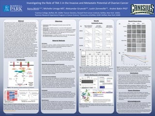
  • 1. RESEARCH POSTER PRESENTATION DESIGN © 2012 www.PosterPresentations.com High-grade serous ovarian cancer (HGS-OvCa) accounts for two-thirds of deaths of OvCa patients. Enhanced capacity to invade surrounding tissues and metastasize is a major characteristic of advanced HGS-OvCa, although molecular pathways that drive OvCa recurrence and metastasis are largely unknown. Several studies have provided significant evidence that TAK1 holds a critical role in tumor invasion, and metastasis of breast cancer. TGF-β- activated kinase 1 (TAK1) is a member of several signaling pathways that are stimulated by cytokines. Our previous studies have indicated that TAK1 controls production of metalloproteinase-9 (MMP-9) that plays an essential role in the invasive and metastatic mechanism. We have reason to believe that TAK1-MMP9 axis maintains a similar role in ovarian cancer since they share similar genetic abnormalities. This study will explore TAK1’s role in ovarian cancer invasion and metastatic potential. Cytotoxicity assays with inhibitors of the TAK1-IKK signaling axis, (5Z)-7-oxozeaenol, CAY10657, and BMS-345541, showed that ovarian cancer cell lines OVCAR-3, OVCAR-4, OVCAR-5, OVCAR-8, and IGROV-1 are growth inhibited by these compounds; this result indicates that growth of these ovarian cancer cell lines is dependent on the TAK1-IKK pathway. Effects of the inhibitors will be further assessed by Cell Cycle Analysis and Anchorage Independent Growth Assays. This should indicate whether TAK1-IKK axis contributes to survival and/or cell- cycle progression. Biochemical analysis of TAK1 signaling pathways and secreted MMP-9 will be carried out using Western Blot assays and whole-cell lysates or conditioned media from ovarian cancer cells lines treated with the TAK1-IKK inhibitors. These assays will provide biochemical proofs and biomarkers of the TAK1 function in ovarian cancer cells. Zymography will be used to assess the activity of MMP-9 in conditioned media. Together these studies will provide molecular evidence for the possible role of TAK1 in the invasive and metastatic potential of OvCa cells. Abstract Introduction Cell Culture: Ovarian cancer cell lines—IGROV1, OVCAR3, OVCAR4, OVCAR5, OVCAR8— were grown in RPMI, 10% FBS, 1% Penicillin/Streptomycin media in a humidified environment at 37 oC with 5% CO2. Cytotoxicity: Cells were seeded at 10,000 cells per well in 96 well plates. The next day, serial dilutions of inhibitors were prepared and added to the wells using a multichannel pipette. Cells were incubated for 48 hours, fixed and stained with 1% Methylene Blue, in 50%Methanol (MeOH), 50% H2O. Wells were washed thoroughly and air-dried overnight. Cells were solubilized in 1% SDS in PBS and absorbance at 650 nm was measured in a Spectramax M2® microplate reader. Data were analyzed in Microsoft Excel and SigmaPlot™ where IC50s were determined. Western Blot and Zymography Sample Preparation: Cells were seeded at 300,000 cells per well in 6-well plates. The next day, cells were washed 3X with PBS, and 600 µL serum-free media alone or with inhibitor was added to each well. Cells were incubated for 48 hours . Conditioned media was collected and cell debris was removed by centrifugation. Conditioned media samples were used for In-Gel Zymography and immunoblotting. Whole-cell lysates were prepared from cells for immunoblot analysis of GAPDH. Western Blot (Immunoblotting): Western blot samples were loaded onto 12.5% SDS-PAGE gels and run at 40 mA until loading buffer ran off. Proteins were transferred to nitrocellulose membranes at 100 V. Protein transfer was validated by Ponceau S staining. Membranes were blocked for 1 h with 5% milk in TBS-Tween and incubated overnight at 4°C with primary antibodies. Membranes were washed with TBS-Tween, incubated with secondary antibodies at room temperature for 1 h then washed with TBS-Tween and TBS. Immune complexes were visualized using the chemiluminescent ECL substrate and exposed to X-Ray film. Zymography: Conditioned media samples were loaded onto 12.5% SDS-PAGE gels containing 2% gelatin and run at 120 V until loading buffer ran off. Gels were washed with 2.5% TritonX-100 then incubated in Development Buffer for 18 h in a 37°C water bath with agitation. Gels were stained with Coomassie Blue staining solution for 2 h and distained with Methanol/Acetic Acid solution. Wound Closure: Cells were seeded at 200,000 cells per well in 12-well plates and grown to complete confluency. Wounds were created in each well with a P200 pipet tip then washed with PBS. RPMI media containing 1% FBS was added to the wells then cytokines and inhibitors were added as necessary. Inhibitors were added to the appropriate wells 1 h prior to cytokines. Pictures of the wound were taken with Spot Software immediately after scratching (0 h) and every 24 h after until the control wounds have closed. Material Methods Figure 3. (A) Graphical representations of cytotoxicity assays. The indicated cell lines were treated with 0.01 µM -100 µM (5Z)-7-oxozeaenol (oxo), 0.005 µM -50 µM CAY 10657 (CAY), or 0.005 µM -50 µM BMS 345541 (BMS). (B)Table shows the IC50 values of the indicated cell lines treated with each inhibitor for 48 hours. Results • Ovarian cancer cell growth is markedly reduced by inhibitors of the TAK1-IKK pathway. Given the increased expression of TAK1 target genes is associated with OvCa recurrence and poor prognosis, this result suggests that TAK1 signaling may drive ovarian cancer progression. • Matrix Metalloproteinase-9 (MMP9) secretion is stimulated by TGF-β and TNF-α in 3 out of 5 tested OvCa lines. MMP9-expressing cells may have greater invasive, metastatic, and angiogenic potentials. • Motility of OVCAR3 cells is increased in response to TNF-α, and decreased by TAK1-IKK pathway inhibitors. This suggests that TAK1-IKK signaling plays an important role in ovarian cancer cell invasion. Acknowledgments This summer research experience was supported in part by DRP-OvCa SPORE grant (to AVB) and by funding from the National Cancer Institute of the National Institutes of Health under award number: 3P30CA016056-37S3. The content is solely the responsibility of the authors and does not necessarily represent the official views of the National Institutes of Health.” • Cytotoxicity (IC50): Is the growth of ovarian cancer cells TAK1 dependent? • Western Blot: Is MMP9 production or secretion TAK1 dependent? • Zymography: Do ovarian cancer cells secrete active MMP9 and can MMP9 secretion be enhanced by cytokine treatment? If so, does MMP9 secretion depend on TAK1, TGFβ-Receptor, or p38 signaling? Can TNF and TGF-β synergistically regulate MMP9 production? • Cell Motility/Invasion: Is motility of ovarian cancer cells TAK1 dependent? Do cytokines increase motility? (Wound Closure Assays) 1Canisius College, Buffalo, NY, 14208; 2Cancer Genetics, Roswell Park Cancer Institute, Buffalo, New York 14263; 3Summer Research Experience Program in Cancer Science; 4University at Buffalo, SUNY, Buffalo, New York 14260 Korry Wirth1,2,3, Michelle Limoge MS2, Aleksandar Gruevski2,4, Justin Zonneville2,4, Andrei Bakin PhD2 Investigating the Role of TAK-1 in the Invasive and Metastatic Potential of Ovarian Cancer Objectives Future Directions • Test in pre-clinical models whether TAK1-IKK inhibitors can improve efficacy of current anti-OvCa therapeutics such as cisplatin, gemcitabine. • Validate the role of TAK1 as a OvCa driver by knockdown (shRNA) or knockout (CRISPR/Cas9) of TAK1 or IKKs in human OvCa cell lines. • Assess the role of TAK1 in tumor dormancy and tumor-stroma interactions. Conclusions IC50 (µM) OXO CAY BMS Cell Line Exp 1 Exp2 Exp 1 Exp 2 Exp 1 Exp 2 IGROV1 0.50 nd 12 4.2 7.4 nd OVCAR3 1.7 1.6 0.058 1.2 13 18 OVCAR4 3.0 9.3 0.24 0.25 23 24 OVCAR5 1.7 1.8 nd 4.0 10 21 OVCAR8 0.88 1.2 nd 4.2 7.4 16 IGROV1+OXO X Data 10-3 10-2 10-1 100 101 102 103 YData 0.0 0.2 0.4 0.6 0.8 1.0 1.2 IGROV1+CAY X Data 10-3 10-2 10-1 100 101 102 103 YData 0.0 0.2 0.4 0.6 0.8 1.0 1.2 1.4 IGROV1+BMS X Data 10-3 10-2 10-1 100 101 102 103 YData 0.0 0.2 0.4 0.6 0.8 1.0 1.2 1.4 1.6 IC50= 0.50 µM IC50= 4.23 µM IC50= 7.42 µM OVCAR8+OXO X Data 10-3 10-2 10-1 100 101 102 103 YData 0.0 0.2 0.4 0.6 0.8 1.0 1.2 1.4 1.6 1.8 OVCAR-5 + BMS X Data 10-3 10-2 10-1 100 101 102 103 YData 0.0 0.2 0.4 0.6 0.8 1.0 1.2 OVCAR5+CAY X Data 10-3 10-2 10-1 100 101 102 103 YData 0.0 0.2 0.4 0.6 0.8 1.0 1.2 OVCAR5+OXO(1) X Data 10-3 10-2 10-1 100 101 102 103 YData 0.0 0.2 0.4 0.6 0.8 1.0 1.2 OVCAR-8 + BMS X Data 10-3 10-2 10-1 100 101 102 103 YData 0.2 0.4 0.6 0.8 1.0 1.2 1.4 1.6 1.8 2.0 2.2 OVCAR8+CAY X Data 10-3 10-2 10-1 100 101 102 103 YData 0.0 0.1 0.2 0.3 0.4 0.5 0.6 IC50= 1.65 µM IC50= 21.3 µM IC50= 15.8 µM IC50= 4.03 µM IC50= 4.18 µMIC50= 1.18 µM (5Z)-7-oxozeaenol (TAK1 inhibitor) BMS 345541 (IKKβ inhibitor) CAY 10657 (TAK1 inhibitor) IGROV1 OVCAR5 OVCAR8 A) B) Figure 4. Zymography gel (upper) and Western blots (lower) of 48-hrs conditioned media. The indicated cell lines were treated with 10 ng/mL TNF- α and 2ng/mL TGF-β. Figure 5. (A) Wound Closure Assay. OVCAR3 cells were treated with 2 ng/mL TGF-β, 10 ng/mL TNF-α, 2 ng/mL TGF-β/ 10 ng/mL TNF-α combination, 5 μM OXO, or 20 μM BMS. (B) Graphical representation of the cell-free space relative to control at each time-point for each treatment. 0% 20% 40% 60% 80% 100% 120% 0 10 20 30 40 50 60 70 80 90 %Cell-FreeSpace Hours B) 0 hr 24 hr 48 hr 80 hr Control TGFβ TNFα TGFβ+TNFα Oxo BMS A)Cytotoxicity Assay : Wound Closure Assay : Western Blotting and In-Gel Zymography: Table 1. IC50 values of TAK1-IKK inhibitors for ovarian cancer cell lines IGROV1 GAPDH MMP9 37 - 50 - 100 - 100 - 75 - MMP9 - + - + - + - + - + TGFβ+TNFα 3 4 5 8 OVCAR kDa Figure 1. TAK1 signaling in cytokine pathways and metastasis. A B C Basal-like Luminal A Luminal B HGS-OvCa GENE 80 cases 209 cases 113 cases 316 cases TP53 * 85.0 11.5 35.4 94.9 EGFR ** 20.0 0.0 0.9 6.7 RB1 *** 45.0 1.0 8.0 19.0 MYC ** 35.0 8.1 16.8 32.3 RELB ** 20.0 1.9 0.9 10.1 FOSL1 ** 17.5 1.4 1.8 5.7 BIRC2 ** 18.8 0.5 0.9 8.0 PTGS2 ** 27.5 4.3 4.4 7.3 VEGFA ** 23.8 2.4 0.0 6.7 IL8 ** 17.5 3.8 2.7 7.7 CCNE1 ** 42.5 1.4 39.8 23.3 IKBKB** 13.8 13.9 24.8 20.0 FADD** 12.5 18.2 25.7 15.7 * Mutations or deletions ** Amplifications or increase in expression *** Mutations, deletions or decrease Cases with Alterations Cases without Alterations Logrank Test P-value: 0.09 TAK1-targets Months Disease Free 0 20 40 60 80 100 120 140 160 180 DiseaseFree HG-Serous Ovarian Cancer (TCGA); Altered in 42 of 158 cases Cases with Alterations Cases without Alterations Logrank Test P-value: 0.005 TAK1-targets Survival 0 20 40 60 80 100 120 140 160 180 Months Survival TAK1 targets: FRA1, JUN, MMP9, NFKB2, RELA Figure 2. Genetics of ovarian and breast cancer. (A) Panel shows percentage of alterations for each gene in Invasive Breast Carcinoma cases (TCGA, Nature, 2012) and Serous OvCa (TCGA, Nature 474, 2011, 609–615). Basal tumors have a higher presentation of alterations in TP53, RB1, and increased expression of TAK1-dependent targets. (B-C) Disease-free and survival data are from TCGA: http://www.cbioportal.org. TAB1 TARGET GENES COX2, IL8, VEGFA, FRA1, JUN, cIAPs, MMP9 TNF-αTGF-β IL1-β TAB2 P AP1 METASTASES Invasion Angiogenesis Survival P P NFκB MAPK (ERK, p38, JNK) IKKα-IKKβ P TAK1 TRAF6 TRAF2,5 cIAP Nemo TRAF4,6 RIP TAK1-Inh (5Z)-7-Oxozeaenol CAY-compound IKKβ-Inh BMS-345541 MEK-Inh (U0126) The prodomain, catalytic domain, fibronectin domains, and hemopexin domain are shown in red, blue, green, and yellow, respectively. Zn2+ ions are indicated in red, and Ca2+ ions are magenta. Asterisk indicates the cleavage site. Cleavage site for u/t-PA; MMP-3; MMP-2; MMP-13. Electrophoresis (protein separation) Incubation in Zn/Ca-supplemented reaction buffer; Coomassie Staining Conditioned Media (Serum-free media) CTR BMS OXO TGFβ TNF+TGFβ TNF I would like to express my gratitude towards the Roswell Park Summer Internship Program, Department of Cancer Genetics, and Andrei Bakin for providing me with this invaluable opportunity. Many thanks go to Michelle Limoge for helping and supporting me throughout this project. Special Acknowledgments go to my colleagues Aleksandar Gruevski, and Justin Zonneville for additional contributions made to this project.