Survey on Automatic Kidney Lesion Detection using Deep Learning
Cheung Ngai-Man and Tai Hong Wen's UROP Poster on Wound Assessment Using Mobile Imaging_clear
1. Advaith Anand1, Casey Hong1, Hong Wen Tai2, Dora Tzeng1
Advised by Professor Ngai-Man Cheung2, Dr. Victor Pomponiu2, Dr. Hossein Nejati2
1: Massachusetts Institute of Technology, Cambridge, MA, USA 2: Singapore University of Technology and Design, Singapore
Background and Motivation
● Diabetes is an affliction affecting a
considerable portion of the world
population
● By 2030, 600,000 Singaporeans and 346
million people worldwide will be affected
by diabetes.
● 10-15% of diabetics will get at least one
foot ulcer in their lifetime, potentially
leading to amputations.
● Foot ulcer care requires recurring visits to
a doctor/nurse to monitor the progress of
a wound. These visits are cumbersome,
especially for elderly patients with
impaired mobility.
● Wound assessment is currently only
monitored qualitatively
● Research goal: A better way for foot
ulcer patients to heal, reducing the
number of total hospital visits.
● Research approach: A suite of
algorithms that can automatically detect
the types of wound tissue present in a
patient’s wound by analyzing a photo of
the wound.
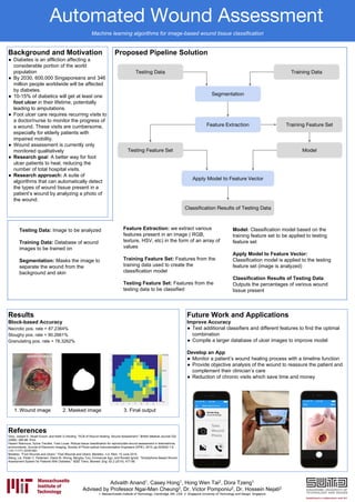
Proposed Pipeline Solution
References
Grey, Joseph E, Stuart Enoch, and Keith G Harding. "ACB of Wound Healing: Wound Assessment." British Medical Journal 332
(2006): 285-88. Print.
Hazem Wannous, Sylvie Treuillet, Yves Lucas. Robust tissue classification for reproducible wound assessment in telemedicine
environments. Journal of Electronic Imaging, Society of Photo-optical Instrumentation Engineers (SPIE), 2010, pp.023002-1-9.
<10.1117/1.3378149>.
Medetec. "Foot Wounds and Ulcers." Foot Wounds and Ulcers. Medetec, n.d. Web. 10 June 2015.
Wang, Lei, Peder C. Pedersen, Diane M. Strong, Bengisu Tulu, Emmanuel Agu, and Ronald Ignotz. "Smartphone-Based Wound
Assessment System for Patients With Diabetes." IEEE Trans. Biomed. Eng. 62.2 (2015): 477-88.
Automated Wound Assessment
Future Work and Applications
Improve Accuracy
● Test additional classifiers and different features to find the optimal
combination
● Compile a larger database of ulcer images to improve model
Develop an App
● Monitor a patient’s wound healing process with a timeline function
● Provide objective analysis of the wound to reassure the patient and
complement their clinician’s care
● Reduction of chronic visits which save time and money
Results
Block-based Accuracy
Necrotic pos. rate = 87.2364%
Sloughy pos. rate = 90.2661%
Granulating pos. rate = 78.3262%
1. Wound image 2. Masked image 3. Final output
Testing Data: Image to be analyzed
Training Data: Database of wound
images to be trained on
Segmentation: Masks the image to
separate the wound from the
background and skin
Feature Extraction: we extract various
features present in an image ( RGB,
texture, HSV, etc) in the form of an array of
values
Training Feature Set: Features from the
training data used to create the
classification model
Testing Feature Set: Features from the
testing data to be classified
Model: Classification model based on the
training feature set to be applied to testing
feature set
Apply Model to Feature Vector:
Classification model is applied to the testing
feature set (image is analyzed)
Classification Results of Testing Data:
Outputs the percentages of various wound
tissue present
Machine learning algorithms for image-based wound tissue classification