
Recomendados
Más contenido relacionado
La actualidad más candente
La actualidad más candente (20)
Destacado
Destacado (19)
Similar a In-Service Project - Dizzy Exam
Similar a In-Service Project - Dizzy Exam (20)
In-Service Project - Dizzy Exam
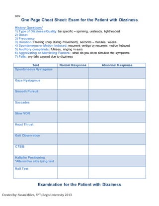
- 1. Created by: Susan Miller, SPT; Regis University 2013 oov One Page Cheat Sheet: Exam for the Patient with Dizziness History Questions* 1) Type of Dizziness/Quality: be specific – spinning, unsteady, lightheaded 2) Onset 3) Frequency 3) Duration: Fleeting (only during movement), seconds – minutes, weeks 4) Spontaneous or Motion Induced: recurrent vertigo or recurrent motion induced 5) Auditory complaints: fullness, ringing in ears 6) Aggravating or Alleviating Factors: what do you do to simulate the symptoms 7) Falls: any falls caused due to dizziness Test Normal Response Abnormal Response Spontaneous Nystagmus Gaze Nystagmus Smooth Pursuit Saccades Slow VOR Head Thrust Gait Observation CTSIB Hallpike Positioning *Alternative side lying test Roll Test Examination for the Patient with Dizziness
- 2. Created by: Susan Miller, SPT; Regis University 2013 GAZE STABILITY ASSESSMENT: SpontaneousNystagmus: Procedure: Ask patient to gaze at tip your finger without head movement with best-corrected vision (glasses or contact lenses in place) Normal Response: Patient will display no nystagmus. Abnormal Response: Spontaneous nystagmus; observe direction. Interpretation: If an abnormal response is present, observe amplitude and direction. A central lesion presents with vertical (down beating) or torsional nystagmus. A peripheral lesion presents as a mixed nystagmus of horizontal and torsional. Gaze Nystagmus: Procedure: Ask patient to gaze at your finger outstretched to the right side of your head (approximately 20° - 30° from central position) without moving their head for 20 seconds. Repeat on left side. Normal Response: Patient will display no nystagmus. Abnormal Response: Gaze-evoked nystagmus; Eyes change in direction, form or intensity; Spontaneous Nystagmus Interpretation: A peripheral dysfunction is present if the gaze provokes nystagmus in a uni-lateral direction, magnitude may increase. Changing direction of the gaze should not change the direction of the nystagmus. A central dysfunction is present if the nystagmus spontaneously changes. Smooth Pursuit: Procedure: Ask patient to follow your finger in an “H” formation without moving their head. Normal Response: Patient will track finger with a smooth movement, no nystagmus and eyes move in a conjugate manner. Abnormal Response: Patient cannot track finger in the horizontal or vertical planes. Interpretation: If abnormal response is present, a dysfunction with central nervous system (CNS) or loss of oculomotor range of motion. Smooth pursuit will not detect a peripheral lesion. If impairment occurs with the vertical portion of the “H”, weakness of the eye muscles may be present. If the impairment occurs with the horizontal portion of the “H”, this is a marker CNS dysfunction.
- 3. Created by: Susan Miller, SPT; Regis University 2013 Saccades: Procedure: Ask patient to look back and forth between your finger (outstretched to the right of your head) and to your nose without moving their head. Repeat on the left. Normal Response: Patient will track finger in quick, accurate and conjugate manner. Abnormal Response: Latency of movement, abnormal speed, accuracy, or disconjugate movement. Interpretation: Abnormal response indicates a dysfunction of central involvement. Slow VOR TEST: Procedure: Ask patient to sit & focus their eyes on your finger as they move their head side to side. Normal Response: Patient maintains gaze (eyes) on your finger. Abnormal Response: Eyes do not stay fixed on your finger and they will need to correct to move back to the focal point. Observe direction of eye movement. Some patients may be resistant to move their head in a smooth manner. Interpretation: A vestibular loss is indicated if a patient cannot fixate their eyes on the target and is an indication of a vestibular peripheral lesion. Head Thrust: Procedure: Ask patient to sit with eyes open & focus on your nose. Grasp patient’s head between your hands high on the skull. Place patient’s head in a chin tuck position, quickly apply a small amplitude rotation of the head to the left or right. Create unpredictable head movements Normal Response: Patient maintains gaze (eyes) on your nose. Abnormal Response: Eyes do not stay fixed on your nose and a correcting saccade takes place back to the focal point Observe if the reflex is hyper/hypo-active. Interpretation: A vestibular loss is present is the patient has a reflex that is hyper/hypoactive. It is an indication of a peripheral loss for hypoactive and central loss for hyperactive.
- 4. Created by: Susan Miller, SPT; Regis University 2013 BALANCE/POSTURALCONTROL TESTING: Gait Observation: Procedure: Ask patient to walk 30 – 50 feet: 1) Normal gait 2) Pitch – move head up & down 3) Yaw – move head left & right Normal Response: No imbalances Abnormal Response: Slow movements, stride length, speed, veering (direction), wobbling, imbalances, muscle weakness, and position of arms. Interpretation: Most patients with a peripheral vestibular loss will veer or wobble towards the dysfunction side and typically do not fall from this disorder. However, this test alone cannot determine inner ear dysfunction. CTSIB (ClinicalTestfor Sensory Interaction for Balance): Procedure: 1) Ask patient to stand with feet as close together as possible – 1st with eyes open; 2nd with eyes closed for 30 sec. each 2) Ask patient to stand on a piece of foam, repeat procedure above. NOTE: PT to stand close to patient for safety. Normal Response: Maintain consistent position throughout each test with no sway. Abnormal Response: minimal, moderate or severe sway. Interpretation: The test is to determine which system (visual, vestibular or somatosensory) the patient is using for balance. The last condition indicates difficulty with the vestibular system. However, this does not tell us specifically which vestibular disorder. HEAD POSITIONING TEST: Dix-HallpikePositioning: Procedure: Have patient long sit on the table. Turn patient’s head 30°-45° to the right and lay them down in a steady movement with their head extended off the table. Once you elicit nystagmus and the symptoms calm down, sit the patient up. Guard the patient in the seated position before repeating on the left side.
- 5. Created by: Susan Miller, SPT; Regis University 2013 Normal Response: No symptoms Abnormal Response: Nystagmus (horizontal, torsional); symptoms of dizziness, room spinning. Interpretation: The direction of the nystagmus directs which canal is causing the dizziness: Posterior Canal: Torsional & rotates toward involved ear Horizontal Canal: Beats horizontal towards involved ear Anterior Canal: Rotates away from involved ear Alternative Test:Side lying Test(Specific for Posterior Canal) *Use the alternative test is a patient has neck or back pain (NOTE: cannot not treat symptoms in this position). Procedure: Have patient sit with legs off in the center of the plinth and a place a pillow to one side of them. If test left posterior canal, turn patients head right (right posterior canal, turn left). Place hands on patient’s head and lay them down on their side with a steady motion maintaining the head position. Have patient keep their eyes open to watch for nystagmus. Bring patient back to starting position with a slight forward flexed position. Guard patient for any instability . Roll Test: Procedure: Ask patient to long sit. Place patient’s head in 30° flexion and turn to left as far as you can. Lay the patient on their back. Observe if any nystagmus occurs. Proceed to roll the patient’s head to the right. Normal Response: No Nystagmus Abnormal Response: observe for horizontal nystagmus Interpretation: Only perform test if nystagmus occurred in a horizontal direction with the Dix-Hallpike test OR if the history & symptoms lead to Benign Paroxysmal Positional vertigo (BPPV) but cannot elicit symptoms with Dix- Hallpike test
- 6. Created by: Susan Miller, SPT; Regis University 2013 *History Algorithm to help direct the exam
- 7. Created by: Susan Miller, SPT; Regis University 2013 References: 1) Fuller, K., Esses, B., Hedenberg, R. My World Is Spinning! What Can I Do? Vestibular Rehabilitation Part 2: Intermediate Module. Evergreen, CO: IRB Solutions: 2004. 2) Walter, J. Bedside and Office Examination of the Vestibular System. Medbridge Education website. Available at: www.medbridgeeducation.com. Accessed: September 2013. 3) Goebel, J. The Ten-Minute Examination of the Dizzy Patient. Seminars in Neurology. 2001; 21: 4; 391- 398.