
Recomendados
Recomendados
Más contenido relacionado
La actualidad más candente
La actualidad más candente (20)
Similar a Natural killer cell activation
Similar a Natural killer cell activation (20)
Más de bestwebsite2008
Más de bestwebsite2008 (20)
Último
Último (20)
Natural killer cell activation
- 1. JOURNAL OF MEDICINAL FOOD J Med Food 10 (3) 2007, 435–441 © Mary Ann Liebert, Inc. and Korean Society of Food Science and Nutrition DOI: 10.1089/jmf.2007.401 Natural Killer Cell Activation and Modulation of Chemokine Receptor Profile In Vitro by an Extract from the Cyanophyta Aphanizomenon flos-aquae Aaron N. Hart,1 Lue Ann Zaske,1 Kelly M. Patterson,1 Christian Drapeau,2 and Gitte S. Jensen3 1NIS Labs, Klamath Falls; 2Desert Lake Technology LLC, Keno, Oregon; and 3Holger NIS, Calgary, Alberta, Canada ABSTRACT The present research was designed to study the effects of an extract from the edible cyanophyta Aphanizomenon flos-aquae on human natural killer (NK) cells. We have previously shown, using a double-blind randomized placebo-con- trolled crossover design, that ingestion of 1.5 g of dried whole A. flos-aquae resulted in a transient reduction in peripheral blood NK cells in 21 healthy human volunteers, suggesting increased NK cell homing into tissue. We have now identified an extract from A. flos-aquae (AFAe) that directly activates NK cells in vitro and modulates the chemokine receptor profile. NK cell activation was evaluated by expression of CD25 and CD69 on CD3ϪCD56ϩ cells after 18 hours. Changes in CXCR3 and CXCR4 chemokine receptor expression after 5–60 minutes were evaluated by immunostaining and flow cytometry. AFAe induced the expression of CD69 on CD3ϪCD56ϩ NK cells, induced CD25 expression on 25% of these cells, and acted in synergy with interleukin 2. NK cells enriched by RosetteSep® (StemCell Technologies Inc., Vancouver, BC, Canada) were not activated by AFAe, indicating that the NK activation was dependent on other cells such as monocytes. The low-molecu- lar-weight fraction Ͻ5,000 of AFAe was responsible for the most robust NK cell activation, suggesting novel compounds dif- ferent from previously reported macrophage-activating large polysaccharides. KEY WORDS: • Aphanizomenon flos-aquae • blue-green algae • human • immunomodulation • low-molecular- weight peptides • trafficking INTRODUCTION Dietary intervention for enhancement of immune sur- veillance and primary defense includes nutritional support T HE EDIBLE CYANOPHYTA Aphanizomenon flos-aquae is known for its bioavailable antioxidants, including phy- cocyanin,1 immunomodulatory polysaccharides,2,3 and a of a healthy composition of the gut flora. Commensal or- ganisms in the gut, including lactobacilli, have been found to possess potent NK-activating properties.10–12 This prop- beneficial lipid profile.4 We have previously reported that erty extends to fixed cells and isolated cell wall fractions of ingestion of A. flos-aquae results in rapid and transient hom- the commensal organisms.13–16 ing of natural killer (NK) cells in vivo.5 The current study Consumption of cyanobacteria as dietary supplements was undertaken to explore several mechanisms that may presents interesting possibilities in terms of NK cell activa- contribute to an induction of NK cell trafficking and acti- tion. Cyanobacteria are primitive organisms resembling bac- vation. teria as well as chloroplasts and are thought to represent or- NK cells play a key role in immune surveillance and the ganisms that gave rise to chloroplasts.17 They share primary defense against viral infections and cancer, as they molecular similarities with bacteria, including bacterial are capable of killing virally infected cells and some tumor DNA and cell wall components such as peptidoglycans, cells without prior sensitization.6 NK cells also contribute which are known to induce NK cell activation.13 In addi- to the defense against some bacteria.7 This involves prior tion, the outer cell wall represents carbon storage and con- phagocytosis of bacteria by macrophages followed by con- sists of large complex polysaccharides with immunomodu- tact between macrophages and NK cells.8 RNAs for Toll- latory activity2,18 and may contribute to the documented like receptor (TLR) 1–6 and TLR9 are expressed in circu- antiviral19 and anticancer20 effects of some blue-green al- lating NK cells,9 allowing NK cells to respond to various gae. Consumption of edible cyanobacteria may thus present bacterial cell wall components, including peptidoglycans similar compounds to the immune system as some com- and lipopolysaccharides, as well as bacterial DNA. mensal gut bacteria. The two most common edible cyanobacteria include Spir- Manuscript received 24 January 2007. Revision accepted 1 May 2007. ulina and A. flos-aquae. Within the class of Cyanophyta, Address reprint requests to: Gitte S. Jensen, Holger NIS, 601 13 Avenue NE, Calgary, these two genera belong to separate orders: Spirulina to the AB T2E 1C7, Canada, E-mail: gitte@holgernis.com Oscillatoriales and Aphanizomenon to the Nostocales. Tax- 435
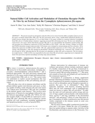
- 2. 436 HART ET AL. onomic re-evaluation of Aphanizomenon strains concluded Reagents and monoclonal antibodies that previous reports on toxin-producing Aphanizomenon The following monoclonal antibodies directly conjugated were based on misclassification of Anabaena, which is with fluorochromes were purchased from Becton-Dickinson toxic.21 During certain seasons, including the late fall, the (San Jose, CA): CD3-peridinin-chlorophyll-protein (PerCP), cyanophyta A. flos-aquae grows in almost complete mono- CD14-phycoerythrin (PE), CD25-fluorescein isothiocyanate culture in the hypereutrophic Upper Klamath Lake, Oregon, (FITC), CD45-FITC, CD56-FITC, CD56-PE, and CD69- during which times the biomass can be harvested for human FITC. Buffers including RPMI 1640 medium, Histopaque and animal consumption.22 1077, and phosphate-buffered saline were purchased from The data presented here show that the extract of A. flos- Sigma-Aldrich. aquae (AFAe) is a potent activator of NK cells in vitro, with the highest activity found in the low-molecular-weight frac- tion. Purification of peripheral blood mononuclear cells (PBMCs) Peripheral venous blood samples were obtained after in- MATERIALS AND METHODS formed consent from healthy human volunteers between the Algal extract Harvested, wild-grown A. flos-aquae biomass was fil- tered, cooled, and dried using Refractance Window™ tech- nology (Desert Lake Technology LLC, Klamath Falls, OR). Dried algal material was heated to 85°C in 10% ethanol for 3 hours. The supernatant was decanted and then precipitated by adding 50% ethanol. The precipitate was dried, giving a pale yellow powder, Migratose™ (Desert Lake Technol- ogy). An aqueous extract was prepared fresh prior to each in vitro experiment by weighing 0.2 g of powder into 2 mL of phosphate-buffered saline. This resulted in a deep orange liquid extract. Solids were removed by centrifugation, and the supernatant was sterile-filtered using a 0.22-m syringe filter. This extract was the stock solution for all in vitro ex- periments and is referred to as AFAe throughout this paper. Serial dilutions were prepared in cell culture medium. In some experiments, AFAe was further separated into high- and low-molecular weight compounds by applying the crude extract to Amicon Ultra centrifugal filter devices (Millipore, Billerica, MA) with a 5-kDa cutoff. Both the low-molecu- lar-weight and the high-molecular-weight fractions were re- suspended in phosphate-buffered saline to the same volume as originally applied to the filter device, and serial dilutions were prepared. Gel electrophoresis The electrophoretic profile of AFAe was evaluated by gel electrophoresis by mixing AFAe 1:1 (vol/vol) in Laemmli sample buffer (Bio-Rad, Hercules, CA) without mercap- toethanol. Sodium dodecyl sulfate-polyacrylamide gel elec- trophoresis was performed on 4–15% precast ReadyGels (Bio-Rad) in Tris/glycine/sodium dodecyl sulfate buffer (Bio-Rad) for 1 hour at 120 V. Staining of the gels for pro- FIG. 1. Comparison of the electrophoretic band pattern of AFAe tein was performed using Coomassie Brilliant Blue G stained with either silver stain (top) or Coomassie Brilliant Blue (bot- (Sigma-Aldrich, St. Louis, MO). Staining for both protein tom). Densitograms are shown to the right of each lane. The silver stain revealed a band of approximately 60 kDa, which did not stain and carbohydrates was performed using a silver staining kit with Coomassie Brilliant Blue (CCblue), indicating a large polysac- (Bio-Rad). Densitograms were prepared using ImageJ ver- charide. Both stains revealed a series of smaller bands below 8 kDa, sion 1.37 software (National Institutes of Health, Bethesda, indicating small peptides in AFAe. The data are representative of three MD). separate experiments.
- 3. NK CELL ACTIVATION BY A. FLOS-AQUAE 437 ages of 20 and 60 years. Heparinized whole blood was lay- medium and exposed to serial dilutions of AFAe for 18 ered onto Histopaque 1077 and centrifuged for 25 minutes hours. For the testing of chemokine receptor CXCR3 and at 400 g. The PBMC-rich interface was harvested and CXCR4 expression, similar cultures were established and washed twice in phosphate-buffered saline without calcium incubated for 5, 15, and 30 minutes. Cells were washed in or magnesium. phosphate-buffered saline containing 1% bovine serum al- bumin and 0.02% sodium azide. Cells were resuspended in Enrichment of NK cells 50 L of buffer. Monoclonal antibodies were added and in- cubated in the dark at room temperature for 10 minutes. An NK cells were enriched by removal of other cell types us- additional 110 L of buffer was added to each well, and the ing the negative depletion kit RosetteSep® (StemCell Tech- plates were washed. Supernatant was discarded, and the cells nologies Inc., Vancouver, BC, Canada), which contains an were resuspended in 50 L of buffer and transferred to 0.4 antibody cocktail towards CD3, CD4, CD19, CD36, CD66, mL of 1% formalin. Samples were stored dark and acquired and glycophorin A. The RosetteSep cocktail was added di- by flow cytometry within 4 hours. Acquisition was per- rectly to whole blood, allowing the antibodies to bind un- formed using a FACScalibur™ flow cytometer and Cell- wanted cells to erythrocytes. The blood was then applied to Quest™ software (both from Becton-Dickinson). Analysis Histopaque 1077 and centrifuged for 25 minutes at 400 g. of fluorescence intensity of each marker was performed by Only cells that were not recognized by the antibodies in the electronic gating on CD3ϪCD56ϩ NK cells as well as on cocktail remained in the interface above the Histopaque. CD3ϩCD56ϩ NKT cells, using the FlowJo software (Tree This allowed the harvest of highly enriched NK cells (90% Star Inc., Ashland, OR). pure). Interferon-␥ (IFN-␥) enzyme-linked immunosorbent Induction of cell surface markers assay (ELISA) For the testing of activation markers CD69 and CD25, The production of IFN-␥ in culture supernatants was freshly purified PBMCs were resuspended in culture evaluated using a commercial ELISA kit (R & D Systems FIG. 2. AFAe induced the expression of the IL-2 receptor CD25 (center panels) and the CD69 activation marker (right panels) on almost all CD3ϪCD56dim NK cells but not on the CD3ϪCD56bright NK cells. (Left panels) Isotype control plots. Contour plots show data from (top row) untreated (UT) cells and (bottom row) AFAe-treated samples. The contour plots display data on CD3ϪCD56ϩ lymphocytes. The two-dimen- sional plot allows evaluation of CD56 intensity versus expression of the activation marker CD69. The data shown are representative of three sep- arate experiments using cells from three different donors.
- 4. 438 HART ET AL. Inc., Minneapolis, MN). Supernatants were tested in tripli- cate from 5-day cultures where PBMCs had been exposed to AFAe and compared to untreated samples (negative con- trols) and phytohemagglutinin-treated samples (positive controls). Microplates were read on a BioTek (Winooski, VT) Powerwave microplate reader. Data were exported into Microsoft Excel (Microsoft Corp., Redmond, WA), where averages and standard deviations for each set of triplicate sample were calculated. Statistical analysis Statistical analysis was performed using Microsoft Excel. Statistical significance was tested using Student’s t test, with a value of P Ͻ .05 indicating a significant difference be- tween treatments. RESULTS Electrophoretic pattern for protein and carbohydrate content in AFAe The extract AFAe was subjected to sodium dodecyl sulfate- polyacrylamide gel electrophoresis, and parallel gels were de- veloped with either the protein stain Coomassie Brilliant Blue or silver stain, which also stains carbohydrates. The band pat- terns and densitometry are shown in Figure 1. A distinct pat- tern was seen, revealing the presence of two different groups of compounds in the extract. A strong band around 60 kDa was visible after silver staining but was completely absent af- ter colloidal Coomassie Brilliant Blue staining, indicating the presence of larger polysaccharides in the extract. Several small compounds stained with both stains in a similar pattern, indi- cating the presence of several smaller peptides in the extract. The crude extract was further separated by centrifugation over a centrifugal filter device with a cutoff at 5 kDa, and the two fractions were tested in parallel to crude AFAe on NK cell ac- tivation in vitro (see Fig. 3). CD69 induction: enhancement of interleukin (IL-2) but partial inhibition of phytohemagglutinin on both NK and NKT cells FIG. 3. Induction of the activation marker CD69 on CD3ϪCD56ϩ The incubation of PBMCs overnight with AFAe resulted cells by either (top panel) crude AFAe or the high- (middle panel) in a strong induction of CD69 and moderate induction of and low- (bottom panel) molecular weight (MW) fractions after sep- CD25 expression on CD3ϪCD56dim NK cells, whereas no aration over a centrifugation filtration device with a 5,000 cutoff. The induction of CD69 or CD25 was seen on CD3ϪCD56bright high MW fraction contained compounds larger than 5,000, which in- NK cells (Fig. 2). cluded the larger polysaccharide seen in Figure 1. The low MW frac- tion contained the smaller peptides. The low MW fraction had the highest NK-activating properties. The data are representative of three TABLE 1. AFAE INDUCTION OF THE CD69 separate experiments. UT, untreated. ACTIVATION MARKER ON NK CELLS CD69 mean fluorescence intensity NK activation by AFAe required the presence of Treatment NK cells from PBMCs Enriched NK cells other cells In order to examine whether the NK activation by AFAe Negative control 5.18 Ϯ 1.13 2.14 Ϯ 0.07 AFAe 8.37 Ϯ 052 2.70 Ϯ 0.08 was a direct effect on NK cells or was dependent on mono- cytes or T cells in the PBMC cultures, we compared the in-
- 5. NK CELL ACTIVATION BY A. FLOS-AQUAE 439 TABLE 2. SYNERGISTIC EFFECTS BY AFAE AND IL-2a Treatment CD69 expression (mean fluorescence intensity) IFN-␥ production (pg/mL) Negative control 2.91 Ϯ 0.11 120 Ϯ 60 AFAe 10.16 Ϯ 0.39 422 Ϯ 118 AFAe ϩ IL-2 27.63 Ϯ 1.76* 4,090 Ϯ 170* IL-2 17.18 Ϯ 0.17 2,690 Ϯ 1,927 aThe dose of IL-2 used was 50 International Units/mL. The increase in response between the treatments with IL-2 alone compared to AFAe ϩ IL-2 was sta- tistically significant with *P Ͻ .05. duction of CD69 expression in parallel cultures of PBMCs and IL-2, the level of IFN-␥ production was higher than with versus enriched NK cells from the same sample of PBMCs. either AFAe or IL-2 alone, indicating that the synergy seen A positive control was used to verify that the functionality between AFAe and IL-2 for CD69 induction extends to IFN- of NK cells was not compromised by the enrichment pro- ␥ production in vitro. tocol. AFAe induced CD69 expression on NK cells from PBMC cultures but not on enriched NK cells (Table 1). Down-regulation of the chemokine receptors CXCR3 and CXCR4 The low-molecular-weight fraction induced strong NK The chemokine receptors CXCR4 and CXCR3 are ex- activation as compared to the high-molecular-weight pressed on a proportion of NK cells. The exposure of cul- fraction of AFAe tures to AFAe for 30 minutes resulted in down-regulation The electrophoretic properties of AFAe showed two ma- of the expression of these two chemokine receptors on both jor groups of compounds: larger polysaccharides and lower- NK cells and NKT cells (Fig. 4). molecular-weight peptides. These were separated using a centrifugation filter device. PBMC cultures were treated with crude AFAe, AFAe high molecular weight, or AFAe low molecular weight and incubated for 18 hours to allow for CD69 induction. The percentage of NK cells expressing CD69 after each treatment was evaluated. Treatment with crude AFAe at 10 mg/mL resulted in activation of 12–14% NK cells, with a dose–response that rapidly decreased to baseline levels (Fig. 3, top panel). The high-molecular- weight fraction of AFAe resulted in a similar level of acti- vation of NK cells (Fig. 3, middle panel). In contrast, the low-molecular-weight fraction of AFAe resulted in a sub- stantially higher activation of NK cells than either crude AFAe or AFAe high molecular weight (Fig. 3, bottom panel). This indicated that the small peptides possess potent NK-activating properties different from the larger polysac- charides with macrophage-activating properties as reported by Pasco and co-workers.2,3 Induction of IFN-␥ production and synergy with IL-2 The induction of the activation marker CD69 on CD3ϪCD56ϩ NK cells by AFAe was assayed in the con- text of IL-2 (50 international units/mL). The intensity of CD69 expression after AFAe treatment alone was signifi- cantly above the baseline expression seen on untreated cells. IL-2 triggered higher expression of CD69 than AFAe alone and acted in synergy with AFAe such that when AFAe was added immediately prior to IL-2, a higher CD69 expression FIG. 4. Expression of the chemokine receptors CXCR3 and CXCR4 level was seen than with IL-2 alone (Table 2). for NK (top histogram) and NKT (bottom histogram) cells. PBMCs Treatment of PBMCs with AFAe for 5 days in vitro re- were incubated for 30 minutes in the presence of AFAe. Untreated (UT) sulted in an increase in IFN-␥ in the culture supernatants cultures served as a control. Flow cytometric analysis included electronic (Table 2). When the cultures were treated with both AFAe gating on CD3ϪCD56ϩ NK cells and CD3ϪCD56ϩ NKT cells.
- 6. 440 HART ET AL. DISCUSSION ACKNOWLEDGMENTS In this study, we have shown that AFAe, an extract from This research was supported by the Merle West Center whole dried A. flos-aquae biomass, rapidly changes the for Medical Research, which is a nonprofit organization sup- chemokine receptor profile of NK cells in vitro. We have porting research into nutrition and complementary therapies, previously shown that consumption of 1.5 g of whole A. and Desert Lake Technologies LLC, a harvester of flos-aquae results in a transient decrease of circulating NK cyanophyta in Upper Klamath Lake, Oregon. cells, using a randomized double-blinded placebo-controlled During the time this study was conducted, the co-author design involving 21 healthy subjects.5 The present study Christian Drapeau held the position of director for Research shows that a specific extract from A. flos-aquae possesses & Development at Desert Lake Technologies LLC, a har- the ability to activate NK cells in vitro, as reflected by the vester of A. flos-aquae. No other co-authors have any fi- induction of CD69 expression. The NK-activating effect ap- nancial interest in the subject matter. pears to be dependent on other cell types, since the effect was not seen on RosetteSep-enriched NK cells. AFAe also produced a significant reduction in the expression of both REFERENCES CXCR3 and CXCR4 on peripheral blood NK cells. The chemokine receptor CXCR4 is specific for stromal cell-de- 1. Benedetti S, Benvenuti F, Pagliarani S, Francogli S, Scoglio S, rived factor-1 and plays a role in recruiting NK cells into Canestrari F: Antioxidant properties of a novel phycocyanin ex- the bone marrow environment. Other chemokines, includ- tract from the blue-green alga Aphanizomenon flos-aquae. Life ing IL-8 and fractalkine, are involved in recruiting CD16ϩ Sci 2004;75:2353–2362. NK cells into nonlymphoid tissue,23 and CCR5 is involved 2. Pugh N, Ross SA, ElSohly HN, ElSohly MA, Pasco DS: Isola- tion of three high molecular weight polysaccharide preparations in homing to the liver.24 The CD16ϩ cells correspond to the with potent immunostimulatory activity from Spirulina platensis, CD56dim cells, which are the cells responding to AFAe in Aphanizomenon flos-aquae and Chlorella pyrenoidosa. Planta our study. We suggest that while the down-regulation of Med 2001;67:737–742. CXCR4 may reduce the NK cell homing to the marrow en- 3. Pugh N, Pasco DS: Characterization of human monocyte activa- vironment, the cells may become more sensitive to other tion by a water soluble preparation of Aphanizomenon flos-aquae. chemotactic signals, thereby possibly increasing homing as Phytomedicine 2001;8:445–453. part of immune surveillance of tissue other than marrow. 4. Kushak, RI, Drapeau C, Van Cott EM, Winter HH: Favorable ef- Given that the effect in vivo is seen after oral consumption, fects of blue-green algae Aphanizomenon flos-aquae on rat plasma it is tempting to suggest that the gut mucosal-associated lym- lipids. JANA 2000;2:59–65. phoid tissue would be affected first, possibly initiating a cas- 5. Jensen GS, Ginsberg DI, Huerta P, Citton M, Drapeau C: Con- cade of events such as macrophage activation and induction sumption of Aphanizomenon flos aquae has rapid effects on the of chemotactic signals. circulation and function of immune cells in humans: a novel ap- Further chemical characterization of the components of proach to nutritional mobilization of the immune system. JANA AFAe is needed; however, the large polysaccharide may 2000;2:50–58. likely be of cell wall origin. The large polysaccharide has a 6. Yokoyama WM, Kim S, French AR: The dynamic life of natural different molecular weight than the complex polysaccharide killer cells. Annu Rev Immunol 2004;22:405–429. isolated from A. flos-aquae by Pasco and co-workers.2,3 The 7. Blach-Olszewska Z: Innate immunity: cells, receptors, and sig- small peptides with NK-activating properties are most likely naling pathways. Arch Immunol Ther Exp (Warsz) to represent cell wall peptidoglycan breakdown products, ca- 2005;53:245–253. pable of triggering signaling via TLRs.25,26 The observation 8. Haller D, Serrant P, Granato D, Schiffrin EJ, Blum S: Activation that NK activation depended on other cells in the cultures of human NK cells by staphylococci and lactobacilli requires cell could suggest that a cross-talk between monocytes and NK contact-dependent costimulation by autologous monocytes. Clin Diagn Lab Immunol 2002;9:649–657. cells is involved. It has been shown that NK cells constitu- 9. Hornung V, Rothenfusser S, Britsch S, Krug A, Jahrsdorfer B, tively express ␣-defensin, which is rapidly released after Giese T, Endres S, Hartmann G: Quantitative expression of Toll- TLR-mediated signaling.27 Interestingly, mammalian de- like receptor 1–10 mRNA in cellular subsets of human peripheral fensins have the ability to act as chemokines and participate blood mononuclear cells and sensitivity to CpG oligodeoxynu- in lymphocyte recruitment.28,29 The in vivo effect of AFAe cleotides. J Immunol 2002;168:4531–4537. consumption on NK cells could involve a cascade of events 10. Matsuzaki T, Chin J: Modulating immune responses with probi- in which TLR-mediated signaling, triggered by proteogly- otic bacteria. Immunol Cell Biol 2000;78:67–73. cans, and A. flos-aquae-derived defensin molecules trigger 11. Haller D, Blum S, Bode C, Hammes WP, Schiffrin EJ: Activa- initial events, leading to rapid amplification locally in the tion of human peripheral blood mononuclear cells by nonpatho- gut. The overall in vivo effect of AFAe consumption must genic bacteria in vitro: evidence of NK cells as primary targets. take into account not only the direct NK-activating proper- Infect Immun 2000;68:752–759. ties, but also the effect of AFAe on other cell types, in- 12. Ginsberg DI, Drapeau C, Jensen GS: Probiotic bacteria and the cluding macrophages. Further work is needed to evaluate to immune system. JANA 2000;3:44–49. what extent consumption of AFAe may contribute to in- 13. Nakao Y, Funami K, Kikkawa S, Taniguchi M, Nishiguchi M, creased protection against viral disease. Fukumori Y, Seya T, Matsumoto M: Surface-expressed TLR6
- 7. NK CELL ACTIVATION BY A. FLOS-AQUAE 441 participates in the recognition of diacylated lipopeptide and pep- evaluation of Aphanizomenon flos-aquae NH-5 based on mor- tidoglycan in human cells. J Immunol 2005;174:1566–1573. phology and 16S rRNA gene sequences. Hydrobiologia 2000; 14. Liu G, Zhai Q, Schaffner D, Popova T, Hayford A, Bailey C, Al- 438:99–105. ibek K: Bacillus alcalophilus peptidoglycan induces IFN-alpha- 22. Carmichael WW, Drapeau C, Anderson DM: Harvesting of Aph- mediated inhibition of vaccinia virus replication. FEMS Immunol anizomenon flos-aquae Ralfs ex Born. & Flah. var. flos-aquae Med Microbiol 2004;42:197–204. (Cyanobacteria) from Klamath Lake for human dietary use. J Appl 15. Cleveland MG, Gorham JD, Murphy TL, Tuomanen E, Murphy Physiol 2000;12:585–595. KM: Lipoteichoic acid preparations of gram-positive bacteria in- 23. Campbell JJ, Qin S, Unutmaz D, Soler D, Murphy KE, Hodge duce interleukin-12 through a CD14-dependent pathway. Infect MR, Wu L, Butcher EC: Unique subpopulations of CD56ϩ NK Immun 1996;64:1906–1912. and NK-T peripheral blood lymphocytes identified by chemokine 16. Paquet A Jr, Raines KM, Brownback PC: Immunopotentiating ac- receptor expression repertoire. J Immunol 2001;166:6477–6482. tivities of cell walls, peptidoglycans, and teichoic acids from two 24. Vermijlen D, Seynaeve C, Luo D, Kruhoffer M, Eizirik DL, strains of Listeria monocytogenes. Infect Immun 1986;54: Orntoft TF, Wisse E: High-density oligonucleotide array analysis 170–176. reveals extensive differences between freshly isolated blood and 17. McFadden GI: Endosymbiosis and evolution of the plant cell. hepatic natural killer cells. Eur J Immunol 2004;34:2529–2540. Curr Opin Plant Biol 1999;2:513–519. 25. Schwandner R, Dziarski R, Wesche H, Rothe M, Kirschning CJ: 18. Hirahashi T, Matsumoto M, Hazeki K, Saeki Y, Ui M, Seya T: Peptidoglycan- and lipoteichoic acid-induced cell activation is Activation of the human innate immune system by Spirulina: aug- mediated by Toll-like receptor 2. J Biol Chem 1999;274: mentation of interferon production and NK cytotoxicity by oral 17406–17409. administration of hot water extract of Spirulina platensis. Int Im- 26. Takeuchi O, Hoshino K, Kawai T, Sanjo H, Takada H, Ogawa T, munopharmacol 2002;2:423–434. Takeda K, Akira S: Differential roles of TLR2 and TLR4 in recog- 19. Hayashi K, Hayashi T, Kojima I: A natural sulfated polysaccha- nition of gram-negative and gram-positive bacterial cell wall com- ride, calcium spirulan, isolated from Spirulina platensis: in vitro ponents. Immunity 1999;11:443–451. and ex vivo evaluation of anti-herpes simplex virus and anti-hu- 27. Chalifour A, Jeannin P, Gauchat JF, Blaecke A, Malissard M, man immunodeficiency virus activities. AIDS Res Hum Retro- N’Guyen T, Thieblemont N, Delneste Y: Direct bacterial protein viruses 1996;12:1463–1471. PAMP recognition by human NK cells involves TLRs and trig- 20. Mishima T, Murata J, Toyoshima M, Fujii H, Nakajima M, gers alpha-defensin production. Blood 2004;104:1778–1783. Hayashi T, Kato T, Saiki I: Inhibition of tumor invasion and 28. Bowdish DM, Davidson DJ, Hancock RE: Immunomodulatory metastasis by calcium spirulan (Ca-SP), a novel sulfated poly- properties of defensins and cathelicidins. Curr Top Microbiol Im- saccharide derived from a blue-green alga, Spirulina platensis. munol 2006;306:27–66. Clin Exp Metastasis 1998;16:541–550. 29. Proud D: The role of defensins in virus-induced asthma. Curr Al- 21. Li R, Carmichael WW, Liu Y, Wanatabe MM: Taxonomic re- lergy Asthma Rep 2006;6:81–85.