

Recomendados
Más contenido relacionado
La actualidad más candente
La actualidad más candente (20)
Destacado
Destacado (20)
Similar a Ankle And Foot
Similar a Ankle And Foot (20)
Más de EM OMSB
Más de EM OMSB (20)
Último
Último (20)
Ankle And Foot
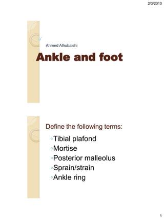
- 1. 2/3/2010 Ahmed Alhubaishi Ankle and foot Define the following terms: ◦Tibial plafond ◦Mortise ◦Posterior malleolus ◦Sprain/strain ◦Ankle ring 1
- 2. 2/3/2010 An emergency physician who applies the Ottawa Ankle Rules correctly would send which of the following ambulatory patients with a chief complaint of “ankle pain” for x-ray? a. A 40-year-old male with tenderness upon palpation of the posterior edge of the medial malleolar tip b. A 25-year-old female with edema, ecchymosis,and tenderness just anterior to the lateral malleolus c. A 60-year-old male with lateral edema, ecchymosis, and a positive anterior drawer test d. A 16-year-old male with posterior ankle tenderness and a positive Thompson test An emergency physician who applies the Ottawa Ankle Rules correctly would send which of the following ambulatory patients with a chief complaint of “ankle pain” for x-ray? a. A 40-year-old male with tenderness upon palpation of the posterior edge of the medial malleolar tip b. A 25-year-old female with edema, ecchymosis,and tenderness just anterior to the lateral malleolus c. A 60-year-old male with lateral edema, ecchymosis, and a positive anterior drawer test d. A 16-year-old male with posterior ankle tenderness and a positive Thompson test 2
- 3. 2/3/2010 Using OAR which of the following not for X-ray: Bone tenderness at med. Malleolus Bone tenderness at lat. Malleolus Bone tenderness of the posterior edge distal 6 cm to the ankle Inability to bear wt on ankle now and immediately after the injury Soft tissue swelling over med. And lat. malleolus Using OAR which of the following not for X-ray: Bone tenderness at med. Malleolus Bone tenderness at lat. Malleolus Bone tenderness of the posterior edge distal 6 cm to the ankle Inability to bear wt on ankle now and immediately after the injury Soft tissue swelling over med. And lat. malleolus 3
- 4. 2/3/2010 Ottawa rules When OAR cannot be applied??? 4
- 5. 2/3/2010 When OAR cannot be applied??? 1. Altered level of consciousness 2. Subacute or chronic injuries 3. Injuries to hindfoot or forefoot 4. Not designed to pick up # < 3 mm One of the following ankle # can be Rx as OPD with close ortho. FU: Fibular # proximal to tibiotalar ( t-t) joint line Lat. Malleolus # below the T-T joint line Lat. Malleolus # with deltoid lig. Rupture Unimalleolar # with syndesmotic diastasis 5
- 6. 2/3/2010 One of the following ankle # can be Rx as OPD with close ortho. FU: Fibular # proximal to tibiotalar ( t-t) joint line Lat. Malleolus # below the T-T joint line Lat. Malleolus # with deltoid lig. Rupture Unimalleolar # with syndesmotic diastasis What is this? 6
- 7. 2/3/2010 The ankle ring consists of the following: tibial plafond, medial malleolus, deltoid ligaments, calcaneus, lateral collateral ligaments, lateral malleolus syndesmotic ligaments. The integrity of this ring determines the stability of the ankle 7
- 8. 2/3/2010 Which of the following is the most commonly injured soft-tissue structure(s) of the ankle? a. Lateral collateral ligaments b. Medial collateral ligaments c. Inferior tibiofibular ligaments d. Achilles tendon Which of the following is the most commonly injured soft-tissue structure(s) of the ankle? a. Lateral collateral ligaments b. Medial collateral ligaments c. Inferior tibiofibular ligaments d. Achilles tendon 8
- 9. 2/3/2010 Each of the following ligaments are part of the ankle syndesmosis except: a. Anterior inferior tibiofibular ligament (AITFL) b. Posterior inferior tibiofibular ligament (PITFL) c. Interosseous ligament (IOL) d. Calcaneofibular ligament (CFL) Each of the following ligaments are part of the ankle syndesmosis except: a. Anterior inferior tibiofibular ligament (AITFL) b. Posterior inferior tibiofibular ligament (PITFL) c. Interosseous ligament (IOL) d. Calcaneofibular ligament (CFL) 9
- 10. 2/3/2010 Ankle ligaments- lateral Ankle ligaments – medial (deltoid) 10
- 11. 2/3/2010 All of the following terms describe a motion of the talus within the mortise except: a. adduction. b. external rotation. c. supination. d. plantar flexion. All of the following terms describe a motion of the talus within the mortise except: a. adduction. b. external rotation. c. supination. d. plantar flexion. 11
- 12. 2/3/2010 Widening of the medial clear space on ankle radiographs suggests injury to each of the following structures except: a. lateral ligament complex. b. deltoid ligament. c. anterior inferior tibiofibular ligament (AITFL). d. posterior inferior tibiofibular ligament (PITFL). Widening of the medial clear space on ankle radiographs suggests injury to each of the following structures except: a. lateral ligament complex. b. deltoid ligament. c. anterior inferior tibiofibular ligament (AITFL). d. posterior inferior tibiofibular ligament (PITFL). 12
- 13. 2/3/2010 Pain at the ankle during squeeze testing is suggestive of injury to which structure(s)? a. Medial collateral ligaments b. Inferior tibiofibular ligaments c. Lateral collateral ligaments d. Peroneal tendons Pain at the ankle during squeeze testing is suggestive of injury to which structure(s)? a. Medial collateral ligaments b. Inferior tibiofibular ligaments c. Lateral collateral ligaments d. Peroneal tendons 13
- 14. 2/3/2010 On a normal AP ankle x-ray, the amount of tibiofibular overlap should be at least: a. 2 mm. b. 4 mm. c. 6 mm. d. 8 mm. On a normal AP ankle x-ray, the amount of tibiofibular overlap should be at least: a. 2 mm. b. 4 mm. c. 6 mm. d. 8 mm. 14
- 15. 2/3/2010 15
- 16. 2/3/2010 -Malleoli superimposed each other -- body of calcaneous visible -Base of 5 th m.t 16
- 17. 2/3/2010 -entire joint space -talar dome No overlap between the previous two -symmetrical joint space -Width of medial space 2-3 mm --T-F ovelap not less than 1-2 mm 17
- 18. 2/3/2010 Widening of the medial clear space or a lesser degree of tibulofibular overlap suggests injury to to the medial ligament,syndesmosis or both 18
- 19. 2/3/2010 The Lauge-Hansen classification of ankle fractures is based on: a. the anatomic location of the fibular fracture with respect to the mortise. b. the mechanism of injury. c. the degree of articular involvement. d. the presence or absence of syndesmotic disruption. The Lauge-Hansen classification of ankle fractures is based on: a. the anatomic location of the fibular fracture with respect to the mortise. b. the mechanism of injury. c. the degree of articular involvement. d. the presence or absence of syndesmotic disruption. 19
- 20. 2/3/2010 Inversion injury. There is a transverse avulsion fracture of the lateral malleolus below the mortise caused by supination-adduction forces (arrow).The lateral ligaments remain intact. This injury is classified as Lauge-Hansen SA grade 1 or Danis- Weber type A. 20
- 21. 2/3/2010 The medial clear space is widened, suggesting deltoid and/or syndesmotic ligament disruption (arrowhead). There is an isolated spiral fracture of the fibula occurring at the level of the mortise caused by supination-external rotation forces.This injury is classified as Lauge-Hansen SE grade 2 or Danis-Weber type B. 21
- 22. 2/3/2010 What is this? What is this? MAISONNEUVE’S FRACTURE 22
- 23. 2/3/2010 Characteristics of a Maisonneuve fracture include all of the following except: a. It occurs in the setting of forceful external rotation. b. It is frequently associated with medial ligament and/or syndesmosis disruption. c. It is highly unstable. d. The diagnosis is readily made on routine ankle x-ray series. Characteristics of a Maisonneuve fracture include all of the following except: a. It occurs in the setting of forceful external rotation. b. It is frequently associated with medial ligament and/or syndesmosis disruption. c. It is highly unstable. d. The diagnosis is readily made on routine ankle x-ray series. 23
- 24. 2/3/2010 What is this? Pilon fracture # of distal tibial metaphysis Due to high energy mechanism Usually comminuted, 20% open Significant soft tissue loss Talus derive into tibial plafond Associated with: # of calcaneus,tibial platue, femoral neck, acetabulum, vertebrae 24
- 25. 2/3/2010 Tillaux Fracture: • Lateral tibia, involving articular surface • Salter-Harris III fracture, mostly in adolescents • Usually requires surgical fixation The best test for Achilles tendon rupture is: a. ability to pronate the foot. b. ability to dorsiflex the foot. c. the Thompson squeeze test. d. the ―wiggle test.‖ 25
- 26. 2/3/2010 The best test for Achilles tendon rupture is: a. ability to pronate the foot. b. ability to dorsiflex the foot. c. the Thompson squeeze test. d. the ―wiggle test.‖ All of the following fractures warrant orthopedic consultation in the ED except: a. unimalleolar fracture. b. bimalleolar fracture. c. trimalleolar fracture. d. triplane fracture. 26
- 27. 2/3/2010 All of the following fractures warrant orthopedic consultation in the ED except: a. unimalleolar fracture. b. bimalleolar fracture. c. trimalleolar fracture. d. triplane fracture. When to consult ortho people to come and see pt with ankle pain ? 27
- 28. 2/3/2010 Unimalleolar Fractures Displaced medial malleolar fracture Medial malleolar fracture with lateral collateral ligament rupture Displaced lateral malleolar fracture Lateral malleolar fracture with deltoid ligament rupture Lateral malleolar fracture with widened medial clear space Unimalleolar fracture with syndesmotic diastasis Fibula fracture at or proximal to the tibiotalar joint line Displaced posterior malleolar fracture Posterior malleolar fracture involving more than 25% of joint surface All Bimalleolar Fractures All Trimalleolar Fractures All Intraarticular Fractures With Step Deformity All Open Fractures All Pilon Fractures Clinical Pathway: Evaluation Of Ankle Injuries 28
- 29. 2/3/2010 29
- 30. 2/3/2010 Clinical Pathway: Management Of Ankle Injuries 30
- 31. 2/3/2010 31
- 32. 2/3/2010 CASE • 24 yo M football player • Another player rolled over his ankle from behind • ANKLE DISLOCATION: • • Usually posterior • • Often associated with fracture and ligamentous injury • Reduction: • • Place one hand behind heel, with other over dorsum of foot. • • Downward and anterior traction, with foot plantar-flexed initially. • • Finally bring ankle back to 90 degrees flexion. • Clinical Pearl: • Put the knee in a slightly flexed position (20-30 degrees) during the reduction to reduce tension at the ankle. • Post-reduction: • • Immobilize in short leg, 3-sided splint, ankle at 90 degrees • • Follow up with Orthopedic surgeon 32
- 33. 2/3/2010 case • 20 year old male twisted his ankle while ―snowboarding‖ • Exam: Ankle is swollen, diffusely tender, and plain films are negative. • Ankle Sprain: • • R.I.C.E. (rest, ice, compression, elevation) • • Functional immobilization • o ACE, AirCast, taping, etc • • Crutches • o Weight-bearing as tolerated • • Follow up exam • o Approximately two weeks after injury • o Repeat physical exam for ligamentous damage • o Most patients will be much improved • o A few may have persistent pain, swelling, and joint effusion, • suggesting the possibility of occult fracture. • When should I consider CT or MRI for occult ankle fracture? • Consider CT or MRI in the setting of negative plain films, and: • o High clinical suspicion • o Persistent pain, swelling, effusion at follow- up • Important occult fractures of the ankle/foot: • o Talar dome • o Tillaux (lateral tibia) • o Calcaneus, Navicular • o Lateral process of the talus 33
- 34. 2/3/2010 • Haapamaki, American Journal of Roentgenology, 2004 • Retrospective study, over 3 years • 344 patients with a fracture on ankle / foot CT • CT’s ordered to delineate fracture, or to r/o occult fracture • Most common occult fx in ankle (not visualized on plain films): • Calcaneus (20) • Talus (15) • Tillaux (7) • Pearls: • 1) CT helpful for: • a. High suspicion (mechanism, exam) • b. Poor recovery • 2) High risk situations: • a. Fall from height—Calcaneus • b. Adolescent—Tillaux • c. Snowboarding—Lat. process of Talus 34
- 35. 2/3/2010 foot Q What is CHOPART’S AND LISFRANCE’S JOINTS? CHOPART: between midfoot and hindfoot LISFRANCE: between midfoot and metatarsals 35
- 36. 2/3/2010 foot What are the foot # need ortho consult in ED ? 36
- 37. 2/3/2010 All talus fractures All calcaneus fractures Significant navicular fractures, especially if intraarticular All cuboid fractures Lisfranc injuries Metatarsal shaft fractures with > 3 mm displacement or 10 degrees angulation Metatarsal head and neck fractures Jones fractures When BOEHLER’S angle < 20 degree means: Navicular fracture Cuboid fracture Lisfrance’s fracture Calcaneal fracture First metatarsal fracture 37
- 38. 2/3/2010 When BOEHLER’S angle < 20 degree means: Navicular fracture Cuboid fracture Lisfrance’s fracture Calcaneal fracture First metatarsal fracture Boehler’s angle 38
- 39. 2/3/2010 What is this? Calcaneus fracture: • Calcaneus fractures most often occur in males (male:female = 5:1) • Peak age: between 30 and 50 years. • Associated injuries (Lumbar spine vertebral compression fractures) • Treatment: Operative vs Casting 39
- 40. 2/3/2010 What is this? Talar body fracture Risk of AVN 40
- 41. 2/3/2010 What is this? Talar neck # 50% of all talar # Extreme dorsiflexion Hawkin’s classification 1-4 41
- 42. 2/3/2010 Talar Dome Fracture: • Osteochondral lesion, articular surface • CT and MRI both excellent to visualize lesion • May be managed by cast (non- weight bearing), or by arthroscopic surgery if loose fragments in joint What is this? 42
- 43. 2/3/2010 Lisfrance’s fracture AP view : ◦ medial margin of the base of the second metatarsal lines up with the medial margin of the middle cuneiform oblique view: ◦ medial margin of the base of the third metatarsal lines up with the medial margin of the lateral cuneiform, and ◦ medial margin of the base of the fourth metatarsal lines up with the medial margin of the cuboid Types of lisfrance’s # 43
- 44. 2/3/2010 What fracture is virtually pathognomonic for a Lisfranc injury? Fracture the base of second metatarsal What are these? 44
- 45. 2/3/2010 What are these? Jones # Pseudojones # 45
- 46. 2/3/2010 Jones’ fracture: transverse fracture at least 15 mm distal to proximal end of 5th metatarsal; high rate of malunion so call ortho Pseudo-Jones’ fracture: avulsion fracture of tuberosity at 5th metatarsal base; treat symptomatically Nonunion and chronic disability may result from inadequate immobilization of: a. lateral malleolar avulsion fractures. b. avulsion fractures of the tuberosity of the fifth metatarsal (pseudo-Jones). c. fifth metatarsal shaft fractures (Jones). d. lateral ligament tears with lateral malleolar avulsion fractures. 46
- 47. 2/3/2010 Nonunion and chronic disability may result from inadequate immobilization of: a. lateral malleolar avulsion fractures. b. avulsion fractures of the tuberosity of the fifth metatarsal (pseudo-Jones). c. fifth metatarsal shaft fractures (Jones). d. lateral ligament tears with lateral malleolar avulsion fractures. What are the indications for reduction of a metatarsal fracture? 47
- 48. 2/3/2010 Greater than 10 degrees angulation or 3 mm displacement Thank you 48