SlideShare una empresa de Scribd logo
1 de 75
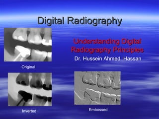
Digital Radiography
Understanding Digital
Radiography Principles
Dr. Hussein Ahmed Hassan
Original

Inverted

Embossed
Purpose: Basic Answers
What is digital radiography?
What components are required?
What types of digital radiography work best in
practice?
 How do we choose the right solution for our
program?
 What are the computer and networking
requirements to support our digital solution?



What is Digital Radiography?
 Sensors or phosphor plates
take the place of traditional
film.
 Radiographic images are
acquired almost instantly
and stored electronically.
 They can be manipulated,
viewed, and transferred with
a software program.
What Components are required for
Digital Radiography?
 Sensors or phosphor plates to replace x-ray film.
 Imaging software that allows image storage and
management.
 Computer system to run imaging software.
 Optional: Practice management software –for
completely paperless patient records
management.
Digital Radiography

 Is a form of x-ray imaging, where digital
X-ray sensors are used instead of traditional
photographic film.
 Advantages include;
1. Time efficiency through bypassing chemical
processing
2.The ability to digitally transfer and enhance
images.
3.Less radiation can be used to produce an
image of similar contrast to conventional
Digital Imaging Technology
Digital methods for processing and displaying x-ray images
were first introduced with the advent of computed tomography
(CT) in 1972.
Continuing advances in computer technology have promoted the
general development of image acquisition in digital form (CCD
cameras), most commonly from image intensifiers (digital
fluoroscopy) or from storage phosphor plates (computed
radiography).
Other detector systems such as ‘flat-panel’ technology for indirect
or direct digital radiography are now available for general purpose
equipment.
Capture technologies used today:

• Screen/film

(S/F) combination system

• Image intensifiers
• Computed radiography (CR) with storage phosphors
• Direct Digital radiography (DDR)
Image Acquisition
Conventional X-ray
Drying
X-ray

fixer

water developer water
Image Acquisition in Digital Imaging
 There are a number of technologies used
for digital imaging in planar radiography
these are;

1. Computed radiography (CR), CR in first
appearance, similar to the used of
film/screen system. The CR plate is in a
cassette, which will fit the table and vertical
Backy trays and can be used with mobile
equipment. The plate is then scanned in a
reading system, this make the change to
computed radiography (CR) easier.
Image Acquisition
CR
X-ray Tube

Latent image

Photomultiplier

Readout

Erasure

ADC

IP Ready

Fluorescent Lamp

Workstation
2.Direct Digital
 Means, instead of using x-ray film, a
sensor connects directly to your
computer - the x-ray image displays
almost immediately after taking it.
 Sensors use either a Charge
Coupled Device (CCD), or a
Complementary Metal Oxide
Semiconductor (CMOS) or thin-film
transistor (TFT) to convert light into
electrons. These are collected &
turned into pixels that show
brightness and contrast - that’s what
makes a digital x-ray image.
 You still use your regular x-ray
machine to take the exposure.
 Both CCD sensors, CMOS sensors
and TFTwork extremely well.
Direct Digital Radiography --cot---Direct Digital Radiography (DDR), DDR
system needs more changes in x-ray couch
and vertical backy design and often
changes to x-ray tube assembly, DR
detector is fully integrated into exposure
equipment. The patient is radiographed and
the image appears on workstation in few
seconds. Here image optimized and then
sent for reporting or repeated if necessary.
Image Acquisition DR
1.X-ray scintillator bonded to readout ray, thin-film transistor (TFT)

X-ray Tube

Workstation

Detectors (TFT)
Why do we need a new technology, what is wrong with
film/screen?
• Absolutely nothing !!!!
• Still the “gold standard” to which all new projection radiography

systems are measured against
• On many accounts (e.g.: mammography) , S/F is still superior than
some new technologies (e.g.: CR)
• The choice of technology depends on clinical, technical, operational
and economic factors
Uses Digital Radiography
 CR is used in all areas where film/screen
systems are currently used, including
mammography.
 Direct Digital Radiography (DDR) can be
used in general radiography and mobile
radiography.
 DDR is very popular in small-field
mammography and being introduced into
full-field mammography.
 DDR detectors are now being used instead
of image intensifiers in fluoroscopy.
Computed Radiography
What is it?
 Computed Radiography, commonly known as CR, is a
digital radiography process that is designed to replace
industrial radiographic film
 The CR process is very similar to the process associated
with film … mainly because of the film-like digital detector
that is used to capture the radiographic image
 The CR film-like digital detector is a flexible phosphor
screen that looks and feels a lot like film
CR Cassettes
Storage phosphor (SP) screens are similar in structure to
conventional intensifying screens
Computed Radiography
Storage Phosphor
Digital X-ray Imaging Systems
Computed Radiography
How does it work?
Step 1: The phosphor screen is inserted into a
soft or hard cassette (with or without lead)
Step 2: A radiation pattern is exposed on the phosphor screen
creating a latent image
Step 3: The phosphor screen is then inserted
into a phosphor scanner to be read
Step 4: The phosphor screen is scanned and the digital
image is displayed on the workstation
monitor for review and evaluation
Step 5: The phosphor screen is then erased and ready to
be reused
Computed Radiography System Process

Differences between Phosphor & Film
Step 1: The phosphor screen is inserted into a soft or hard
cassette (with or without lead screens)
Do not need a light-tight darkroom for phosphor imaging
Step 2: A radiation pattern is exposed on the phosphor screen
creating a latent image
Phosphor is faster and much more forgiving (wide latitude)
Step 3: The phosphor screen is then inserted into a phosphor
scanner to be read No chemical.
Film processor (8 minutes) vs. Phosphor (1 to 2 minutes)
Step 4: The phosphor screen is scanned and the digital image is
displayed on the workstation monitor for review
Digital image can be enhanced increasing OD
Step 5: The phosphor screen is then erased and ready to be reused
The phosphor screens are reusable
Computed Radiography System Process

Similarities between Phosphor & Film
Step 1: The phosphor screen is inserted into a soft or hard
cassette (with or without lead screens)
Just like film
Step 2: A radiation pattern is exposed on the phosphor screen
creating a latent image
Shot set-up and technique are basically the same
Step 3: The phosphor screen is then inserted into a phosphor
scanner to be read
Very similar to film being put into a film processor
Step 4: The phosphor screen is scanned and the digital image
is displayed on the workstation monitor for review
Film is put on a lightbox to view
Step 5: The phosphor screen is then erased and ready to be
reused
Computed Radiography System
Components Overview
Computed Radiography System
consists of the following components:

WORKSTATION
& SOFTWARE

PHOSPHOR
SCANNER
& ERASER

PHOSPHOR
SCREENS
Computed Radiography System
Workstation and Software
Computed Radiography System
Workstation and Software
 Operates the Phosphor Scanner
 Manages your images and
associated data: drawings, word
documents, digital photos, procedures,
reference images, techniques, etc.

 Image enhancement capabilities:
zoom, gray-scale manipulation, pan & scroll,
density & measurements tools (histograms, point profiles, line
profiles, line and area measurements), image processing,
annotation, etc.

 Ability to save and archive your digital images and
associated data
Computed Radiography System
Phosphor Scanners
Two different types of Phosphor Scanners
Computed Radiography Systems

Table-top
CR100

CR Tower
Computed Radiography System
(Table-top Unit)
consists of the following components:

WORKSTATION
& SOFTWARE

PHOSPHOR
SCANNER
Phosphor Screen is placed
directly into the scanner

PHOSPHOR
SCREENS
Computed Radiography System
(CR Tower Free-Standing Unit)
consists of the following components:

WORKSTATION
& SOFTWARE

PHOSPHOR
SCANNER
Hard-cassette
based system

PHOSPHOR
SCREENS
Phosphor Scanners
How is the phosphor
screen scanned?
He-Ne laser

rotating mirror

light-guide
PMT
storage phosphor plate
Overall mechanism
Computed Radiography System
Phosphor Imaging Screens
and customized Cassettes
Computed Radiography
Phosphor Screens
Manufactured in similar sizes as film
Utilizes the same cassettes as film
Utilizes the same x-ray sources as film
Set-up in the field is the same as film
Typically faster exposures than film
Very wide latitude which makes it much
more forgiving than film / less reshots
 No chemicals required for processing
 Image quality good match for aircraft
applications






PSP- PhotoStimulated Phosphor
 Reusable plastic plate  Laser scanning - the
coated with phosphor
trapped electrons are
released  It stores the energy of
photostimulated
the remnant x-ray - xluminescence
ray photons excites the
 The emitted light is
electrons in the
phosphor.
detected by
photomultiplier tube,
and is digitized to form
an image
Computed Radiography
How is the phosphor screen
manufactured (very similar to film)?

phosphor
layer

phosphor
grains

protective
layer

substrate
Computed Radiography Technology
The active phosphor layer of a CR plate usually comprises a
layer of europium-doped barium fluorobromide, which is coated
on to a semi-rigid or flexible polyester base.

phosphor
layer

phosphor
grains

protective
layer

substrate

X-ray photons are absorbed by the phosphor layer, the phosphor
electron become excited and raised to high energy level, where they
can stay trapped in semi-stable high energy state.
The trapped electron represent a latent image in the phosphor plate in
form of stored energy.
The stored energy is released by adding energy to the trapped electron
This done by stimulation with laser beam, the trap electrons then escap
from the traps to fall back to their equilibrium state.
Computed Radiography Technology---cot----

As the electrons fall back, the electrons release energy in
form of light.
This phenomenon is known as photostimulable
luminescence (PSL).
The emitted light intensity is proportional to original x-ray
intensity.
The light energy detected and signal is digitized and
processed digitally to produce a visible diagnostic radiograph
 on a monitor.
The phosphor plate is then erased with a bright white light t
remove any remaining trapped electrons, and the plate is
then ready for the next examination.
Directed Digital Radiography
(DDR)
Directed digital radiography, a
term used to describe total
electronic imaging capturing.
Eliminates the need for an
image plate altogether.
Amorphous Selenium detector technology for
DR Direct Radiography
IMAGE CAPTURE
CR
– PSP – photostimulable phosphor plate
– REPLACES FILM IN THE CASSETTE

DR – NO CASSETTE – PHOTONS
– CAPTURED DIRECTLY
– ONTO A TRANSISTOR
– SENT DIRECTLY TO A MONITOR
Densities of the IMAGE
 The light is proportional to
amount of light received
 digital values are then equivalent
(not exactly the same) to a value
of optical density (OD) from a
film, at that location of the image
CR VS DR
– CR -Indirect capture where the image
is first captured on plate and stored =
then converted to digital signal
– DDR -Direct capture where the image is
acquired immediately as a matrix of
pixels – sent to a monitor
DIRECT RADIOGRAPHY






uses a transistor receiver (like bucky)
that captures and converts x-ray energy
directly into digital signal
seen immediately on monitor
then sent to PACS/ printer/ other
workstations FOR VIEWING
CR vs DR
CR
 imaging plate
 processed in a Digital
Reader

DR
 transistor receiver (like
bucky)
 directly into digital
signal

 Signal sent to computer
 Viewed on a monitor

 seen immediately on
monitor –
ADVANTAGE OF CR/DR
 Can optimize image quality
 by manipulating digital data
 to improve visualization of anatomy
and pathology
 AFTER EXPOSURE TO PATIENT
ADVANTAGE OF CR/DR
 CHANGES MADE TO IMAGE

 AFTER THE EXPOSURE
 CAN ELIMINATE THE NEED TO
REPEAT THE EXPOSURE
ADVANTAGE OF CR/DR vs FS
Rapid storage
retrieval of images NO LOST FILMS!
PAC (storage management)
Teleradiology - long distance transmission
of image information
 Economic advantage - at least in the long
run?




CR/DR VS FILM/SCREEN
 FILM these can not be modified
once processed
 If copied – lose quality
 DR/CR – print from file – no loss
of quality
“no fault” TECHNIQUES
F/S: RT must choose technical factors
(mAs & kvp) to optimally visualize anatomic detail
CR: the selection of processing algorithms and
anatomical regions controls how the acquired
latent image is presented for display
 HOW THE IMAGE LOOKS CAN BE ALTERED BY
THE COMPUTER – EVEN WHEN “BAD”
TECHNIQUES ARE SET
DR
Initial expense high
very low dose to pt –
image quality of 100s using a 400s
technique
 Therfore ¼ the dose needed to make the
image



Storage /Archiving
FILM/SCREEN
 films: bulky
 deteriorates over time
 requires large storage
& expense
 environmental
concerns

 CR & DR
 8000 images stored
on CD-R
 Jukebox CD storage
 no deterioration of
images
 easy access
Computed Radiography System
Compatibility & Growth Opportunities
Most of the CR Workstations also have the ability to
operate a wide variety of:
- printers
- document scanners
- archiving hardware
- film digitizers
- flat panel arrays
- and other digital
detectors
Transmission of Images
 PACS - Picture Archiving &
Communications
System
 DICOM - Digital Images & Communication
in Medicine
 TELERADIOGRAPHY -Remote
Transmission of Images
Digital imaging technology (cont)
The technique of digital subtraction
angiography (DSA), based on digital
image processing, allows enhanced
visualization of blood vessels by
electronically subtracting unwanted
parts of the image.
Digital subtraction angiography.
(a) Mask ,immediately prior to contrast injection a preliminary digitized image
known as the “mask” is performed. Note pelvic bone, bowel gas and arterial catheter.
(b) Contrast image. Contrast is injected through the catheter producing opacification
of arteries.
(c) Subtracted image. The computer subtract the mask from the contrast image leaving
an image of contrast fill blood vessels unobscured by overlying bone and bowel.
Note a tight localized stenosis of the right common illiac artery (arrow)
Digital imaging technology
At this time, there is no consensus on the best
technology for balancing dose and image quality. Digital
imaging potentially can provide lower doses than the
film-intensifying screen method.
However, through post-exposure manipulation of the data,
satisfactory diagnostic images can be produced even
when unnecessarily high patient radiation doses are
used. Proper quality assurance procedures are
essential.
Main Characteristics of an Image Receptor
The selection of an imaging system should
involve a thorough evaluation and analysis of its
complete characteristics together with
consideration of the technical and human
environment in which the system will be used.
The main characteristics to be considered when selecting
an image receptor are:spatial resolution; contrast resolution; dose efficiency;
Modulation Transfer Function; detector size;
possibilities of image storage and transfer; and
qualities such as weight, robustness, fast image
access, etc).
CR - ? Which shows
“better detail” Why/how?

8 x 10 cassette

14 x 17 cassette
To Produce Quality Images
For Conventional Projection
or CR Radiography:
The same rules, theories, and laws still
apply and can not be overlooked

FFD/OFD (SID/SOD)
Inverse Square Law
Beam Alignment Tube-Part-Film Alignment
Collimation
Grids
Exposure Factors: KVP, MaS

Patient Positioning
90 KVP

GE DR
BURNING
0˚ Angle

30˚ Angle

50˚ Angle
 towel that was used
to help in
positioning a child

 CR is MORE
sensitive to
 ARTIFACTS

NEW IMAGE
CR image – NEW IMAGE
 Line caused
from dirt
collected in a
CR Reader
Double
exposure
Child
120 KVP

Fuji CR
?

Hands over upper
abdomen
High resolution with digital
imaging
Vertical patterns of hyperintense signal


usually represent foreign materials
that are stuck to the light path
assembly that acquires the
photostimulated luminescence
signal from the CR imaging plate as
it is being scanned by a laser beam.
As the light is blocked at the same
spot as the plate translated through
the optical stage, the artifact occurs
perpendicular to the laser beam
readout, in the plate translation
(slow-scan) direction. The stripe
appears bright, since the image
undergoes a reverse grayscale
transformation to make the image
appear similar to a screen-film
image with processing.
A lateral chest image with an unusual
superimposed pattern on the anatomy.
 This is an example of a CR
image obtained with
cassette reversed, where
the tube side of cassette is
pointed away from the x-ray
tube source and toward the
patient. Cassette plastic

structural patterns are
projected onto the imaging
plate (particularly
noticeable in the arms and
anterior part of the patient.
‫المأذن حرابنا والقباب خوزاتنا مساجدنا ثكناتنا والمصلون جنودنا وهذا الجيش المقدس يحمي ديننا‬

‫محمد عاكف شاعر تركى‬
THANK YOU!!

Question?

Más contenido relacionado

La actualidad más candente

Difference Between Single Slice and Multi Slice CT Scanner
Difference Between Single Slice and Multi Slice CT ScannerDifference Between Single Slice and Multi Slice CT Scanner
Difference Between Single Slice and Multi Slice CT ScannerAbdul Rehman (R)(CT)(MR)
 
Computed radiography and digital radiography- CR/DR
Computed radiography and digital radiography- CR/DRComputed radiography and digital radiography- CR/DR
Computed radiography and digital radiography- CR/DRAshim Budhathoki
 
CT ARTIFACTS.pptx
CT ARTIFACTS.pptxCT ARTIFACTS.pptx
CT ARTIFACTS.pptxdypradio
 
Introduction to digital radiography and pacs
Introduction to digital radiography and pacsIntroduction to digital radiography and pacs
Introduction to digital radiography and pacsRad Tech
 
computed and digital radiography(2)
computed and digital radiography(2)computed and digital radiography(2)
computed and digital radiography(2)SHASHI BHUSHAN
 
Digital radiography
Digital radiographyDigital radiography
Digital radiographyharibudke
 
Ct instrumentation and types of detector configuration
Ct instrumentation and types of detector configurationCt instrumentation and types of detector configuration
Ct instrumentation and types of detector configurationSUJAN KARKI
 
Computed radiography
Computed radiographyComputed radiography
Computed radiographyLirih Kuu
 
Exposure factors2
Exposure factors2Exposure factors2
Exposure factors2mr_koky
 
Computed Radiography and digital radiography
Computed Radiography and  digital radiographyComputed Radiography and  digital radiography
Computed Radiography and digital radiographyDurga Singh
 
Digital Fluoroscopy Imaging System
Digital Fluoroscopy Imaging SystemDigital Fluoroscopy Imaging System
Digital Fluoroscopy Imaging SystemMuhammad Arif Afridi
 
MRI Coil and Gradient power-point slide pk
MRI Coil and Gradient power-point slide pkMRI Coil and Gradient power-point slide pk
MRI Coil and Gradient power-point slide pkDr pradeep Kumar
 
Helical and Multislice CT
Helical and Multislice CTHelical and Multislice CT
Helical and Multislice CTManojzz Bhatta
 

La actualidad más candente (20)

Difference Between Single Slice and Multi Slice CT Scanner
Difference Between Single Slice and Multi Slice CT ScannerDifference Between Single Slice and Multi Slice CT Scanner
Difference Between Single Slice and Multi Slice CT Scanner
 
Computed Radiography
Computed RadiographyComputed Radiography
Computed Radiography
 
CR and DR ppt
CR and DR pptCR and DR ppt
CR and DR ppt
 
Computed radiography and digital radiography- CR/DR
Computed radiography and digital radiography- CR/DRComputed radiography and digital radiography- CR/DR
Computed radiography and digital radiography- CR/DR
 
CT ARTIFACTS.pptx
CT ARTIFACTS.pptxCT ARTIFACTS.pptx
CT ARTIFACTS.pptx
 
Introduction to digital radiography and pacs
Introduction to digital radiography and pacsIntroduction to digital radiography and pacs
Introduction to digital radiography and pacs
 
computed and digital radiography(2)
computed and digital radiography(2)computed and digital radiography(2)
computed and digital radiography(2)
 
Digital radiography
Digital radiographyDigital radiography
Digital radiography
 
Mri physics
Mri physicsMri physics
Mri physics
 
Ct instrumentation and types of detector configuration
Ct instrumentation and types of detector configurationCt instrumentation and types of detector configuration
Ct instrumentation and types of detector configuration
 
soft tissue radiography
soft tissue radiographysoft tissue radiography
soft tissue radiography
 
COMPUTED RADIOGRAPHY
COMPUTED RADIOGRAPHYCOMPUTED RADIOGRAPHY
COMPUTED RADIOGRAPHY
 
Computed radiography
Computed radiographyComputed radiography
Computed radiography
 
digital radiography
digital radiographydigital radiography
digital radiography
 
Exposure factors2
Exposure factors2Exposure factors2
Exposure factors2
 
Mri gradient coils
Mri gradient coilsMri gradient coils
Mri gradient coils
 
Computed Radiography and digital radiography
Computed Radiography and  digital radiographyComputed Radiography and  digital radiography
Computed Radiography and digital radiography
 
Digital Fluoroscopy Imaging System
Digital Fluoroscopy Imaging SystemDigital Fluoroscopy Imaging System
Digital Fluoroscopy Imaging System
 
MRI Coil and Gradient power-point slide pk
MRI Coil and Gradient power-point slide pkMRI Coil and Gradient power-point slide pk
MRI Coil and Gradient power-point slide pk
 
Helical and Multislice CT
Helical and Multislice CTHelical and Multislice CT
Helical and Multislice CT
 

Destacado

Current applications of interventional radiology 97
Current applications of interventional radiology 97Current applications of interventional radiology 97
Current applications of interventional radiology 97Arun Jagannathan
 
Principles of radiographic interpretations
Principles of radiographic interpretationsPrinciples of radiographic interpretations
Principles of radiographic interpretationsShweta Meeee
 
INTRAORAL RADIOGRAPHY
INTRAORAL RADIOGRAPHYINTRAORAL RADIOGRAPHY
INTRAORAL RADIOGRAPHYshamika147
 
Radiographic cassettes
Radiographic cassettesRadiographic cassettes
Radiographic cassettesSudil Paudyal
 
modulation transfer function (MTF)
modulation transfer function (MTF)modulation transfer function (MTF)
modulation transfer function (MTF)AJAL A J
 
Intraoral radiographic techniques/prosthodontic courses
Intraoral radiographic techniques/prosthodontic coursesIntraoral radiographic techniques/prosthodontic courses
Intraoral radiographic techniques/prosthodontic coursesIndian dental academy
 
Artifact and errors in intraoral periapical radiograph.ppt
Artifact and errors in intraoral periapical radiograph.pptArtifact and errors in intraoral periapical radiograph.ppt
Artifact and errors in intraoral periapical radiograph.pptjyoti sharma
 
radio-graphic-techniques-bisecting-and-occlusal
 radio-graphic-techniques-bisecting-and-occlusal radio-graphic-techniques-bisecting-and-occlusal
radio-graphic-techniques-bisecting-and-occlusalParth Thakkar
 
CR, DR and recent advances
CR, DR and recent advancesCR, DR and recent advances
CR, DR and recent advancesVishal Sankpal
 
Intra oral radiographs
Intra oral radiographsIntra oral radiographs
Intra oral radiographsmelbia shine
 
Computer radiography and digital radiography
Computer radiography and digital radiographyComputer radiography and digital radiography
Computer radiography and digital radiographyAnand Rk
 

Destacado (15)

Radiography
RadiographyRadiography
Radiography
 
Advancd imagiology /dental courses
Advancd imagiology /dental coursesAdvancd imagiology /dental courses
Advancd imagiology /dental courses
 
Pioneers of Radiology
Pioneers of RadiologyPioneers of Radiology
Pioneers of Radiology
 
Current applications of interventional radiology 97
Current applications of interventional radiology 97Current applications of interventional radiology 97
Current applications of interventional radiology 97
 
Principles of radiographic interpretations
Principles of radiographic interpretationsPrinciples of radiographic interpretations
Principles of radiographic interpretations
 
INTRAORAL RADIOGRAPHY
INTRAORAL RADIOGRAPHYINTRAORAL RADIOGRAPHY
INTRAORAL RADIOGRAPHY
 
historical perspective of radiology
historical perspective of radiologyhistorical perspective of radiology
historical perspective of radiology
 
Radiographic cassettes
Radiographic cassettesRadiographic cassettes
Radiographic cassettes
 
modulation transfer function (MTF)
modulation transfer function (MTF)modulation transfer function (MTF)
modulation transfer function (MTF)
 
Intraoral radiographic techniques/prosthodontic courses
Intraoral radiographic techniques/prosthodontic coursesIntraoral radiographic techniques/prosthodontic courses
Intraoral radiographic techniques/prosthodontic courses
 
Artifact and errors in intraoral periapical radiograph.ppt
Artifact and errors in intraoral periapical radiograph.pptArtifact and errors in intraoral periapical radiograph.ppt
Artifact and errors in intraoral periapical radiograph.ppt
 
radio-graphic-techniques-bisecting-and-occlusal
 radio-graphic-techniques-bisecting-and-occlusal radio-graphic-techniques-bisecting-and-occlusal
radio-graphic-techniques-bisecting-and-occlusal
 
CR, DR and recent advances
CR, DR and recent advancesCR, DR and recent advances
CR, DR and recent advances
 
Intra oral radiographs
Intra oral radiographsIntra oral radiographs
Intra oral radiographs
 
Computer radiography and digital radiography
Computer radiography and digital radiographyComputer radiography and digital radiography
Computer radiography and digital radiography
 

Similar a Oct.2013 c.r

Similar a Oct.2013 c.r (20)

Dental Digital Radiography in Easy to Understand Steps
Dental Digital  Radiography in Easy to Understand StepsDental Digital  Radiography in Easy to Understand Steps
Dental Digital Radiography in Easy to Understand Steps
 
Advances in digital imaging
Advances in digital imagingAdvances in digital imaging
Advances in digital imaging
 
Digital xray
Digital xrayDigital xray
Digital xray
 
Radiology lecture 2 CR and DR .pptx
Radiology lecture 2 CR and DR  .pptxRadiology lecture 2 CR and DR  .pptx
Radiology lecture 2 CR and DR .pptx
 
Digitalradiography
Digitalradiography Digitalradiography
Digitalradiography
 
Teleradiology.ppt
Teleradiology.pptTeleradiology.ppt
Teleradiology.ppt
 
Ndtm 5
Ndtm 5Ndtm 5
Ndtm 5
 
Radiology lecture 2 CR and DR .pptx
Radiology lecture 2 CR and DR  .pptxRadiology lecture 2 CR and DR  .pptx
Radiology lecture 2 CR and DR .pptx
 
file004736.ppt
file004736.pptfile004736.ppt
file004736.ppt
 
CR & DR
CR & DRCR & DR
CR & DR
 
Digital imaging system
Digital  imaging systemDigital  imaging system
Digital imaging system
 
Dr,system abhishek
Dr,system abhishekDr,system abhishek
Dr,system abhishek
 
CR and DR.ppt
CR and DR.pptCR and DR.ppt
CR and DR.ppt
 
Ee 417 Senior Design
Ee 417 Senior DesignEe 417 Senior Design
Ee 417 Senior Design
 
Recent advances in diagnostic aids /certified fixed orthodontic courses by In...
Recent advances in diagnostic aids /certified fixed orthodontic courses by In...Recent advances in diagnostic aids /certified fixed orthodontic courses by In...
Recent advances in diagnostic aids /certified fixed orthodontic courses by In...
 
Recent advances in diagnostic aid
Recent advances in diagnostic aidRecent advances in diagnostic aid
Recent advances in diagnostic aid
 
Application of image processing
Application of image processingApplication of image processing
Application of image processing
 
digital imaging ppt.ppt
digital imaging ppt.pptdigital imaging ppt.ppt
digital imaging ppt.ppt
 
Presentation for Scanner .pptx
Presentation for Scanner .pptxPresentation for Scanner .pptx
Presentation for Scanner .pptx
 
Digital radiography/cosmetic dentistry courses
Digital radiography/cosmetic dentistry coursesDigital radiography/cosmetic dentistry courses
Digital radiography/cosmetic dentistry courses
 

Más de mr_koky

Digestive system radiography
Digestive system radiographyDigestive system radiography
Digestive system radiographymr_koky
 
Biliary tract
Biliary tractBiliary tract
Biliary tractmr_koky
 
Angiography {arteriography}
Angiography {arteriography}Angiography {arteriography}
Angiography {arteriography}mr_koky
 
Myelography
MyelographyMyelography
Myelographymr_koky
 
Positioning and radiographic anatomy of the skull
Positioning and radiographic anatomy of the skullPositioning and radiographic anatomy of the skull
Positioning and radiographic anatomy of the skullmr_koky
 
M 1 spine overlays-web-ss & unknowns
M 1 spine overlays-web-ss & unknownsM 1 spine overlays-web-ss & unknowns
M 1 spine overlays-web-ss & unknownsmr_koky
 
Chest overlays for web ss and unknowns 2012
Chest overlays for web ss and unknowns 2012Chest overlays for web ss and unknowns 2012
Chest overlays for web ss and unknowns 2012mr_koky
 
Advanced radiographic positions for the lower extremities
Advanced radiographic positions for the lower extremitiesAdvanced radiographic positions for the lower extremities
Advanced radiographic positions for the lower extremitiesmr_koky
 
2011 processig 1
2011 processig 12011 processig 1
2011 processig 1mr_koky
 
Storage of exposed film new microsoft office powerpoint 97 20
Storage of exposed film new microsoft office powerpoint 97 20Storage of exposed film new microsoft office powerpoint 97 20
Storage of exposed film new microsoft office powerpoint 97 20mr_koky
 
Storage of exposed film new microsoft office powerpoint 9 (1)
Storage of exposed film new microsoft office powerpoint 9 (1)Storage of exposed film new microsoft office powerpoint 9 (1)
Storage of exposed film new microsoft office powerpoint 9 (1)mr_koky
 
Some special imaging process
Some special imaging processSome special imaging process
Some special imaging processmr_koky
 
Sensitometry3
Sensitometry3Sensitometry3
Sensitometry3mr_koky
 
Revision lecture 1
Revision lecture 1Revision lecture 1
Revision lecture 1mr_koky
 
Radiographic image4
Radiographic image4Radiographic image4
Radiographic image4mr_koky
 
Photoflurography new microsoft office powerpoint 97 2003 presentation
Photoflurography new microsoft office powerpoint 97 2003 presentationPhotoflurography new microsoft office powerpoint 97 2003 presentation
Photoflurography new microsoft office powerpoint 97 2003 presentationmr_koky
 
Invisible x ray-image
Invisible x ray-imageInvisible x ray-image
Invisible x ray-imagemr_koky
 
Floroscopy new microsoft office powerpoint 97 2003 presentation (2)
Floroscopy new microsoft office powerpoint 97 2003 presentation (2)Floroscopy new microsoft office powerpoint 97 2003 presentation (2)
Floroscopy new microsoft office powerpoint 97 2003 presentation (2)mr_koky
 

Más de mr_koky (20)

I.v.u
I.v.uI.v.u
I.v.u
 
H.s.g
H.s.gH.s.g
H.s.g
 
Digestive system radiography
Digestive system radiographyDigestive system radiography
Digestive system radiography
 
Biliary tract
Biliary tractBiliary tract
Biliary tract
 
Angiography {arteriography}
Angiography {arteriography}Angiography {arteriography}
Angiography {arteriography}
 
Myelography
MyelographyMyelography
Myelography
 
Positioning and radiographic anatomy of the skull
Positioning and radiographic anatomy of the skullPositioning and radiographic anatomy of the skull
Positioning and radiographic anatomy of the skull
 
M 1 spine overlays-web-ss & unknowns
M 1 spine overlays-web-ss & unknownsM 1 spine overlays-web-ss & unknowns
M 1 spine overlays-web-ss & unknowns
 
Chest overlays for web ss and unknowns 2012
Chest overlays for web ss and unknowns 2012Chest overlays for web ss and unknowns 2012
Chest overlays for web ss and unknowns 2012
 
Advanced radiographic positions for the lower extremities
Advanced radiographic positions for the lower extremitiesAdvanced radiographic positions for the lower extremities
Advanced radiographic positions for the lower extremities
 
2011 processig 1
2011 processig 12011 processig 1
2011 processig 1
 
Storage of exposed film new microsoft office powerpoint 97 20
Storage of exposed film new microsoft office powerpoint 97 20Storage of exposed film new microsoft office powerpoint 97 20
Storage of exposed film new microsoft office powerpoint 97 20
 
Storage of exposed film new microsoft office powerpoint 9 (1)
Storage of exposed film new microsoft office powerpoint 9 (1)Storage of exposed film new microsoft office powerpoint 9 (1)
Storage of exposed film new microsoft office powerpoint 9 (1)
 
Some special imaging process
Some special imaging processSome special imaging process
Some special imaging process
 
Sensitometry3
Sensitometry3Sensitometry3
Sensitometry3
 
Revision lecture 1
Revision lecture 1Revision lecture 1
Revision lecture 1
 
Radiographic image4
Radiographic image4Radiographic image4
Radiographic image4
 
Photoflurography new microsoft office powerpoint 97 2003 presentation
Photoflurography new microsoft office powerpoint 97 2003 presentationPhotoflurography new microsoft office powerpoint 97 2003 presentation
Photoflurography new microsoft office powerpoint 97 2003 presentation
 
Invisible x ray-image
Invisible x ray-imageInvisible x ray-image
Invisible x ray-image
 
Floroscopy new microsoft office powerpoint 97 2003 presentation (2)
Floroscopy new microsoft office powerpoint 97 2003 presentation (2)Floroscopy new microsoft office powerpoint 97 2003 presentation (2)
Floroscopy new microsoft office powerpoint 97 2003 presentation (2)
 

Último

Finology Group – Insurtech Innovation Award 2024
Finology Group – Insurtech Innovation Award 2024Finology Group – Insurtech Innovation Award 2024
Finology Group – Insurtech Innovation Award 2024The Digital Insurer
 
08448380779 Call Girls In Diplomatic Enclave Women Seeking Men
08448380779 Call Girls In Diplomatic Enclave Women Seeking Men08448380779 Call Girls In Diplomatic Enclave Women Seeking Men
08448380779 Call Girls In Diplomatic Enclave Women Seeking MenDelhi Call girls
 
Histor y of HAM Radio presentation slide
Histor y of HAM Radio presentation slideHistor y of HAM Radio presentation slide
Histor y of HAM Radio presentation slidevu2urc
 
Strategies for Landing an Oracle DBA Job as a Fresher
Strategies for Landing an Oracle DBA Job as a FresherStrategies for Landing an Oracle DBA Job as a Fresher
Strategies for Landing an Oracle DBA Job as a FresherRemote DBA Services
 
Driving Behavioral Change for Information Management through Data-Driven Gree...
Driving Behavioral Change for Information Management through Data-Driven Gree...Driving Behavioral Change for Information Management through Data-Driven Gree...
Driving Behavioral Change for Information Management through Data-Driven Gree...Enterprise Knowledge
 
08448380779 Call Girls In Friends Colony Women Seeking Men
08448380779 Call Girls In Friends Colony Women Seeking Men08448380779 Call Girls In Friends Colony Women Seeking Men
08448380779 Call Girls In Friends Colony Women Seeking MenDelhi Call girls
 
Data Cloud, More than a CDP by Matt Robison
Data Cloud, More than a CDP by Matt RobisonData Cloud, More than a CDP by Matt Robison
Data Cloud, More than a CDP by Matt RobisonAnna Loughnan Colquhoun
 
Presentation on how to chat with PDF using ChatGPT code interpreter
Presentation on how to chat with PDF using ChatGPT code interpreterPresentation on how to chat with PDF using ChatGPT code interpreter
Presentation on how to chat with PDF using ChatGPT code interpreternaman860154
 
Apidays Singapore 2024 - Building Digital Trust in a Digital Economy by Veron...
Apidays Singapore 2024 - Building Digital Trust in a Digital Economy by Veron...Apidays Singapore 2024 - Building Digital Trust in a Digital Economy by Veron...
Apidays Singapore 2024 - Building Digital Trust in a Digital Economy by Veron...apidays
 
ProductAnonymous-April2024-WinProductDiscovery-MelissaKlemke
ProductAnonymous-April2024-WinProductDiscovery-MelissaKlemkeProductAnonymous-April2024-WinProductDiscovery-MelissaKlemke
ProductAnonymous-April2024-WinProductDiscovery-MelissaKlemkeProduct Anonymous
 
[2024]Digital Global Overview Report 2024 Meltwater.pdf
[2024]Digital Global Overview Report 2024 Meltwater.pdf[2024]Digital Global Overview Report 2024 Meltwater.pdf
[2024]Digital Global Overview Report 2024 Meltwater.pdfhans926745
 
Axa Assurance Maroc - Insurer Innovation Award 2024
Axa Assurance Maroc - Insurer Innovation Award 2024Axa Assurance Maroc - Insurer Innovation Award 2024
Axa Assurance Maroc - Insurer Innovation Award 2024The Digital Insurer
 
Automating Google Workspace (GWS) & more with Apps Script
Automating Google Workspace (GWS) & more with Apps ScriptAutomating Google Workspace (GWS) & more with Apps Script
Automating Google Workspace (GWS) & more with Apps Scriptwesley chun
 
What Are The Drone Anti-jamming Systems Technology?
What Are The Drone Anti-jamming Systems Technology?What Are The Drone Anti-jamming Systems Technology?
What Are The Drone Anti-jamming Systems Technology?Antenna Manufacturer Coco
 
Partners Life - Insurer Innovation Award 2024
Partners Life - Insurer Innovation Award 2024Partners Life - Insurer Innovation Award 2024
Partners Life - Insurer Innovation Award 2024The Digital Insurer
 
presentation ICT roal in 21st century education
presentation ICT roal in 21st century educationpresentation ICT roal in 21st century education
presentation ICT roal in 21st century educationjfdjdjcjdnsjd
 
How to Troubleshoot Apps for the Modern Connected Worker
How to Troubleshoot Apps for the Modern Connected WorkerHow to Troubleshoot Apps for the Modern Connected Worker
How to Troubleshoot Apps for the Modern Connected WorkerThousandEyes
 
How to Troubleshoot Apps for the Modern Connected Worker
How to Troubleshoot Apps for the Modern Connected WorkerHow to Troubleshoot Apps for the Modern Connected Worker
How to Troubleshoot Apps for the Modern Connected WorkerThousandEyes
 
Exploring the Future Potential of AI-Enabled Smartphone Processors
Exploring the Future Potential of AI-Enabled Smartphone ProcessorsExploring the Future Potential of AI-Enabled Smartphone Processors
Exploring the Future Potential of AI-Enabled Smartphone Processorsdebabhi2
 
CNv6 Instructor Chapter 6 Quality of Service
CNv6 Instructor Chapter 6 Quality of ServiceCNv6 Instructor Chapter 6 Quality of Service
CNv6 Instructor Chapter 6 Quality of Servicegiselly40
 

Último (20)

Finology Group – Insurtech Innovation Award 2024
Finology Group – Insurtech Innovation Award 2024Finology Group – Insurtech Innovation Award 2024
Finology Group – Insurtech Innovation Award 2024
 
08448380779 Call Girls In Diplomatic Enclave Women Seeking Men
08448380779 Call Girls In Diplomatic Enclave Women Seeking Men08448380779 Call Girls In Diplomatic Enclave Women Seeking Men
08448380779 Call Girls In Diplomatic Enclave Women Seeking Men
 
Histor y of HAM Radio presentation slide
Histor y of HAM Radio presentation slideHistor y of HAM Radio presentation slide
Histor y of HAM Radio presentation slide
 
Strategies for Landing an Oracle DBA Job as a Fresher
Strategies for Landing an Oracle DBA Job as a FresherStrategies for Landing an Oracle DBA Job as a Fresher
Strategies for Landing an Oracle DBA Job as a Fresher
 
Driving Behavioral Change for Information Management through Data-Driven Gree...
Driving Behavioral Change for Information Management through Data-Driven Gree...Driving Behavioral Change for Information Management through Data-Driven Gree...
Driving Behavioral Change for Information Management through Data-Driven Gree...
 
08448380779 Call Girls In Friends Colony Women Seeking Men
08448380779 Call Girls In Friends Colony Women Seeking Men08448380779 Call Girls In Friends Colony Women Seeking Men
08448380779 Call Girls In Friends Colony Women Seeking Men
 
Data Cloud, More than a CDP by Matt Robison
Data Cloud, More than a CDP by Matt RobisonData Cloud, More than a CDP by Matt Robison
Data Cloud, More than a CDP by Matt Robison
 
Presentation on how to chat with PDF using ChatGPT code interpreter
Presentation on how to chat with PDF using ChatGPT code interpreterPresentation on how to chat with PDF using ChatGPT code interpreter
Presentation on how to chat with PDF using ChatGPT code interpreter
 
Apidays Singapore 2024 - Building Digital Trust in a Digital Economy by Veron...
Apidays Singapore 2024 - Building Digital Trust in a Digital Economy by Veron...Apidays Singapore 2024 - Building Digital Trust in a Digital Economy by Veron...
Apidays Singapore 2024 - Building Digital Trust in a Digital Economy by Veron...
 
ProductAnonymous-April2024-WinProductDiscovery-MelissaKlemke
ProductAnonymous-April2024-WinProductDiscovery-MelissaKlemkeProductAnonymous-April2024-WinProductDiscovery-MelissaKlemke
ProductAnonymous-April2024-WinProductDiscovery-MelissaKlemke
 
[2024]Digital Global Overview Report 2024 Meltwater.pdf
[2024]Digital Global Overview Report 2024 Meltwater.pdf[2024]Digital Global Overview Report 2024 Meltwater.pdf
[2024]Digital Global Overview Report 2024 Meltwater.pdf
 
Axa Assurance Maroc - Insurer Innovation Award 2024
Axa Assurance Maroc - Insurer Innovation Award 2024Axa Assurance Maroc - Insurer Innovation Award 2024
Axa Assurance Maroc - Insurer Innovation Award 2024
 
Automating Google Workspace (GWS) & more with Apps Script
Automating Google Workspace (GWS) & more with Apps ScriptAutomating Google Workspace (GWS) & more with Apps Script
Automating Google Workspace (GWS) & more with Apps Script
 
What Are The Drone Anti-jamming Systems Technology?
What Are The Drone Anti-jamming Systems Technology?What Are The Drone Anti-jamming Systems Technology?
What Are The Drone Anti-jamming Systems Technology?
 
Partners Life - Insurer Innovation Award 2024
Partners Life - Insurer Innovation Award 2024Partners Life - Insurer Innovation Award 2024
Partners Life - Insurer Innovation Award 2024
 
presentation ICT roal in 21st century education
presentation ICT roal in 21st century educationpresentation ICT roal in 21st century education
presentation ICT roal in 21st century education
 
How to Troubleshoot Apps for the Modern Connected Worker
How to Troubleshoot Apps for the Modern Connected WorkerHow to Troubleshoot Apps for the Modern Connected Worker
How to Troubleshoot Apps for the Modern Connected Worker
 
How to Troubleshoot Apps for the Modern Connected Worker
How to Troubleshoot Apps for the Modern Connected WorkerHow to Troubleshoot Apps for the Modern Connected Worker
How to Troubleshoot Apps for the Modern Connected Worker
 
Exploring the Future Potential of AI-Enabled Smartphone Processors
Exploring the Future Potential of AI-Enabled Smartphone ProcessorsExploring the Future Potential of AI-Enabled Smartphone Processors
Exploring the Future Potential of AI-Enabled Smartphone Processors
 
CNv6 Instructor Chapter 6 Quality of Service
CNv6 Instructor Chapter 6 Quality of ServiceCNv6 Instructor Chapter 6 Quality of Service
CNv6 Instructor Chapter 6 Quality of Service
 

Oct.2013 c.r

  • 1. Digital Radiography Understanding Digital Radiography Principles Dr. Hussein Ahmed Hassan Original Inverted Embossed
  • 2. Purpose: Basic Answers What is digital radiography? What components are required? What types of digital radiography work best in practice?  How do we choose the right solution for our program?  What are the computer and networking requirements to support our digital solution?   
  • 3. What is Digital Radiography?  Sensors or phosphor plates take the place of traditional film.  Radiographic images are acquired almost instantly and stored electronically.  They can be manipulated, viewed, and transferred with a software program.
  • 4. What Components are required for Digital Radiography?  Sensors or phosphor plates to replace x-ray film.  Imaging software that allows image storage and management.  Computer system to run imaging software.  Optional: Practice management software –for completely paperless patient records management.
  • 5. Digital Radiography  Is a form of x-ray imaging, where digital X-ray sensors are used instead of traditional photographic film.  Advantages include; 1. Time efficiency through bypassing chemical processing 2.The ability to digitally transfer and enhance images. 3.Less radiation can be used to produce an image of similar contrast to conventional
  • 6. Digital Imaging Technology Digital methods for processing and displaying x-ray images were first introduced with the advent of computed tomography (CT) in 1972. Continuing advances in computer technology have promoted the general development of image acquisition in digital form (CCD cameras), most commonly from image intensifiers (digital fluoroscopy) or from storage phosphor plates (computed radiography). Other detector systems such as ‘flat-panel’ technology for indirect or direct digital radiography are now available for general purpose equipment.
  • 7. Capture technologies used today: • Screen/film (S/F) combination system • Image intensifiers • Computed radiography (CR) with storage phosphors • Direct Digital radiography (DDR)
  • 9. Image Acquisition in Digital Imaging  There are a number of technologies used for digital imaging in planar radiography these are; 1. Computed radiography (CR), CR in first appearance, similar to the used of film/screen system. The CR plate is in a cassette, which will fit the table and vertical Backy trays and can be used with mobile equipment. The plate is then scanned in a reading system, this make the change to computed radiography (CR) easier.
  • 10. Image Acquisition CR X-ray Tube Latent image Photomultiplier Readout Erasure ADC IP Ready Fluorescent Lamp Workstation
  • 11. 2.Direct Digital  Means, instead of using x-ray film, a sensor connects directly to your computer - the x-ray image displays almost immediately after taking it.  Sensors use either a Charge Coupled Device (CCD), or a Complementary Metal Oxide Semiconductor (CMOS) or thin-film transistor (TFT) to convert light into electrons. These are collected & turned into pixels that show brightness and contrast - that’s what makes a digital x-ray image.  You still use your regular x-ray machine to take the exposure.  Both CCD sensors, CMOS sensors and TFTwork extremely well.
  • 12. Direct Digital Radiography --cot---Direct Digital Radiography (DDR), DDR system needs more changes in x-ray couch and vertical backy design and often changes to x-ray tube assembly, DR detector is fully integrated into exposure equipment. The patient is radiographed and the image appears on workstation in few seconds. Here image optimized and then sent for reporting or repeated if necessary.
  • 13. Image Acquisition DR 1.X-ray scintillator bonded to readout ray, thin-film transistor (TFT) X-ray Tube Workstation Detectors (TFT)
  • 14. Why do we need a new technology, what is wrong with film/screen? • Absolutely nothing !!!! • Still the “gold standard” to which all new projection radiography systems are measured against • On many accounts (e.g.: mammography) , S/F is still superior than some new technologies (e.g.: CR) • The choice of technology depends on clinical, technical, operational and economic factors
  • 15. Uses Digital Radiography  CR is used in all areas where film/screen systems are currently used, including mammography.  Direct Digital Radiography (DDR) can be used in general radiography and mobile radiography.  DDR is very popular in small-field mammography and being introduced into full-field mammography.  DDR detectors are now being used instead of image intensifiers in fluoroscopy.
  • 16. Computed Radiography What is it?  Computed Radiography, commonly known as CR, is a digital radiography process that is designed to replace industrial radiographic film  The CR process is very similar to the process associated with film … mainly because of the film-like digital detector that is used to capture the radiographic image  The CR film-like digital detector is a flexible phosphor screen that looks and feels a lot like film
  • 17. CR Cassettes Storage phosphor (SP) screens are similar in structure to conventional intensifying screens
  • 19. Computed Radiography How does it work? Step 1: The phosphor screen is inserted into a soft or hard cassette (with or without lead) Step 2: A radiation pattern is exposed on the phosphor screen creating a latent image Step 3: The phosphor screen is then inserted into a phosphor scanner to be read Step 4: The phosphor screen is scanned and the digital image is displayed on the workstation monitor for review and evaluation Step 5: The phosphor screen is then erased and ready to be reused
  • 20. Computed Radiography System Process Differences between Phosphor & Film Step 1: The phosphor screen is inserted into a soft or hard cassette (with or without lead screens) Do not need a light-tight darkroom for phosphor imaging Step 2: A radiation pattern is exposed on the phosphor screen creating a latent image Phosphor is faster and much more forgiving (wide latitude) Step 3: The phosphor screen is then inserted into a phosphor scanner to be read No chemical. Film processor (8 minutes) vs. Phosphor (1 to 2 minutes) Step 4: The phosphor screen is scanned and the digital image is displayed on the workstation monitor for review Digital image can be enhanced increasing OD Step 5: The phosphor screen is then erased and ready to be reused The phosphor screens are reusable
  • 21. Computed Radiography System Process Similarities between Phosphor & Film Step 1: The phosphor screen is inserted into a soft or hard cassette (with or without lead screens) Just like film Step 2: A radiation pattern is exposed on the phosphor screen creating a latent image Shot set-up and technique are basically the same Step 3: The phosphor screen is then inserted into a phosphor scanner to be read Very similar to film being put into a film processor Step 4: The phosphor screen is scanned and the digital image is displayed on the workstation monitor for review Film is put on a lightbox to view Step 5: The phosphor screen is then erased and ready to be reused
  • 23. Computed Radiography System consists of the following components: WORKSTATION & SOFTWARE PHOSPHOR SCANNER & ERASER PHOSPHOR SCREENS
  • 25. Computed Radiography System Workstation and Software  Operates the Phosphor Scanner  Manages your images and associated data: drawings, word documents, digital photos, procedures, reference images, techniques, etc.  Image enhancement capabilities: zoom, gray-scale manipulation, pan & scroll, density & measurements tools (histograms, point profiles, line profiles, line and area measurements), image processing, annotation, etc.  Ability to save and archive your digital images and associated data
  • 26. Computed Radiography System Phosphor Scanners Two different types of Phosphor Scanners
  • 28. Computed Radiography System (Table-top Unit) consists of the following components: WORKSTATION & SOFTWARE PHOSPHOR SCANNER Phosphor Screen is placed directly into the scanner PHOSPHOR SCREENS
  • 29. Computed Radiography System (CR Tower Free-Standing Unit) consists of the following components: WORKSTATION & SOFTWARE PHOSPHOR SCANNER Hard-cassette based system PHOSPHOR SCREENS
  • 30. Phosphor Scanners How is the phosphor screen scanned? He-Ne laser rotating mirror light-guide PMT storage phosphor plate
  • 32. Computed Radiography System Phosphor Imaging Screens and customized Cassettes
  • 33. Computed Radiography Phosphor Screens Manufactured in similar sizes as film Utilizes the same cassettes as film Utilizes the same x-ray sources as film Set-up in the field is the same as film Typically faster exposures than film Very wide latitude which makes it much more forgiving than film / less reshots  No chemicals required for processing  Image quality good match for aircraft applications      
  • 34. PSP- PhotoStimulated Phosphor  Reusable plastic plate  Laser scanning - the coated with phosphor trapped electrons are released  It stores the energy of photostimulated the remnant x-ray - xluminescence ray photons excites the  The emitted light is electrons in the phosphor. detected by photomultiplier tube, and is digitized to form an image
  • 35. Computed Radiography How is the phosphor screen manufactured (very similar to film)? phosphor layer phosphor grains protective layer substrate
  • 36. Computed Radiography Technology The active phosphor layer of a CR plate usually comprises a layer of europium-doped barium fluorobromide, which is coated on to a semi-rigid or flexible polyester base. phosphor layer phosphor grains protective layer substrate X-ray photons are absorbed by the phosphor layer, the phosphor electron become excited and raised to high energy level, where they can stay trapped in semi-stable high energy state. The trapped electron represent a latent image in the phosphor plate in form of stored energy. The stored energy is released by adding energy to the trapped electron This done by stimulation with laser beam, the trap electrons then escap from the traps to fall back to their equilibrium state.
  • 37. Computed Radiography Technology---cot---- As the electrons fall back, the electrons release energy in form of light. This phenomenon is known as photostimulable luminescence (PSL). The emitted light intensity is proportional to original x-ray intensity. The light energy detected and signal is digitized and processed digitally to produce a visible diagnostic radiograph  on a monitor. The phosphor plate is then erased with a bright white light t remove any remaining trapped electrons, and the plate is then ready for the next examination.
  • 38. Directed Digital Radiography (DDR) Directed digital radiography, a term used to describe total electronic imaging capturing. Eliminates the need for an image plate altogether.
  • 39. Amorphous Selenium detector technology for DR Direct Radiography
  • 40.
  • 41. IMAGE CAPTURE CR – PSP – photostimulable phosphor plate – REPLACES FILM IN THE CASSETTE DR – NO CASSETTE – PHOTONS – CAPTURED DIRECTLY – ONTO A TRANSISTOR – SENT DIRECTLY TO A MONITOR
  • 42. Densities of the IMAGE  The light is proportional to amount of light received  digital values are then equivalent (not exactly the same) to a value of optical density (OD) from a film, at that location of the image
  • 43.
  • 44. CR VS DR – CR -Indirect capture where the image is first captured on plate and stored = then converted to digital signal – DDR -Direct capture where the image is acquired immediately as a matrix of pixels – sent to a monitor
  • 45. DIRECT RADIOGRAPHY      uses a transistor receiver (like bucky) that captures and converts x-ray energy directly into digital signal seen immediately on monitor then sent to PACS/ printer/ other workstations FOR VIEWING
  • 46. CR vs DR CR  imaging plate  processed in a Digital Reader DR  transistor receiver (like bucky)  directly into digital signal  Signal sent to computer  Viewed on a monitor  seen immediately on monitor –
  • 47. ADVANTAGE OF CR/DR  Can optimize image quality  by manipulating digital data  to improve visualization of anatomy and pathology  AFTER EXPOSURE TO PATIENT
  • 48. ADVANTAGE OF CR/DR  CHANGES MADE TO IMAGE  AFTER THE EXPOSURE  CAN ELIMINATE THE NEED TO REPEAT THE EXPOSURE
  • 49. ADVANTAGE OF CR/DR vs FS Rapid storage retrieval of images NO LOST FILMS! PAC (storage management) Teleradiology - long distance transmission of image information  Economic advantage - at least in the long run?    
  • 50. CR/DR VS FILM/SCREEN  FILM these can not be modified once processed  If copied – lose quality  DR/CR – print from file – no loss of quality
  • 51. “no fault” TECHNIQUES F/S: RT must choose technical factors (mAs & kvp) to optimally visualize anatomic detail CR: the selection of processing algorithms and anatomical regions controls how the acquired latent image is presented for display  HOW THE IMAGE LOOKS CAN BE ALTERED BY THE COMPUTER – EVEN WHEN “BAD” TECHNIQUES ARE SET
  • 52. DR Initial expense high very low dose to pt – image quality of 100s using a 400s technique  Therfore ¼ the dose needed to make the image   
  • 53. Storage /Archiving FILM/SCREEN  films: bulky  deteriorates over time  requires large storage & expense  environmental concerns  CR & DR  8000 images stored on CD-R  Jukebox CD storage  no deterioration of images  easy access
  • 54. Computed Radiography System Compatibility & Growth Opportunities Most of the CR Workstations also have the ability to operate a wide variety of: - printers - document scanners - archiving hardware - film digitizers - flat panel arrays - and other digital detectors
  • 55. Transmission of Images  PACS - Picture Archiving & Communications System  DICOM - Digital Images & Communication in Medicine  TELERADIOGRAPHY -Remote Transmission of Images
  • 56.
  • 57. Digital imaging technology (cont) The technique of digital subtraction angiography (DSA), based on digital image processing, allows enhanced visualization of blood vessels by electronically subtracting unwanted parts of the image.
  • 58. Digital subtraction angiography. (a) Mask ,immediately prior to contrast injection a preliminary digitized image known as the “mask” is performed. Note pelvic bone, bowel gas and arterial catheter. (b) Contrast image. Contrast is injected through the catheter producing opacification of arteries. (c) Subtracted image. The computer subtract the mask from the contrast image leaving an image of contrast fill blood vessels unobscured by overlying bone and bowel. Note a tight localized stenosis of the right common illiac artery (arrow)
  • 59. Digital imaging technology At this time, there is no consensus on the best technology for balancing dose and image quality. Digital imaging potentially can provide lower doses than the film-intensifying screen method. However, through post-exposure manipulation of the data, satisfactory diagnostic images can be produced even when unnecessarily high patient radiation doses are used. Proper quality assurance procedures are essential.
  • 60. Main Characteristics of an Image Receptor The selection of an imaging system should involve a thorough evaluation and analysis of its complete characteristics together with consideration of the technical and human environment in which the system will be used. The main characteristics to be considered when selecting an image receptor are:spatial resolution; contrast resolution; dose efficiency; Modulation Transfer Function; detector size; possibilities of image storage and transfer; and qualities such as weight, robustness, fast image access, etc).
  • 61. CR - ? Which shows “better detail” Why/how? 8 x 10 cassette 14 x 17 cassette
  • 62. To Produce Quality Images For Conventional Projection or CR Radiography: The same rules, theories, and laws still apply and can not be overlooked FFD/OFD (SID/SOD) Inverse Square Law Beam Alignment Tube-Part-Film Alignment Collimation Grids Exposure Factors: KVP, MaS Patient Positioning
  • 66.  towel that was used to help in positioning a child  CR is MORE sensitive to  ARTIFACTS NEW IMAGE
  • 67. CR image – NEW IMAGE  Line caused from dirt collected in a CR Reader
  • 71. High resolution with digital imaging
  • 72. Vertical patterns of hyperintense signal  usually represent foreign materials that are stuck to the light path assembly that acquires the photostimulated luminescence signal from the CR imaging plate as it is being scanned by a laser beam. As the light is blocked at the same spot as the plate translated through the optical stage, the artifact occurs perpendicular to the laser beam readout, in the plate translation (slow-scan) direction. The stripe appears bright, since the image undergoes a reverse grayscale transformation to make the image appear similar to a screen-film image with processing.
  • 73. A lateral chest image with an unusual superimposed pattern on the anatomy.  This is an example of a CR image obtained with cassette reversed, where the tube side of cassette is pointed away from the x-ray tube source and toward the patient. Cassette plastic structural patterns are projected onto the imaging plate (particularly noticeable in the arms and anterior part of the patient.
  • 74. ‫المأذن حرابنا والقباب خوزاتنا مساجدنا ثكناتنا والمصلون جنودنا وهذا الجيش المقدس يحمي ديننا‬ ‫محمد عاكف شاعر تركى‬

Notas del editor

  1. PATIENT POSITIONING Accounts for 85% of the total number of repeat exposures. Has a direct affect on exposure technique.