Skeletol system advanced
•
14 recomendaciones•1,398 vistas

For the candidates who are preparing for N.E.E.T UG exams and PUC.

Recomendados
Más contenido relacionado
La actualidad más candente
La actualidad más candente (20)
Destacado
Destacado (20)
Similar a Skeletol system advanced
Similar a Skeletol system advanced (20)
Más de Srinivasreddy Patil
Más de Srinivasreddy Patil (15)
Último
Último (20)
Skeletol system advanced
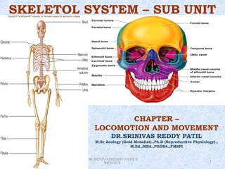
- 1. SKELETOL SYSTEM – SUB UNIT CHAPTER – LOCOMOTION AND MOVEMENT DR.SRINIVAS REDDY PATIL M.Sc Zoology (Gold Medalist).,Ph.D (Reproductive Physiology)., M.Ed.,MBA.,PGDBA.,FMSPI Saturday, February 9, 2013 DR.SRINIVASREDDY PATIL'S 1 BIOLOGY
- 2. Fig. 6.10
- 4. Functions of the Bones 1.Support 2.Protection 3.Movement 4.Storage 5.Hematopoiesis
- 5. Table. 6.1
- 7. AXIAL SKELETON I. SKULL = skeleton --- head & face = flattened & irregular = united by joints (sutures) Cranium -- skull minus mandible Calvarium -- skull after the bones of the face have been removed Cavities: a. Cranial - contains the brain b. Orbital - contains eyeball & accessory organs c. Nasal
- 9. Bones of the Skull Figure 5.11 Copyright © 2003 Pearson Education, Inc. publishing as Benjamin Cummings
- 10. Divisions of the bones of the skull a. Cerebral / cranial bones / brain case (8 bones) unpaired (4) paired (4) 1. occipital 1. parietal 2. frontal 2. temporal 3. sphenoid 4. ethmoid b. Facial or visceral cranium paired (12) unpaired (2) a. Nasal a. Vomer b. Lacrimal b. Mandible c. Maxilla d. Zygomatic / malar / cheek bones e. Palatine f. Inferior nasal concha or turbinate
- 12. Fig. 6.13
- 13. Figure 5.10 Copyright © 2003 Pearson Education, Inc. publishing as Benjamin Cummings
- 14. Fontanelle = membrane filled spaces found in the skull of newborn infants ex. 1. anterior = largest 2. posterior 3. anterolateral (sphenoidal) 4. posterolateral (mastoid)
- 17. Fig. 6.37
- 18. AXIAL SKELETON I. HYOID BONE = small U-shape; lies in front of the neck = base of the tongue is attached = lies between mandible & thyroid cartilage II. OSSICLES = small bones of the ear a. Stapes (stirrup) 2 b. Incus (anvil) 2 c. Malleus (hammer) 2
- 19. Fig. 6.16
- 21. AXIAL SKELETON I. VERTEBRAL COLUMN = long, curved, slightly movable pillar = united together by cartilage & ligaments = 71 – 75 cm. long = formed by series of bones -- vertebrae FUNCTION: 1. support of the trunk 2. contains & protects the spinal cord & nerves
- 22. VERTEBRAL COLUMN Classification of vertebra young adult cervical 07 07 thoracic 12 12 lumbar 05 05 sacral 05 01 coccygeal 04 01 33 26 Intervertebral discs = flattened plates of fibrocartilage that are interposed between the adjacent surfaces of the bodies of vertebrae Function: 1. uniting medium between vertebrae 2. main shock absorber 3. give flexibility & movement to the whole vertebral column
- 23. VERTEBRAL COLUMN General parts of vertebrae 1. body 2. arch 3. pedicle or root 4. lamina 5. transverse process 6. articular process 7. spinous process 8. spinal or vertebral foramen
- 24. Special characteristics of individual vertebrae a. Cervical vertebrae (7) = forms the skeleton of the neck, all have transverse foramen atypical cervical vertebrae: 1. atlas -- 1st 2. axis or epistropheus = 2nd 3. 7th cervical vertebrae = spinous process not bifid, small transverse foramen b. Thoracic vertebrae (12) = costal pits - rib attachment = circular vertebral canal
- 25. c. Lumbar vertebrae (5) = presence of mamillary & accessory processes = triangular vertebral foramen d. Sacrum = inverted triangular bone situated between hip bones e. Coccygeal vertebrae (1) = 4 small incomplete vertebrae fused to form the coccyx / tail bone; triangular
- 29. Fig. 6.17
- 30. Fig. 6.18
- 32. Fig. 6.20
- 33. AXIAL SKELETON I. STERNUM (breast bone) = flat bone, found -- anterior thoracic wall = composed of 2 plates of compact bone with a layer of spongy bone in between containing red bone marrow. PARTS: a. Manubrium b. Corpus or body c. Xiphoid process
- 34. AXIAL SKELETON I. RIBS (12 pairs) = narrow arched flat bones with 2 ends. 1. vertebral - posterior; attaches with thoracic 2. sternal - anterior; attaches with costal cartilages Classification of ribs: a. Sternal or true ribs (1st to 7th) - ribs whose costal cartilages are directly attached to sternum b. Asternal or false ribs (8th to 12th) - ribs whose costal cartilages are not attached directly to the sternum but to 7th subdivisions: 1. false rib proper - 8th, 9th, 10th ribs 2. floating or hanging ribs – 11th & 12th
- 35. Fig. 6.21
- 36. APPENDICULAR SKELETON BONES of the UPPER EXTREMITY (UE) 1. Clavicle (collar bone) 2. Scapula (shoulder blade) – articulates with humerus & clavicle 3. Humerus (arm bone) - longest & largest bone of UE articulates with scapula (above) radius & ulna (below) 4. Radius - lateral bone of the forearm; cup- shaped head 5. Ulna - principal bone of the forearm; longer & larger than radius
- 37. BONES of the UPPER EXTREMITY (UE) 6. Carpals (wrist bone) - 8 bones arranged into 2 rows - proximal & distal rows 7. Metacarpals (bones of the hand) - 5 long bones placed between carpals & phalanges - numbered from lateral to medial Phalanges (bones of the fingers) = 14 long bones of the fingers -- 3 bones except thumb - 2 bones
- 40. Fig. 6.26
- 41. Fig. 6.22
- 42. Fig. 6.27
- 45. APPENDICULAR SKELETON BONES of the LOWER EXTREMITY (LE) 1. Hip bone (innominate bone) right & left hip bones + sacrum = pelvic girdle 3 bones: 1. ilium* 2. ischium* *Converge on acetabulum a concave fossa -- articulates with 3. pubis* head of femur form hip joint 2. Femur (thigh) = longest, strongest, largest bone in the body 3. Tibia (shin bone) = long bone; anterior, medial, & larger of the 2 bones of the leg 4. Fibula (peroneal bone) = long slender bone placed parallel with the tibia but located laterally
- 46. Fig. 6.29
- 47. Fig. 6.30
- 48. Fig. 6.31
- 49. Fig. 6.32
- 50. Fig. 6.33
- 51. Fig. 6.34
- 52. BONES of the LOWER EXTREMITY 5. Tarsals (ankle bone) = short bones; 2 rows: internal & external rows 6. Metatarsals (bones of foot) = 5 long bones numbered from medial to lateral 7. Phalanges (bones of toes) = similar to bones of the fingers
- 54. Fig. 6.35
- 55. Common Fractures greenstick fracture = the bone does not break all of the way through. simple, or closed = when the bone breaks but the skin does not. compound, or open = when the broken bone tears through the skin, introducing the dangerous possibility of infection. The area around a break swells and discolors, but some fractures can be detected only by X-ray. The weakened bones of the elderly are
- 57. Fig. 6B
- 58. Fig. 6C
- 59. Bone Deformation Rickets can result from insufficient vitamin D in the diet or from insufficient amounts of ultraviolet radiation from the sun. It can lead to skeletal deformation, such as vertebral or leg curvature.
- 60. JOINTS
- 61. JOINTS = a site where 2 or more bones come together whether with movement or none ARTHROLOGY
- 62. JOINTS CLASSIFICATION: 1. Fibrous joints = articulating bone surfaces = sutures of skull, inferior tibiofibular joints = very little movement possible
- 64. JOINT CLASSIFICATION: cont’n. 2. Cartilagenous joints 2 types: a. Primary - united by a plate or bar of hyaline cartilage b. Secondary - united by a plate of fibrocartilage = articular surfaces of bones -- covered by a thin layer of hyaline cartilage = small amount of movement
- 65. JOINT CLASSIFICATION: cont’n. 1. Synovial joints = articular surfaces of bones covered by thin layer of hyaline cartilage separated by a joint cavity = permits great degree of movement
- 66. JOINT CLASSIFICATION ACCORDING to DEGREE of MOVEMENT I. Synarthroses = immovable joints = articulating surface is in direct contact = uniting medium: fibrous tissue hyaline cartilage fibrocartilage
- 67. JOINT Synarthroses a. Sutures - bones of the skull b. Schindylosis - bony plate inserted into a cleft or fissure e.g.: vomer into maxilla, palatine bones c. Gomphosis - a conical process received into corresponding socket e.g.: root of teeth into alveolus of maxilla or mandible d. Synchondrosis - a cartilagenous medium which later may ossify e.g.: between epiphysis & diaphysis of long bone
- 68. JOINT CLASSIFICATION ACCORDING to DEGREE of MOVEMENT (cont’n) I. Amphiarthroses = slightly movable joints = articulating surfaces connected by a wide disc of fibrocartilage a. Symphysis - uniting medium: fibrocartilage e.g.: symphysis pubis b. Syndesmosis - large amount of fibrous connective tissue wide membrane e.g.: interosseous membrane between radius & ulna
- 69. JOINT CLASSIFICATION ACCORDING to DEGREE of MOVEMENT (cont’n) I. Diarthroses = freely movable joints Types: a. Articular surfaces covered by hyaline cartilage e.g.: sternoclavicular, acromioclavicular joints b. Hinge joints (ginglymus) = flexion & extension movements possible e.g.: elbow, knee, ankle
- 70. Diarthroses c. Pivot joints (trochoid) = central body pivot surrounded by a bony ligamentous ring = possible movement is rotation only e.g.: atlantoaxial & superior radioulnar joints d. Condyloid = have 2 distinct convex surfaces that articulate with 2 concave surfaces = flexion, extension, adduction, abduction possible = small amount of rotation e.g.: metacarpophalangeal metatarsophalangeal joints
- 71. Diarthroses e. Ellipsoid = elliptical convex articular surface that fit into an elliptical concave articular surface = F, E, add., abd. possible = rotation impossible e.g.: wrist joint f. Saddle joints = articular surfaces are reciprocally concavoconvex, resembling saddle on a horse’s back = F, E, add., abd., rotation possible e.g.: carpometacarpal joint of thumb
- 72. Diarthroses g. Ball & socket joints = ball – shaped head of one bone fits into a socket – like concavity of another = free movements possible: F, E, add., abd., medial rotation, lateral rotation, circumduction e.g.: acetabulum of hip bone with head of thigh bone
- 73. Fig. 6.39a
- 74. Fig. 6.39b
- 75. POSSIBLE MOVEMENTS of JOINTS 1. Gliding - simple slipping or rubbing of the apposed flat surfaces - no angular or rotary movement e.g.: in between vertebral bodies 2. Angular - generally found in long bones a. Flexion - movement that forms an acute angulation between 2 approximating joints = angle is decreased b. Extension - movement that form an obtuse angulation between 2 parts = angle is increased
- 76. POSSIBLE MOVEMENTS of JOINTS (cont’n) 1. Angular (cont’n) c. Abduction - movement that carries extremity away from the median plane of the body d. Adduction - movement that carries extremity towards the median plane of the body 4. Circumduction - circular motion 5. Rotation - movement along a central axis without the bones being displaced from such axis - directed medially or laterally
- 77. POSSIBLE MOVEMENTS of JOINTS (cont’n) 6. Peculiar movements & positions forearm & hand a. Supination b. Pronation foot a. Inversion - plantar surface of the foot directed towards the median plane b. Eversion - plantar surface of the foot directed away from the median plane
- 78. QUALITY IS NEVER AN ACCIDENT.IT IS ALWAYS THE RESULT OF HIGH AIM,SINCERE EFFORT,INTELLIGENT DIRECTION AND PERFECT EXECUTION