
Recomendados
Más contenido relacionado
La actualidad más candente
La actualidad más candente (20)
Similar a C08 Mitosis[1]
Similar a C08 Mitosis[1] (20)
Más de swalden01
C08 Mitosis[1]
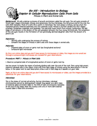
- 1. Bio 103 - Introduction to Biology Chapter 8: Cellular Reproduction: Cells from Cells Mitosis in Plant and Animal Cells Background: All cells undergo a process of growth and division called the cell cycle. The cell cycle consists of three major stages-interphase, mitosis, and cytokinesis. During interphase, the cell grows and, at the end, the cell's DNA replicates. Most of a cell's life is spent in interphase. The next phase is mitosis, during which the replicated genetic material separates into two separate nuclei. Mitosis is further divided into four stages: prophase, metaphase, anaphase, and telophase. Two identical nuclei result from mitosis. Cytokinesis, the last stage of cell division, is the division of the cell cytoplasm between the two newly forming cells. The completion of the cell cycle results in the formation of two genetically identical daughter cells from the division of a parent cell. Objectives: • Identify cells undergoing the process of mitosis. • Compare the stages of mitosis in plant cells with these stages in animal cells. Materials: • prepared slides of onion or garlic root tips (longitudinal sections) • compound light microscope As this is an online class and you don’t have access to microscopes or slides, the images as one would see through a compound light microscope are included for your observations. Procedure: PART 1: Mitosis in Plant Cells 1. Observe a prepared slide of a longitudinal section of onion or garlic root tip. Use low power to locate the region of actively dividing cells near the end of the root. Then using high power, examine individual cells in the region. Find a cell from each stage of mitosis using the descriptions in the list below. In the space next to each description, draw a cell in that stage as it appears on your slide. Again, as this is an online class and you don’t have access to microscopes or slides, use the images provided as a substitute for your observations. Interphase This is the phase of normal cell activity. During interphase, individual chromosomes cannot be distinguished. Instead, they appear as a dark mass of material called chromatin (label C). The DNA of each chromosome replicates at the end of this stage. Note the nucleus with one or more dark-stained nucleoli (label I) filled with chromatin. 1 success = preparation + execution
- 2. Prophase In this stage, the chromatin appears as a mass of thick threads. These threads are the replicated chromosomes, which have coiled up and shortened. Each chromosome now consists of a pair of chromatids, which are duplicates of the original chromosome. The chromatids are held together by a centromere. In late prophase, the nuclear membrane and nucleoli cannot be seen, but the chromosomes are distinctly visible as pairs of chromatids in the central region of the cell. Metaphase In this stage, the chromosomes line up across the equator of the cell (label Ch). A mass of fibers called a spindle (label S) has formed between the poles of the cell and the mass of chromosomes. A spindle fiber from each pole attaches to each chromosome (pair of chromatids). Anaphase In this stage, the centromere of each chromatid pair divides. The chromatids move along the spindle fibers toward the poles of the cell. Each chromatid in the pair of chromatids moves toward opposite poles of the cell. 2 success = preparation + execution
- 3. Telophase In this stage, the chromatids (now called chromosomes) have formed distinctive clumps at each pole. A new nuclear membrane forms around each clump of chromosomes, which uncoil and return to the chromatin network seen in interphase. The nucleoli reappear. In plants, the new cell walls (the arrows in both pictures) grow to form the two new, identical daughter cells. Analysis: Complete the following questions using information from this laboratory activity. 1. What is the shape of the cells? 2. What color are the chromosomes stained? 3. What happens during cytokinesis? 4. How many new cells have formed from the parent cell? Procedure: PART 2: Mitosis in Animal Cells 2. Observe the prepared slide of the whitefish blastula. Locate the blastula using low power, then examine it under high power. Find a cell from each stage, using the list below. In the space beside each description, draw a cell in that stage as it appears on your slide. Label the parts of the cell that are visible. Interphase A distinct nucleus and a nucleolus (label C) are visible. The genetic material appears as chromatin. 3 success = preparation + execution
- 4. Early Prophase Astral rays have formed around the centrioles, and the spindle (label A) has formed between them. The chromosomes (paired chromatids) are becoming visible, and the nuclear membrane has disappeared. Late Prophase The chromosomes are shortened and thickened and are distinct in the central region of the cell. Metaphase The chromosomes line up at the center of the cell, along the equator (label C). The astral rays are also visible (label A). Anaphase The chromatids (label B) separate at their centromere and are pulled to opposite poles along the spindle fibers (label A). 4 success = preparation + execution
- 5. Telophase The chromosomes appear in clusters at the poles. The parent cell begins elongating, and the nuclear membranes re-form around the chromosome clusters. The spindle and chromosomes become less distinct. The cytoplasm pinches (label A) in until the two daughter cells separate during late telophase. Analysis: 5. What is the shape of the cells? 6. Describe the shape of the chromosomes as they are pulled to the poles in anaphase. 7. At the completion of cytokinesis, in what phase are the two daughter cells? 8. How does mitosis differ in plant and animal cells? 9. Which phase of mitosis shows the greatest difference between animal and plant cells? Explain your choice. 10. What role do you think mitosis plays in living things? Justify your answer. 11. Mitosis in onion root tip cells takes about 80 minutes. If you view a slide of a root tip and count the number of cells in each stage of mitosis, you can then calculate the amount of time each stage takes. This is because the percentage of the cells in a particular stage of mitosis is equal to the percentage of 80 minutes that the stage takes. Using this information, devise a method for calculating the amount of time each stage of mitosis takes. 5 success = preparation + execution