Sturge-Weber synbdrome
•
2 likes•1,385 views

Sturge-Weber synbdrome http://yassermetwally.com http://yassermetwally.net

Recommended
More Related Content
What's hot
What's hot (20)
Viewers also liked
Viewers also liked (12)
Similar to Sturge-Weber synbdrome
Similar to Sturge-Weber synbdrome (20)
More from Professor Yasser Metwally
More from Professor Yasser Metwally (20)
Recently uploaded
Recently uploaded (20)
Sturge-Weber synbdrome
- 1. Online newspaper of professor Yasser M etwally Encephalotrigeminal angiomatosis (Sturge- Weber Syndrome) The author: Professor Yasser Metwally http://yassermetwally.com INTRODUCTION February 29, 2012 — Encephalotrigeminal angiomatosis classically includes the presence of cutaneous vascular portwine nevus of the face, contralateral hemiparesis or hemiatrophy, glaucoma, seizures and mental retardation. The syndrome is a neurocutaneous syndrome (phakomatosis) characterized by a facial port-wine nevus (a capillary malformation), seizures, and mental retardation (1). Central nervous system manifestations include a leptomeningeal venous malformation (“ angioma” ) and cerebral hemiatrophy, which are typically, though not always, ipsilateral to the facial nevus. Calcifications are seen in the atrophic cortex deep to the venous malformation. Absence of cortical veins with centripetal venous drainage into enlarged medullary veins or anomalous deep veins in Sturge-Weber syndrome has been described in the angiographic, computed tomographic, and MR literature (2–10). These enlarged deep veins typically drain into the galenic venous system and likely represent collateral veins rather than primary vascular abnormalities. Dural venous sinus and galenic venous occlusions have also been documented with conventional and MR angiography in patients with Sturge-Weber syndrome (3, 6–10), and they may modify the pattern of centripetal venous drainage. Pathology This syndrome is also called encephalotrigeminal or encephalofacial angiomatosis, and typically consists of capillary hemangioma in the upper portion of the face, Leptomeningeal angiomatosis, and cerebral calcification. The brain has an anomalous proliferation of leptomeningeal veins, showing clustering of veins with a somewhat irregular thickened wall. The underlying cerebral cortex shows numerous calcospherites, particularly along its superficial layers. Two abutting calcified cortices around a sulcus may exhibit a tram track-like radiologic appearance. The involved cortex is frequently atrophic, showing neuronal loss and gliosis. As in tuberous sclerosis, seizures are the most common manifestation in Sturge-Weber syndrome, occurring in 71 % to 89% of patients. Follow “Online
- 2. Figure 1. Sturge- Weber Syndrome: The brain has an anomalous proliferation of leptomeningeal veins, showing clustering of veins with a somewhat irregular thickened wall. The underlying cerebral cortex shows numerous calcospherites, particularly along its superficial layers. Figure 2. Plain x ray skull in two patients with sturge weber syndrome, notice the trolley-track sign with curvilinear calcification which is due to cortical or vascular calcification It has been suggested that stepwise clinical deterioration in patients with Sturge-Weber syndrome might be caused by cerebral venous hypertension resulting from sluggish venous drainage through the leptomeningeal angioma (9). Probst (9) hypothesized that the latter might be further exacerbated by progressive occlusion of deep veins with resultant centrifugal rerouting of deep cerebral venous drainage into the already overburdened angioma. Slowing and diminution of regional cerebral blood flow has been demonstrated by using radionuclide and xenon inhalation techniques in these patients (9, 11). On the basis of the postulate that these findings might be caused by progressive venous thrombosis, antiplatelet therapy was administered in one series of patients and resulted in stabilization of neurologic function (12). Follow “Online newspaper of
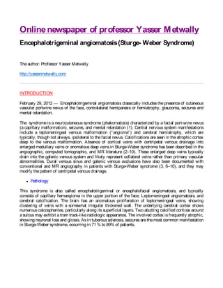
- 3. Figure 3. Sturge-Weber syndrome, venous drainage modified by deep venous occlusion. A, Axial enhanced T1-weighted MR image shows enlarged deep medullary veins (small black arrowheads) draining centripetally from the cortical surface into a longitudinal caudate vein (white arrowhead). Note leptomeningeal “ angioma” (large black arrowheads). B, More inferiorly at the level of the cavum velum interpositum, the anomalous deep vein can be seen traveling in a vertical orientation (large arrowhead). The internal cerebral veins are not visible. Note prominent glomus of the left choroid plexus (small arrowheads) and the left occipital leptomeningeal Isolated nonvisualization of cortical veins, deep veins, and dural venous sinuses on conventional or MR angiography in patients with Sturge-Weber syndrome does not establish whether these veins are aplastic, hypoplastic, thrombosed, or occluded by processes such as intimal hyperplasia or other nonthrombotic diseases. Without follow-up, these findings also do not prove a progressive venoocclusive process in Sturge-Weber syndrome. Figure 4. Sturge-Weber Syndrome (or disease) is a congenital vascular malformation affecting the head, face, and brain. The primary process appears to be faulty development of the venous drainage for the cerebral capillary bed. A similar process affects the skin, eye, and the soft-tissues of the head. Development of the brain usually proceeds to a normal size, but after birth, there is progressive atrophy of the affected hemisphere(s). The disease is usually unilateral, but bilateral cases can occur. Typically the patient presents at birth with a "Port Wine Nevus" – a reddish-brown or pink discoloration of the face, often following the distribution of the trigeminal nerve. Intracranially, ipsilateral to the facial nevus, there is abnormal circulation that leads to 1) cerebral dysfunction; 2) electrical instability Follow (seizures); and 3) cerebral cortical atrophy. Seizures usually present within the first two years of life. Typically the occipital lobes are affected first, and most severely, but the disease may also involve the parietal and temporal lobes, and (rarely) the frontal lobe Follow “Online newspaper of
- 4. The observation of venous abnormalities in this neonate suggests that the venous pathology in Sturge- Weber syndrome might commence in utero and progress postnatally. We could not identify intraluminal venous thrombus as the cause of progressive nonvisualization of veins in this patient, suggesting that nonthrombotic venoocclusive processes might play a role in the pathophysiology of Sturge-Weber syndrome. Indeed, abnormal veins with walls thickened by a layer of hyalinized connective tissue or fibrosis have been noted at pathologic examination within the leptomeninges and subcortical white matter of patients with Sturge-Weber syndrome (13). Figure 5. Sturge Weber syndrome: Right parietal occipital pial angioma that enhances with contrast administration (A). The underlying cortex is atrophic and MR imaging demonstrates decreased signal intensity throughout the atrophic cortical mantle, which may be due to hemosiderin deposition. calcification, or both (B). Given the noninvasive nature of conventional MR and MR venography, it should be feasible to study a larger group of patients with Sturge-Weber syndrome to determine if there is a relationship between clinical deterioration and progressive venous occlusion, and to what extent venous thrombosis contributes to venous occlusion in these patients. If thrombosis is established as a significant contributor to the disease process, clinical trials that use antiplatelet medications could be guided by periodic MR venographic evaluation. References 1. Braffman B, Naidich TP. The phakomatoses: part II. Neuroimaging Clin North Am 1994;4:330–332 2. Elster AD, Chen MYM. MR imaging of Sturge-Weber syndrome: role of gadopentetate dimeglumine and gradient echo sequences. AJNR Am J Neuroradiol 1990;11:685–689 3. Terdjman P, Aicardi J, SainteRose C, Brunelle F. Neuroradiological findings in Sturge-Weber syndrome and isolated pial angiomatosis. Neuropediatrics 1990;22:115–120 4. Marti´-Bonmati´ L, Menor F, Poyatos C, Cortina H. Diagnosis of Sturge-Weber syndrome: comparison of the efficacy of CT and MR imaging in 14 cases. AJR Am J Roentgenol 1992;158:867– 871 Follow “Online newspaper of
- 5. Encephalotrigeminal angiomatosis (Sturge- Weber Syndrome) « Online newspaper of pro... Page 5 of 10 5. Benedikt RA, Brown DC, Walker R, Ghaed VN, Mitchell M, Geyer CA. Sturge-Weber syndrome: cranial MR imaging with Gd-DTPA. AJNR Am J Neuroradiol 1993;14:409–415 6. Vogl TJ, Stemmler J, Bergman C, Pfluger T, Egger E, Lissner J. MR and MR angiography in Sturge- Weber syndrome. AJNR Am J Neuroradiol 1993;14:417–425 7. Bentson JR, Wilson GH, Newton TH. Cerebral venous drainage pattern of the Sturge-Weber syndrome. Radiology 1971;101:111– 118 8. Poser CM, Taveras JM. Cerebral angiography in encephalotrigeminal angiomatosis. Radiology 1957;68:327–336 9. Probst FP. Vascular morphology and angiographic flow patterns in Sturge-Weber angiomatosis: facts, thoughts, and suggestions. Neuroradiology 1980;20:73–78 10. Segall HD, Ahmadi J, McComb JG, Zee C, Becker TS, Han HS. Computed tomographic observations pertinent to intracranial occlusive disease in childhood. Radiology 1982;143:441–449 11. Garcia JC, Roach ES, McLean WT. Recurrent thrombotic deterioration in the Sturge-Weber syndrome. Childs Nerv Syst 1981;8: 427–433 12. Roach ES, Riela AR, McLean WT, Stump DA. Aspirin therapy for Sturge-Weber syndrome. Ann Neurol 1985;18:387 13. Wohlwill FJ, Yakovlev PL. Histopathology of meningofacial angiomatosis (Sturge-Weber disease): report of four cases. J Neuropathol Exp Neurol 1957;16:341–364 Please log in using one of these methods to post your comment: Follow Follow “Online Notify me of follow-up comments via email. newspaper of