SlideShare una empresa de Scribd logo
1 de 9
thorough knowledge of surgical anatomy is essential for any nasal surgeon embarking on endoscopic, intranasal or external surgery. This helps not only in keeping the complication rate down, but also in understanding the pathology and achieving an optimum surgical result. The relevant anatomy and physiology of the nose and paranasal sinuses is discussed, especially in relation to endoscopic sino-nasal surgery. <br />Introduction<br />The paranasal sinuses are a group of air containing spaces that surround the nasal cavity. At birth they are mostly rudimentary and their development continues during childhood and into early adult life. These sinuses are formed as a result of a process wherein solid facial skeletal elements surrounding the nose are invaded by respiratory mucosa and subsequently pneumatized. Since they are actually pneumatic diverticuli from the primitive nasal cavity, their lining mucosa is similar to that of the nasal cavity, as a result of which, any pathological changes affecting the nasal mucosa can spread to the paranasal sinuses. The drainage of the sinuses is through their respective ostia into the nose. <br />The paranasal sinuses derive their nomenclature from the bones that they pneumatize. They comprise the maxillary, the frontal, the ethmoid and the sphenoid sinuses. An accurate knowledge of the anatomy of the sinuses becomes essential in order to understand the pathophysiology of the diseases affecting them and their subsequent management. This cannot be adequately stressed especially in the era of endoscopic sinus surgery wherein lack of familiarity with sinus anatomy can lead to catastrophic complications.<br />ANATOMY<br />Maxillary sinus<br />These pyramidal shaped sinuses occupy a central position in the facial skeleton and are formed as a result of pneumatisation of the maxilla. The base of the maxillary sinus is directed medially and the apex superolaterally. Its anterior and posterior surface is formed by the corresponding surfaces of the maxilla, its roof by the orbital surface and floor by the palatine and alveolar processes of the maxilla. Occasionally, dehiscences may be present in the floor of the sinus (2%) wherein the roots of the teeth come to lie in close relation to the mucosa facilitating spread of infection. <br />The maxillary sinus though present at birth continues to increase in all dimensions postnatally. Vertical growth of the sinus occurs in stages. In young children, the floor of the sinus is approximately 4 mm above the level of the nasal floor, following which it subsequently extends 1 to 5.5 mm below the nasal floor after the age of 12 years. It is following this that the sinus can now be accessible through the inferior meatus. The sinus derives its nerve supply from branches of the infra-orbital and the posterior superior alveolar nerve, whereas the arterial supply is from branches of the internal maxillary artery.<br />The anterior wall of the sinus corresponds to the anterior surface of the maxilla extending superiorly from the orbital rim above to the teeth below. It is thinnest just above the root of the canine tooth and it is from this area that the sinus can be approached via canine puncture techniques or through the classical Caldwell-Luc approach. The posterior wall is formed by the corresponding surface of the maxilla superiorly, and part of the palatine bone inferiorly. The medial wall of the sinus is shared with the nasal cavity and forms part of the lateral nasal wall within which is present the nasolacrimal duct. The sinus drains into the nasal cavity through its ostium that is present high up in its medial wall and empties in the posterior aspect of the hiatus semilunaris situated in the middle meatus. The normal ostium of the sinus is 7-11 mm in length and 2-6 mm high. Oedema of mucosa surrounding it can markedly block the ostium. Accessory ostia are present in 28%.<br />Ethmoid sinus<br />The word ‘Ethmoid’ in Greek means sieve-like. This sinus comprises a group of air cells, which form one of the most complex structures in the body. Hence the sinus is rightly named the ethmoid ‘labyrinth’. This complex arrangement of air cells and their particular anatomical location makes them play a pivotal role in the pathophysiology of sinus inflammatory disease. Mosher has rightly stated that ‘Had the Ethmoidal sinus been placed in any other part of the body, it would be an insignificant and harmless collection of bony cells. In the place where nature has put it, it has major relationships, so that diseases and surgery in this region often lead to disaster’. The Ethmoid bone per se comprises a horizontal plate - the cribriform plate, a vertical plate - the perpendicular plate and two lateral ethmoidal sinuses. The ethmoidal sinuses occupy a position between the orbit laterally and the nasal cavity medially (Figs. 1, 2). They are related to the anterior cranial fossa superiorly and the maxillary sinus inferiorly. The entire ethmoid sinus structure measures 4-5 cm in length and 2.5-3 cm in height. The ethmoid air cells have been classified according to their location in relation to specific anatomic structures. The air cells are divided into anterior and posterior groups by the lateral (or basal) lamella termed the ‘ground lamella’. The anterior ethmoidal cells which lie anterior to it open into the middle meatus and the posterior group opens into the superior meatus. The anterior ethmoidal cells tend to be smaller than the posterior group of cells, their number varying between 4-17. The posterior ethmoid cells number between 2-6. The air cells are separated from each other by thin intervening septae of bone that may be complete or incomplete. The agger nasi (1-4 cells) are the anterior most group of cells located anterior to the uncinate process. Exenteration of these cells is often necessary to expose the frontal recess. The lateral wall of each sinus separates it from the orbit. This is formed by the solid lacrimal bone anteriorly and a paper thin bone, aptly named the lamina papyracea situated behind it. The lamina papyracea may occasionally have dehiscences within it. The optic canal with the optic nerve comes in close relation with the lateral wall of the ethmoid sinus posteriorly. The roof of the sinus is formed by a horizontal plate of the frontal bone termed the fovea ethmoidalis that separates the air cells from the anterior cranial fossa. The medial most part of the fovea ethmoidalis is relatively thin and slopes downwards to join the cribriform plate. The anterior ethmoidal artery in its bony canal runs along the roof of the anterior group of air cells and is a useful landmark for the superior limit of dissection. <br />A few ethmoid air cells are located near the area of the fronto-nasal duct, and are termed the fronto ethmoidal cells. The Haller cell is an ethmoidal cell in relation with the floor of the orbit (Fig. 3). Occasionally, a posterior ethmoid air cell may invade the anterior-superior aspect of the sphenoid sinus and come to lie in close relation with the optic nerve. It is then called the cell of Onodi and could be mistaken for the sphenoid sinus during endoscopic surgery.<br /> Fig 1:Coronal CT Scan through anterior ethmoidal air cells. BE-Bulla ethmoidalis; CB-Concha bullosa;Cp-Cribriform plate; FE- Fovea ethoidalis; In- infundibulum; IT-inferior turbinate; Lp-Lamina papyracea; MM-Middle meatus; MS- Maxillary sinus; S- septum; UP-Uncinate process Fig 2:Coronal CT Scan through posterior ethmoidal air cells. FE- Fovea ethoidalis ; IOF-Inferior turbinate ;MS- Maxillary sinus; ON- Optic nerve ; PE- Posterior ethmoid air cell ;ST- Superior turbinate.<br /> <br />Frontal Sinus<br />The frontal sinus is the last sinus to develop, its complete development takes place by 14 - 16 years. The size of this sinus is highly variable and can range from a few cubic centimeters to a size that occupies most of the area of the frontal bone. The average height is about 24.3 mm. The floor of the sinus which forms the orbital roof and the posterior wall which is related to the anterior cranial fossa are considerably thinner than the anterior wall which is formed by the thick bone of the skull. A mid-line septum is present within the sinus. Incomplete septations extending from the roof are commonly present, giving the sinus a scalloped configuration. The sinus opens in the upper part of the infundibulum into the fronto-nasal recess. <br />Fig:3Coronal CT Scan showing an infected right Haller cell<br /> <br />Sphenoid Sinus<br />The body of the sphenoid can get pneumatized to a variable extent. Thus according to Congdon one may encounter a conchal (5%), a pre-sellar (23.5%) or a post sellar (67%) pneumatisation.<br />On an average the adult sphenoid sinus measures 13.5 mm in width superiorly, 16.9 mm in the middle, 18.7 mm in its lowest segment. The length averages 19.4 mm in the upper part, 4-8 mm in the middle and 18.5 mm in the lower part. The ostium of this sinus is located in the upper 1/3 of its anterior wall and it drains into the spheno-ethmoidal recess. An inter-sinus septum is generally present which is almost always deviated to one side making the interior of the sinus asymmetrical (Fig. 4). Accessory septations may be occasionally seen within the sinus. The sinus may sometimes extend superior or inferior to the optic nerve making this anatomical variation the most dangerous. In the lateral wall of the sphenoid sinus, a superior bulge corresponds to the optic nerve whereas the infero-lateral bulge is formed by the internal carotid artery. Bulges corresponding to the maxillary nerve, and vidian nerves may also be present. The pituitary gland lies above the roof of the sphenoid sinus and can thus be approached trans-sphenoidally.<br />Anatomy of the lateral nasal wall<br />The lateral wall of the nose has a characteristic complex structure as a result of scrolls and projections being present on it. These projections are the superior, middle and inferior turbinates and occasionally when present, the supreme turbinate. Underlying these shelves of bone are the superior middle and inferior meati respectively. The inferior turbinate is a separate bone, while the superior and middle turbinates are parts of the ethmoid bone. The casual observer must be wary of not mistaking a hypertrophied inferior turbinate for a nasal mass or polyp. The superior turbinate is the smallest turbinate and overlies the superior meatus into which the posterior ethmoidal air cells drain. The nasolacrimal duct opens into the inferior meatus. The middle turbinate forms an important landmark from the point of view of endoscopic sinus surgery and should be preserved. <br />The ostiomeatal complex is a word coined by Naumann and comprises the region of the middle meatus with the anterior air cells. This is the most important area for normal sinus functioning and any pathology in this area will disrupt physiology and lead to sinus dysfunction. In the middle meatus are several important structures, which play a vital role in the pathogenesis of sino-nasal inflammatory disease. These structures also form important landmarks in sinus endoscopic surgery. Anteriorly, the first landmark is a hook shaped bone or the uncinate process. Posterior to the uncinate process is present a groove known as the hiatus semilunaris which leads into the ethmoidal infundibulum. The ethmoidal bulla is a bulge posterior to the hiatus, which hitherto was referred to as the middle ethmoidal air cell system. Present nomenclature however groups the ethmoids into only anterior and posterior air cells, and the bulla ethmoidalis is included as part of the anterior cells. The frontal sinus opens into the superior most aspect of the ethmoidal infundibulum into the fronto-nasal recess while the anterior ethmoidal cells open anteriorly into the infundibulum. The ostium of the maxillary sinus opens postero-inferiorly into the infundibulum and can be located just above the inferior turbinate. Occasionally accessory ostia of the maxillary sinus may be present. Behind the posterior end of the middle turbinate is the sphenopalatine foramen leading into the pterygopalatine fossa. <br />Certain anatomical variations may exist which predispose to development of recurrent infections by narrowing the middle meatus space and hampering the mucociliary drainage. Some of these which is almost always deviated to one side making the interior of the sinus asymmetrical (Fig. 4). Accessory septations may be occasionally seen within the sinus. The sinus may sometimes extend superior or inferior to the optic nerve making this anatomical variation the most dangerous. In the lateral wall of the sphenoid sinus, a superior bulge corresponds to the optic nerve whereas the infero-lateral bulge is formed by the internal carotid artery. Bulges corresponding to the maxillary nerve, and vidian nerves may also be present. The pituitary gland lies above the roof of the sphenoid sinus and can thus be approached trans-sphenoidally.<br /> <br />Fig:4 :Coronal CT Scan through the sphenoid sinusOC- optic canal; PC- Choana; SOF-superior orbital fissure; SS- sphenoid sinus.<br /> <br />Anatomy of the lateral nasal wall<br />The lateral wall of the nose has a characteristic complex structure as a result of scrolls and projections being present on it. These projections are the superior, middle and inferior turbinates and occasionally when present, the supreme turbinate. Underlying these shelves of bone are the superior middle and inferior meati respectively. The inferior turbinate is a separate bone, while the superior and middle turbinates are parts of the ethmoid bone. The casual observer must be wary of not mistaking a hypertrophied inferior turbinate for a nasal mass or polyp. The superior turbinate is the smallest turbinate and overlies the superior meatus into which the posterior ethmoidal air cells drain. The nasolacrimal duct opens into the inferior meatus. The middle turbinate forms an important landmark from the point of view of endoscopic sinus surgery and should be preserved. <br />The ostiomeatal complex is a word coined by Naumann and comprises the region of the middle meatus with the anterior air cells. This is the most important area for normal sinus functioning and any pathology in this area will disrupt physiology and lead to sinus dysfunction. In the middle meatus are several important structures, which play a vital role in the pathogenesis of sino-nasal inflammatory disease. These structures also form important landmarks in sinus endoscopic surgery. Anteriorly, the first landmark is a hook shaped bone or the uncinate process. Posterior to the uncinate process is present a groove known as the hiatus semilunaris which leads into the ethmoidal infundibulum. The ethmoidal bulla is a bulge posterior to the hiatus, which hitherto was referred to as the middle ethmoidal air cell system. Present nomenclature however groups the ethmoids into only anterior and posterior air cells, and the bulla ethmoidalis is included as part of the anterior cells. The frontal sinus opens into the superior most aspect of the ethmoidal infundibulum into the fronto-nasal recess while the anterior ethmoidal cells open anteriorly into the infundibulum. The ostium of the maxillary sinus opens postero-inferiorly into the infundibulum and can be located just above the inferior turbinate. Occasionally accessory ostia of the maxillary sinus may be present. Behind the posterior end of the middle turbinate is the sphenopalatine foramen leading into the pterygopalatine fossa. <br />Certain anatomical variations may exist which predispose to development of recurrent infections by narrowing the middle meatus space and hampering the mucociliary drainage. Some of thesewhich is almost always deviated to one side making the interior of the sinus asymmetrical (Fig. 4). Accessory septations may be occasionally seen within the sinus. The sinus may sometimes extend superior or inferior to the optic nerve making this anatomical variation the most dangerous. In the lateral wall of the sphenoid sinus, a superior bulge corresponds to the optic nerve whereas the infero-lateral bulge is formed by the internal carotid artery. Bulges corresponding to the maxillary nerve, and vidian nerves may also be present. The pituitary gland lies above the roof of the sphenoid sinus and can thus be approached trans-sphenoidally.<br />Anatomy of the lateral nasal wall<br />The lateral wall of the nose has a characteristic complex structure as a result of scrolls and projections being present on it. These projections are the superior, middle and inferior turbinates and occasionally when present, the supreme turbinate. Underlying these shelves of bone are the superior middle and inferior meati respectively. The inferior turbinate is a separate bone, while the superior and middle turbinates are parts of the ethmoid bone. The casual observer must be wary of not mistaking a hypertrophied inferior turbinate for a nasal mass or polyp. The superior turbinate is the smallest turbinate and overlies the superior meatus into which the posterior ethmoidal air cells drain. The nasolacrimal duct opens into the inferior meatus. The middle turbinate forms an important landmark from the point of view of endoscopic sinus surgery and should be preserved. <br />The ostiomeatal complex is a word coined by Naumann and comprises the region of the middle meatus with the anterior air cells. This is the most important area for normal sinus functioning and any pathology in this area will disrupt physiology and lead to sinus dysfunction. In the middle meatus are several important structures, which play a vital role in the pathogenesis of sino-nasal inflammatory disease. These structures also form important landmarks in sinus endoscopic surgery. Anteriorly, the first landmark is a hook shaped bone or the uncinate process. Posterior to the uncinate process is present a groove known as the hiatus semilunaris which leads into the ethmoidal infundibulum. The ethmoidal bulla is a bulge posterior to the hiatus, which hitherto was referred to as the middle ethmoidal air cell system. Present nomenclature however groups the ethmoids into only anterior and posterior air cells, and the bulla ethmoidalis is included as part of the anterior cells. The frontal sinus opens into the superior most aspect of the ethmoidal infundibulum into the fronto-nasal recess while the anterior ethmoidal cells open anteriorly into the infundibulum. The ostium of the maxillary sinus opens postero-inferiorly into the infundibulum and can be located just above the inferior turbinate. Occasionally accessory ostia of the maxillary sinus may be present. Behind the posterior end of the middle turbinate is the sphenopalatine foramen leading into the pterygopalatine fossa. <br />Certain anatomical variations may exist which predispose to development of recurrent infections by narrowing the middle meatus space and hampering the mucociliary drainage. Some of thesewhich is almost always deviated to one side making the interior of the sinus asymmetrical (Fig. 4). Accessory septations may be occasionally seen within the sinus. The sinus may sometimes extend superior or inferior to the optic nerve making this anatomical variation the most dangerous. In the lateral wall of the sphenoid sinus, a superior bulge corresponds to the optic nerve whereas the infero-lateral bulge is formed by the internal carotid artery. Bulges corresponding to the maxillary nerve, and vidian nerves may also be present. The pituitary gland lies above the roof of the sphenoid sinus and can thus be approached trans-sphenoidally.<br />Anatomy of the lateral nasal wall<br />The lateral wall of the nose has a characteristic complex structure as a result of scrolls and projections being present on it. These projections are the superior, middle and inferior turbinates and occasionally when present, the supreme turbinate. Underlying these shelves of bone are the superior middle and inferior meati respectively. The inferior turbinate is a separate bone, while the superior and middle turbinates are parts of the ethmoid bone. The casual observer must be wary of not mistaking a hypertrophied inferior turbinate for a nasal mass or polyp. The superior turbinate is the smallest turbinate and overlies the superior meatus into which the posterior ethmoidal air cells drain. The nasolacrimal duct opens into the inferior meatus. The middle turbinate forms an important landmark from the point of view of endoscopic sinus surgery and should be preserved. <br />The ostiomeatal complex is a word coined by Naumann and comprises the region of the middle meatus with the anterior air cells. This is the most important area for normal sinus functioning and any pathology in this area will disrupt physiology and lead to sinus dysfunction. In the middle meatus are several important structures, which play a vital role in the pathogenesis of sino-nasal inflammatory disease. These structures also form important landmarks in sinus endoscopic surgery. Anteriorly, the first landmark is a hook shaped bone or the uncinate process. Posterior to the uncinate process is present a groove known as the hiatus semilunaris which leads into the ethmoidal infundibulum. The ethmoidal bulla is a bulge posterior to the hiatus, which hitherto was referred to as the middle ethmoidal air cell system. Present nomenclature however groups the ethmoids into only anterior and posterior air cells, and the bulla ethmoidalis is included as part of the anterior cells. The frontal sinus opens into the superior most aspect of the ethmoidal infundibulum into the fronto-nasal recess while the anterior ethmoidal cells open anteriorly into the infundibulum. The ostium of the maxillary sinus opens postero-inferiorly into the infundibulum and can be located just above the inferior turbinate. Occasionally accessory ostia of the maxillary sinus may be present. Behind the posterior end of the middle turbinate is the sphenopalatine foramen leading into the pterygopalatine fossa. <br />Certain anatomical variations may exist which predispose to development of recurrent infections by narrowing the middle meatus space and hampering the mucociliary drainage. Some of these predisposing factors are a large concha bullosa (Fig. 5), which is an enlarged middle turbinate due to pneumatisation, a paradoxically curved middle turbinate with a convexity laterally, and a medially rotated uncinate process<br />fig: 5Endoscopic photograph of concha bullosa middle turbinate.<br /> <br />Anatomy of the nasal septum<br />The nasal septum divides the nose into two halves. It comprises an anterior membranous and cartilaginous portion and a posterior bony part. The anterior most part or the columella is formed by the medial crurae of the two alar cartilages and are connected to the free caudal border of the septal cartilage by the membranous septum. The quadrilateral or the septal cartilage articulates postero-superiorly with the perpendicular plate of the ethmoid and postero-inferiorly with the vomer. The latter two form the bony part of the nasal septum. The perpendicular plate of the ethmoid articulates anteriorly with the nasal bone and the nasal spine of the frontal bone, superiorly with the cribriform plate of the ethmoid and posteriorly with the crest of the sphenoid. The vomer articulates inferiorly with the nasal crest of the maxilla and the palatine bone. The rostrum of the sphenoid is a wedge of bone projecting from the body of the sphenoid in between the perpendicular plate of ethmoid and the vomer and always lies in the mid-line. Hence it acts as a landmark for trans-sphenoidal hypophysectomy. The nasal septum is very rarely present in the mid-line and more often than not is deviated to one side. Thus one can have a ‘C’ or an ‘S’ shaped deviation of the nasal septum. Occasionally sharp angulations or ‘spurs’ may be present at the chondro-vomerine junction. All the above contribute to the symptom of nasal obstruction.<br />The nasal cavity derives its blood supply from branches of both the external, as well as the internal carotid arteries. The antero-superior quadrant of the nose is supplied by the anterior and posterior ethmoidal arteries, which are branches of the ophthalmic artery which is a branch of the internal carotid artery. The facial artery and the superior labial artery supply the vestibular area. The sphenopalatine artery supplies the posterior and inferior quadrants. It is a branch of the internal maxillary artery, which in turn is a branch of the external carotid artery. An area of anastomosis between all these arteries is present on the antero-inferior part of the nasal septum which is termed the Little’s area or the Kiesselbach’s plexus and is the commonest site of epistaxis.<br />The nasal nerve supply is via the general sensory, parasympathetic and sympathetic innervation. The nasociliary nerve (branch of the ophthalmic division of the trigeminal nerve) supplies the anterosuperior part of the nose. The sphenopalatine, infra-orbital and superior alveolar nerves, which are branches of the maxillary division of the trigeminal nerve, innervate the rest of the nasal cavity.<br />The greater superficial petrosal nerve, which contains the post-ganglionic parasympathetic secretomotor fibres, joins the deep petrosal nerve, which in turn contains sympathetic fibres from the plexus around the internal carotid artery. The two together form the vidian nerve, which enters the pterygo-palatine fossa and synapses in the pterygo-palatine ganglion. The maxillary nerve too enters this space. Branches of the pterygo-palatine ganglion enter the nose to innervate it. The one of primary importances being the spheno-palatine nerve which innervates the major part of the nose.<br />Physiology of the nose and the paranasal sinuses<br />The chief functions of the nose are respiratory and olfactory. In addition to being the sensory organ of smell, the nose also plays an important role in the cleansing and conditioning of inspired air and contributing to a small extent in altering the quality of speech. The inspired air is first filtered wherein particulate matter is trapped by the vibrissae in the vestibule which prevent it from reaching the lower airway. The air is then humidified with the help of secretions of various serous and mucous glands present in the respiratory epithelium. The temperature of the inspired air is brought to the level of the body temperature by way of heat exchange. Nasal secretions apart from conditioning the air, also have an immunological function. It contains substances like IgA and IgE, which are involved in local mucosal defense mechanisms, and enzymes like lysozymes, lactoferrin, and complement and C reactive proteins. Sensory nerve endings present in the nose can mediate various reflexes. Receptors present in the nose are sensitive to chemical irritation, temperature changes, and physical stimuli. Stimulation of these receptors brings about widespread cardiovascular and respiratory responses. For example the sneezing reflex can cause a change in the respiratory rate, closure of the glottis, and a cardiovascular response. Several receptors for various neurotransmitters are also present in the nose, which significantly alter the physiology. <br />As the sensory organ of olfaction, the nose allows perception of different odours. Air containing these odouriferous compounds reaches the olfactory mucosa situated in the uppermost part of the nose. This mucosa bears sensory hair cells, which are stimulated by these compounds. Action potentials are then generated and the impulses are transmitted along axons of the sensory hair cells to the olfactory area in the brain thus enabling one to perceive smell. Local pathological conditions like allergic rhinitis, atrophic rhinitis, nasal polyposis and some hereditary disorders can cause alteration or loss of the sensation of smell. The nose modifies speech by allowing some amount of air to escape from it thereby adding resonance to the sound produced and improving the quality of speech.<br />The physiological functions of the paranasal sinuses are controversial. Some believe that the sinuses play a role in conditioning of the inspired air, pressure damping, heat insulation and voice resonance. Others however are of the opinion that the sinuses may indeed have no function at all and may merely be vestigeal structures assuming a significant role when diseased.<br />
Nasal 2
Nasal 2
Nasal 2
Nasal 2
Nasal 2
Nasal 2
Nasal 2
Nasal 2

Más contenido relacionado

La actualidad más candente

Paranasal sinus-2002-01-slides
Paranasal sinus-2002-01-slidesParanasal sinus-2002-01-slides
Paranasal sinus-2002-01-slides
Bhishm Dubey
 
Anatomy of Paranasal Sinuses
Anatomy of Paranasal SinusesAnatomy of Paranasal Sinuses
Anatomy of Paranasal Sinuses
Meghna Rai
 

La actualidad más candente (20)

ANATOMICAL VARIANTS OF CT PNS
ANATOMICAL VARIANTS OF CT PNSANATOMICAL VARIANTS OF CT PNS
ANATOMICAL VARIANTS OF CT PNS
 
Endoscopic anatomy of Nose, PNS and anterior skull base
Endoscopic anatomy of Nose, PNS and anterior skull base Endoscopic anatomy of Nose, PNS and anterior skull base
Endoscopic anatomy of Nose, PNS and anterior skull base
 
Endoscopic anatomy of paranasal sinuses
Endoscopic anatomy of paranasal sinusesEndoscopic anatomy of paranasal sinuses
Endoscopic anatomy of paranasal sinuses
 
Endoscopic anatomy of nose and PNS
Endoscopic anatomy of nose and PNSEndoscopic anatomy of nose and PNS
Endoscopic anatomy of nose and PNS
 
Surgical anatomy of osteomeatal complex
Surgical anatomy of osteomeatal complexSurgical anatomy of osteomeatal complex
Surgical anatomy of osteomeatal complex
 
Anatomy of pns
Anatomy of pnsAnatomy of pns
Anatomy of pns
 
Endoscopic anatomy of lateral wall of sphenoid sinus
Endoscopic anatomy of lateral wall of sphenoid sinusEndoscopic anatomy of lateral wall of sphenoid sinus
Endoscopic anatomy of lateral wall of sphenoid sinus
 
Paranasal air sinus
Paranasal air sinusParanasal air sinus
Paranasal air sinus
 
PNS (Para-nasal-sinuses) anatomy and variants
PNS (Para-nasal-sinuses) anatomy and variantsPNS (Para-nasal-sinuses) anatomy and variants
PNS (Para-nasal-sinuses) anatomy and variants
 
Paranasal sinus-2002-01-slides
Paranasal sinus-2002-01-slidesParanasal sinus-2002-01-slides
Paranasal sinus-2002-01-slides
 
CT anatomy of the paranasal sinuses
CT anatomy of the paranasal sinusesCT anatomy of the paranasal sinuses
CT anatomy of the paranasal sinuses
 
Ossicles - Arjun Antony Graison
Ossicles - Arjun Antony GraisonOssicles - Arjun Antony Graison
Ossicles - Arjun Antony Graison
 
PNS
PNSPNS
PNS
 
Pns New
Pns NewPns New
Pns New
 
Anatomy of Internal Nose
Anatomy of Internal Nose  Anatomy of Internal Nose
Anatomy of Internal Nose
 
Paranasal sinuses anatomy and pathology dr ashok
Paranasal  sinuses  anatomy and pathology dr ashokParanasal  sinuses  anatomy and pathology dr ashok
Paranasal sinuses anatomy and pathology dr ashok
 
Anatomy of paranasal sinuses
Anatomy of paranasal sinusesAnatomy of paranasal sinuses
Anatomy of paranasal sinuses
 
Paranasal sinuses
Paranasal sinusesParanasal sinuses
Paranasal sinuses
 
Endoscopic anatomy of nose ,paranasal sinus and anterior skull base
Endoscopic anatomy of nose ,paranasal sinus and anterior skull baseEndoscopic anatomy of nose ,paranasal sinus and anterior skull base
Endoscopic anatomy of nose ,paranasal sinus and anterior skull base
 
Anatomy of Paranasal Sinuses
Anatomy of Paranasal SinusesAnatomy of Paranasal Sinuses
Anatomy of Paranasal Sinuses
 

Destacado

Men vs Women. Social media.
Men vs Women. Social media. Men vs Women. Social media.
Men vs Women. Social media.
Elizaveta Leonova
 
Presentation ubrique
Presentation ubriquePresentation ubrique
Presentation ubrique
lucytogo
 
Math testvocab
Math testvocabMath testvocab
Math testvocab
Anna L.
 
How to teach social media #2
How to teach social media #2How to teach social media #2
How to teach social media #2
Elizaveta Leonova
 
How to teach social media #1
How to teach social media #1How to teach social media #1
How to teach social media #1
Elizaveta Leonova
 
Александр Левитас. Партизанский маркетинг.
Александр Левитас. Партизанский маркетинг.Александр Левитас. Партизанский маркетинг.
Александр Левитас. Партизанский маркетинг.
Elizaveta Leonova
 

Destacado (17)

CEOs on social media
CEOs on social mediaCEOs on social media
CEOs on social media
 
Why people follow brands
Why people follow brandsWhy people follow brands
Why people follow brands
 
Smart phones
Smart phonesSmart phones
Smart phones
 
B-MET Airport Transfers Opportunities
B-MET Airport Transfers OpportunitiesB-MET Airport Transfers Opportunities
B-MET Airport Transfers Opportunities
 
Men vs Women. Social media.
Men vs Women. Social media. Men vs Women. Social media.
Men vs Women. Social media.
 
Presentation ubrique
Presentation ubriquePresentation ubrique
Presentation ubrique
 
Smart phones
Smart phonesSmart phones
Smart phones
 
Artist slide show
Artist slide show Artist slide show
Artist slide show
 
Non-profits and Twitter
Non-profits and TwitterNon-profits and Twitter
Non-profits and Twitter
 
Be remarkarble!
Be remarkarble!Be remarkarble!
Be remarkarble!
 
Math testvocab
Math testvocabMath testvocab
Math testvocab
 
Boomerang Creative Agency_Presentation
Boomerang Creative Agency_PresentationBoomerang Creative Agency_Presentation
Boomerang Creative Agency_Presentation
 
TEDx infographic
TEDx infographicTEDx infographic
TEDx infographic
 
Boomerag Creative Agency_Presentation
Boomerag Creative Agency_PresentationBoomerag Creative Agency_Presentation
Boomerag Creative Agency_Presentation
 
How to teach social media #2
How to teach social media #2How to teach social media #2
How to teach social media #2
 
How to teach social media #1
How to teach social media #1How to teach social media #1
How to teach social media #1
 
Александр Левитас. Партизанский маркетинг.
Александр Левитас. Партизанский маркетинг.Александр Левитас. Партизанский маркетинг.
Александр Левитас. Партизанский маркетинг.
 

Similar a Nasal 2

PNS- radiological anatomy (wecompress.com) (1).pptx
PNS- radiological anatomy (wecompress.com) (1).pptxPNS- radiological anatomy (wecompress.com) (1).pptx
PNS- radiological anatomy (wecompress.com) (1).pptx
VishnuDutt40
 
anatomyofparanasalsinuses-100511005544-phpapp01.pdf
anatomyofparanasalsinuses-100511005544-phpapp01.pdfanatomyofparanasalsinuses-100511005544-phpapp01.pdf
anatomyofparanasalsinuses-100511005544-phpapp01.pdf
UmaMaheshwariJ3
 
ANATOMY OF NOSE AND PNS.pptx
ANATOMY OF NOSE AND PNS.pptxANATOMY OF NOSE AND PNS.pptx
ANATOMY OF NOSE AND PNS.pptx
Satishray9
 
Anatomy and physiology of ENT organs
Anatomy and physiology of ENT organsAnatomy and physiology of ENT organs
Anatomy and physiology of ENT organs
Kapil Dhital
 
Anatomy and physiology of ENT organs
Anatomy and physiology of ENT organsAnatomy and physiology of ENT organs
Anatomy and physiology of ENT organs
Kapil Dhital
 
Allergic Rhinities 1st Class
Allergic Rhinities  1st ClassAllergic Rhinities  1st Class
Allergic Rhinities 1st Class
Sumit Prajapati
 
Middle ear by arifa
Middle      ear by arifaMiddle      ear by arifa
Middle ear by arifa
arifahussain
 

Similar a Nasal 2 (20)

Cross Sectional Anatomy of Paranasal sinus
Cross Sectional Anatomy of Paranasal sinus Cross Sectional Anatomy of Paranasal sinus
Cross Sectional Anatomy of Paranasal sinus
 
PNS- radiological anatomy (wecompress.com) (1).pptx
PNS- radiological anatomy (wecompress.com) (1).pptxPNS- radiological anatomy (wecompress.com) (1).pptx
PNS- radiological anatomy (wecompress.com) (1).pptx
 
Surgical anatomy of nose
Surgical anatomy of noseSurgical anatomy of nose
Surgical anatomy of nose
 
anatomyofparanasalsinuses-100511005544-phpapp01.pdf
anatomyofparanasalsinuses-100511005544-phpapp01.pdfanatomyofparanasalsinuses-100511005544-phpapp01.pdf
anatomyofparanasalsinuses-100511005544-phpapp01.pdf
 
Nose
NoseNose
Nose
 
Imaging of paranasal sinuses
Imaging of paranasal sinusesImaging of paranasal sinuses
Imaging of paranasal sinuses
 
Anatomy of lateral wall of nose
Anatomy of lateral wall of noseAnatomy of lateral wall of nose
Anatomy of lateral wall of nose
 
EXTERNAL ANATOMY OF NOS by dr gangaE .pptx
EXTERNAL ANATOMY OF NOS by dr gangaE .pptxEXTERNAL ANATOMY OF NOS by dr gangaE .pptx
EXTERNAL ANATOMY OF NOS by dr gangaE .pptx
 
Maxillary sinus presentation
Maxillary sinus presentationMaxillary sinus presentation
Maxillary sinus presentation
 
Ct anatomy of paranasal sinuses( PNS) pk.pdf ppt
Ct anatomy of paranasal sinuses( PNS) pk.pdf pptCt anatomy of paranasal sinuses( PNS) pk.pdf ppt
Ct anatomy of paranasal sinuses( PNS) pk.pdf ppt
 
Anatomy of Lateral wall of nose
Anatomy of Lateral wall of noseAnatomy of Lateral wall of nose
Anatomy of Lateral wall of nose
 
ANATOMY OF NOSE AND PNS.pptx
ANATOMY OF NOSE AND PNS.pptxANATOMY OF NOSE AND PNS.pptx
ANATOMY OF NOSE AND PNS.pptx
 
Anatomy and physiology of ENT organs
Anatomy and physiology of ENT organsAnatomy and physiology of ENT organs
Anatomy and physiology of ENT organs
 
Anatomy and physiology of ENT organs
Anatomy and physiology of ENT organsAnatomy and physiology of ENT organs
Anatomy and physiology of ENT organs
 
1 nose
1 nose1 nose
1 nose
 
Allergic Rhinities 1st Class
Allergic Rhinities  1st ClassAllergic Rhinities  1st Class
Allergic Rhinities 1st Class
 
Nose and pns 25.04.16 dr.sithandandhakumar
Nose and pns 25.04.16 dr.sithandandhakumarNose and pns 25.04.16 dr.sithandandhakumar
Nose and pns 25.04.16 dr.sithandandhakumar
 
Middle ear by arifa
Middle      ear by arifaMiddle      ear by arifa
Middle ear by arifa
 
CT ANATOMY OF PARA NASAL SINUSES
CT ANATOMY OF PARA NASAL SINUSESCT ANATOMY OF PARA NASAL SINUSES
CT ANATOMY OF PARA NASAL SINUSES
 
Proptosis by Dr.Ashwin Menon
Proptosis by Dr.Ashwin MenonProptosis by Dr.Ashwin Menon
Proptosis by Dr.Ashwin Menon
 

Nasal 2

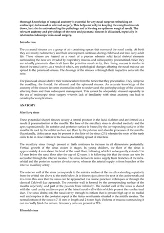
  • 1. thorough knowledge of surgical anatomy is essential for any nasal surgeon embarking on endoscopic, intranasal or external surgery. This helps not only in keeping the complication rate down, but also in understanding the pathology and achieving an optimum surgical result. The relevant anatomy and physiology of the nose and paranasal sinuses is discussed, especially in relation to endoscopic sino-nasal surgery. <br />Introduction<br />The paranasal sinuses are a group of air containing spaces that surround the nasal cavity. At birth they are mostly rudimentary and their development continues during childhood and into early adult life. These sinuses are formed as a result of a process wherein solid facial skeletal elements surrounding the nose are invaded by respiratory mucosa and subsequently pneumatized. Since they are actually pneumatic diverticuli from the primitive nasal cavity, their lining mucosa is similar to that of the nasal cavity, as a result of which, any pathological changes affecting the nasal mucosa can spread to the paranasal sinuses. The drainage of the sinuses is through their respective ostia into the nose. <br />The paranasal sinuses derive their nomenclature from the bones that they pneumatize. They comprise the maxillary, the frontal, the ethmoid and the sphenoid sinuses. An accurate knowledge of the anatomy of the sinuses becomes essential in order to understand the pathophysiology of the diseases affecting them and their subsequent management. This cannot be adequately stressed especially in the era of endoscopic sinus surgery wherein lack of familiarity with sinus anatomy can lead to catastrophic complications.<br />ANATOMY<br />Maxillary sinus<br />These pyramidal shaped sinuses occupy a central position in the facial skeleton and are formed as a result of pneumatisation of the maxilla. The base of the maxillary sinus is directed medially and the apex superolaterally. Its anterior and posterior surface is formed by the corresponding surfaces of the maxilla, its roof by the orbital surface and floor by the palatine and alveolar processes of the maxilla. Occasionally, dehiscences may be present in the floor of the sinus (2%) wherein the roots of the teeth come to lie in close relation to the mucosa facilitating spread of infection. <br />The maxillary sinus though present at birth continues to increase in all dimensions postnatally. Vertical growth of the sinus occurs in stages. In young children, the floor of the sinus is approximately 4 mm above the level of the nasal floor, following which it subsequently extends 1 to 5.5 mm below the nasal floor after the age of 12 years. It is following this that the sinus can now be accessible through the inferior meatus. The sinus derives its nerve supply from branches of the infra-orbital and the posterior superior alveolar nerve, whereas the arterial supply is from branches of the internal maxillary artery.<br />The anterior wall of the sinus corresponds to the anterior surface of the maxilla extending superiorly from the orbital rim above to the teeth below. It is thinnest just above the root of the canine tooth and it is from this area that the sinus can be approached via canine puncture techniques or through the classical Caldwell-Luc approach. The posterior wall is formed by the corresponding surface of the maxilla superiorly, and part of the palatine bone inferiorly. The medial wall of the sinus is shared with the nasal cavity and forms part of the lateral nasal wall within which is present the nasolacrimal duct. The sinus drains into the nasal cavity through its ostium that is present high up in its medial wall and empties in the posterior aspect of the hiatus semilunaris situated in the middle meatus. The normal ostium of the sinus is 7-11 mm in length and 2-6 mm high. Oedema of mucosa surrounding it can markedly block the ostium. Accessory ostia are present in 28%.<br />Ethmoid sinus<br />The word ‘Ethmoid’ in Greek means sieve-like. This sinus comprises a group of air cells, which form one of the most complex structures in the body. Hence the sinus is rightly named the ethmoid ‘labyrinth’. This complex arrangement of air cells and their particular anatomical location makes them play a pivotal role in the pathophysiology of sinus inflammatory disease. Mosher has rightly stated that ‘Had the Ethmoidal sinus been placed in any other part of the body, it would be an insignificant and harmless collection of bony cells. In the place where nature has put it, it has major relationships, so that diseases and surgery in this region often lead to disaster’. The Ethmoid bone per se comprises a horizontal plate - the cribriform plate, a vertical plate - the perpendicular plate and two lateral ethmoidal sinuses. The ethmoidal sinuses occupy a position between the orbit laterally and the nasal cavity medially (Figs. 1, 2). They are related to the anterior cranial fossa superiorly and the maxillary sinus inferiorly. The entire ethmoid sinus structure measures 4-5 cm in length and 2.5-3 cm in height. The ethmoid air cells have been classified according to their location in relation to specific anatomic structures. The air cells are divided into anterior and posterior groups by the lateral (or basal) lamella termed the ‘ground lamella’. The anterior ethmoidal cells which lie anterior to it open into the middle meatus and the posterior group opens into the superior meatus. The anterior ethmoidal cells tend to be smaller than the posterior group of cells, their number varying between 4-17. The posterior ethmoid cells number between 2-6. The air cells are separated from each other by thin intervening septae of bone that may be complete or incomplete. The agger nasi (1-4 cells) are the anterior most group of cells located anterior to the uncinate process. Exenteration of these cells is often necessary to expose the frontal recess. The lateral wall of each sinus separates it from the orbit. This is formed by the solid lacrimal bone anteriorly and a paper thin bone, aptly named the lamina papyracea situated behind it. The lamina papyracea may occasionally have dehiscences within it. The optic canal with the optic nerve comes in close relation with the lateral wall of the ethmoid sinus posteriorly. The roof of the sinus is formed by a horizontal plate of the frontal bone termed the fovea ethmoidalis that separates the air cells from the anterior cranial fossa. The medial most part of the fovea ethmoidalis is relatively thin and slopes downwards to join the cribriform plate. The anterior ethmoidal artery in its bony canal runs along the roof of the anterior group of air cells and is a useful landmark for the superior limit of dissection. <br />A few ethmoid air cells are located near the area of the fronto-nasal duct, and are termed the fronto ethmoidal cells. The Haller cell is an ethmoidal cell in relation with the floor of the orbit (Fig. 3). Occasionally, a posterior ethmoid air cell may invade the anterior-superior aspect of the sphenoid sinus and come to lie in close relation with the optic nerve. It is then called the cell of Onodi and could be mistaken for the sphenoid sinus during endoscopic surgery.<br /> Fig 1:Coronal CT Scan through anterior ethmoidal air cells. BE-Bulla ethmoidalis; CB-Concha bullosa;Cp-Cribriform plate; FE- Fovea ethoidalis; In- infundibulum; IT-inferior turbinate; Lp-Lamina papyracea; MM-Middle meatus; MS- Maxillary sinus; S- septum; UP-Uncinate process Fig 2:Coronal CT Scan through posterior ethmoidal air cells. FE- Fovea ethoidalis ; IOF-Inferior turbinate ;MS- Maxillary sinus; ON- Optic nerve ; PE- Posterior ethmoid air cell ;ST- Superior turbinate.<br /> <br />Frontal Sinus<br />The frontal sinus is the last sinus to develop, its complete development takes place by 14 - 16 years. The size of this sinus is highly variable and can range from a few cubic centimeters to a size that occupies most of the area of the frontal bone. The average height is about 24.3 mm. The floor of the sinus which forms the orbital roof and the posterior wall which is related to the anterior cranial fossa are considerably thinner than the anterior wall which is formed by the thick bone of the skull. A mid-line septum is present within the sinus. Incomplete septations extending from the roof are commonly present, giving the sinus a scalloped configuration. The sinus opens in the upper part of the infundibulum into the fronto-nasal recess. <br />Fig:3Coronal CT Scan showing an infected right Haller cell<br /> <br />Sphenoid Sinus<br />The body of the sphenoid can get pneumatized to a variable extent. Thus according to Congdon one may encounter a conchal (5%), a pre-sellar (23.5%) or a post sellar (67%) pneumatisation.<br />On an average the adult sphenoid sinus measures 13.5 mm in width superiorly, 16.9 mm in the middle, 18.7 mm in its lowest segment. The length averages 19.4 mm in the upper part, 4-8 mm in the middle and 18.5 mm in the lower part. The ostium of this sinus is located in the upper 1/3 of its anterior wall and it drains into the spheno-ethmoidal recess. An inter-sinus septum is generally present which is almost always deviated to one side making the interior of the sinus asymmetrical (Fig. 4). Accessory septations may be occasionally seen within the sinus. The sinus may sometimes extend superior or inferior to the optic nerve making this anatomical variation the most dangerous. In the lateral wall of the sphenoid sinus, a superior bulge corresponds to the optic nerve whereas the infero-lateral bulge is formed by the internal carotid artery. Bulges corresponding to the maxillary nerve, and vidian nerves may also be present. The pituitary gland lies above the roof of the sphenoid sinus and can thus be approached trans-sphenoidally.<br />Anatomy of the lateral nasal wall<br />The lateral wall of the nose has a characteristic complex structure as a result of scrolls and projections being present on it. These projections are the superior, middle and inferior turbinates and occasionally when present, the supreme turbinate. Underlying these shelves of bone are the superior middle and inferior meati respectively. The inferior turbinate is a separate bone, while the superior and middle turbinates are parts of the ethmoid bone. The casual observer must be wary of not mistaking a hypertrophied inferior turbinate for a nasal mass or polyp. The superior turbinate is the smallest turbinate and overlies the superior meatus into which the posterior ethmoidal air cells drain. The nasolacrimal duct opens into the inferior meatus. The middle turbinate forms an important landmark from the point of view of endoscopic sinus surgery and should be preserved. <br />The ostiomeatal complex is a word coined by Naumann and comprises the region of the middle meatus with the anterior air cells. This is the most important area for normal sinus functioning and any pathology in this area will disrupt physiology and lead to sinus dysfunction. In the middle meatus are several important structures, which play a vital role in the pathogenesis of sino-nasal inflammatory disease. These structures also form important landmarks in sinus endoscopic surgery. Anteriorly, the first landmark is a hook shaped bone or the uncinate process. Posterior to the uncinate process is present a groove known as the hiatus semilunaris which leads into the ethmoidal infundibulum. The ethmoidal bulla is a bulge posterior to the hiatus, which hitherto was referred to as the middle ethmoidal air cell system. Present nomenclature however groups the ethmoids into only anterior and posterior air cells, and the bulla ethmoidalis is included as part of the anterior cells. The frontal sinus opens into the superior most aspect of the ethmoidal infundibulum into the fronto-nasal recess while the anterior ethmoidal cells open anteriorly into the infundibulum. The ostium of the maxillary sinus opens postero-inferiorly into the infundibulum and can be located just above the inferior turbinate. Occasionally accessory ostia of the maxillary sinus may be present. Behind the posterior end of the middle turbinate is the sphenopalatine foramen leading into the pterygopalatine fossa. <br />Certain anatomical variations may exist which predispose to development of recurrent infections by narrowing the middle meatus space and hampering the mucociliary drainage. Some of these which is almost always deviated to one side making the interior of the sinus asymmetrical (Fig. 4). Accessory septations may be occasionally seen within the sinus. The sinus may sometimes extend superior or inferior to the optic nerve making this anatomical variation the most dangerous. In the lateral wall of the sphenoid sinus, a superior bulge corresponds to the optic nerve whereas the infero-lateral bulge is formed by the internal carotid artery. Bulges corresponding to the maxillary nerve, and vidian nerves may also be present. The pituitary gland lies above the roof of the sphenoid sinus and can thus be approached trans-sphenoidally.<br /> <br />Fig:4 :Coronal CT Scan through the sphenoid sinusOC- optic canal; PC- Choana; SOF-superior orbital fissure; SS- sphenoid sinus.<br /> <br />Anatomy of the lateral nasal wall<br />The lateral wall of the nose has a characteristic complex structure as a result of scrolls and projections being present on it. These projections are the superior, middle and inferior turbinates and occasionally when present, the supreme turbinate. Underlying these shelves of bone are the superior middle and inferior meati respectively. The inferior turbinate is a separate bone, while the superior and middle turbinates are parts of the ethmoid bone. The casual observer must be wary of not mistaking a hypertrophied inferior turbinate for a nasal mass or polyp. The superior turbinate is the smallest turbinate and overlies the superior meatus into which the posterior ethmoidal air cells drain. The nasolacrimal duct opens into the inferior meatus. The middle turbinate forms an important landmark from the point of view of endoscopic sinus surgery and should be preserved. <br />The ostiomeatal complex is a word coined by Naumann and comprises the region of the middle meatus with the anterior air cells. This is the most important area for normal sinus functioning and any pathology in this area will disrupt physiology and lead to sinus dysfunction. In the middle meatus are several important structures, which play a vital role in the pathogenesis of sino-nasal inflammatory disease. These structures also form important landmarks in sinus endoscopic surgery. Anteriorly, the first landmark is a hook shaped bone or the uncinate process. Posterior to the uncinate process is present a groove known as the hiatus semilunaris which leads into the ethmoidal infundibulum. The ethmoidal bulla is a bulge posterior to the hiatus, which hitherto was referred to as the middle ethmoidal air cell system. Present nomenclature however groups the ethmoids into only anterior and posterior air cells, and the bulla ethmoidalis is included as part of the anterior cells. The frontal sinus opens into the superior most aspect of the ethmoidal infundibulum into the fronto-nasal recess while the anterior ethmoidal cells open anteriorly into the infundibulum. The ostium of the maxillary sinus opens postero-inferiorly into the infundibulum and can be located just above the inferior turbinate. Occasionally accessory ostia of the maxillary sinus may be present. Behind the posterior end of the middle turbinate is the sphenopalatine foramen leading into the pterygopalatine fossa. <br />Certain anatomical variations may exist which predispose to development of recurrent infections by narrowing the middle meatus space and hampering the mucociliary drainage. Some of thesewhich is almost always deviated to one side making the interior of the sinus asymmetrical (Fig. 4). Accessory septations may be occasionally seen within the sinus. The sinus may sometimes extend superior or inferior to the optic nerve making this anatomical variation the most dangerous. In the lateral wall of the sphenoid sinus, a superior bulge corresponds to the optic nerve whereas the infero-lateral bulge is formed by the internal carotid artery. Bulges corresponding to the maxillary nerve, and vidian nerves may also be present. The pituitary gland lies above the roof of the sphenoid sinus and can thus be approached trans-sphenoidally.<br />Anatomy of the lateral nasal wall<br />The lateral wall of the nose has a characteristic complex structure as a result of scrolls and projections being present on it. These projections are the superior, middle and inferior turbinates and occasionally when present, the supreme turbinate. Underlying these shelves of bone are the superior middle and inferior meati respectively. The inferior turbinate is a separate bone, while the superior and middle turbinates are parts of the ethmoid bone. The casual observer must be wary of not mistaking a hypertrophied inferior turbinate for a nasal mass or polyp. The superior turbinate is the smallest turbinate and overlies the superior meatus into which the posterior ethmoidal air cells drain. The nasolacrimal duct opens into the inferior meatus. The middle turbinate forms an important landmark from the point of view of endoscopic sinus surgery and should be preserved. <br />The ostiomeatal complex is a word coined by Naumann and comprises the region of the middle meatus with the anterior air cells. This is the most important area for normal sinus functioning and any pathology in this area will disrupt physiology and lead to sinus dysfunction. In the middle meatus are several important structures, which play a vital role in the pathogenesis of sino-nasal inflammatory disease. These structures also form important landmarks in sinus endoscopic surgery. Anteriorly, the first landmark is a hook shaped bone or the uncinate process. Posterior to the uncinate process is present a groove known as the hiatus semilunaris which leads into the ethmoidal infundibulum. The ethmoidal bulla is a bulge posterior to the hiatus, which hitherto was referred to as the middle ethmoidal air cell system. Present nomenclature however groups the ethmoids into only anterior and posterior air cells, and the bulla ethmoidalis is included as part of the anterior cells. The frontal sinus opens into the superior most aspect of the ethmoidal infundibulum into the fronto-nasal recess while the anterior ethmoidal cells open anteriorly into the infundibulum. The ostium of the maxillary sinus opens postero-inferiorly into the infundibulum and can be located just above the inferior turbinate. Occasionally accessory ostia of the maxillary sinus may be present. Behind the posterior end of the middle turbinate is the sphenopalatine foramen leading into the pterygopalatine fossa. <br />Certain anatomical variations may exist which predispose to development of recurrent infections by narrowing the middle meatus space and hampering the mucociliary drainage. Some of thesewhich is almost always deviated to one side making the interior of the sinus asymmetrical (Fig. 4). Accessory septations may be occasionally seen within the sinus. The sinus may sometimes extend superior or inferior to the optic nerve making this anatomical variation the most dangerous. In the lateral wall of the sphenoid sinus, a superior bulge corresponds to the optic nerve whereas the infero-lateral bulge is formed by the internal carotid artery. Bulges corresponding to the maxillary nerve, and vidian nerves may also be present. The pituitary gland lies above the roof of the sphenoid sinus and can thus be approached trans-sphenoidally.<br />Anatomy of the lateral nasal wall<br />The lateral wall of the nose has a characteristic complex structure as a result of scrolls and projections being present on it. These projections are the superior, middle and inferior turbinates and occasionally when present, the supreme turbinate. Underlying these shelves of bone are the superior middle and inferior meati respectively. The inferior turbinate is a separate bone, while the superior and middle turbinates are parts of the ethmoid bone. The casual observer must be wary of not mistaking a hypertrophied inferior turbinate for a nasal mass or polyp. The superior turbinate is the smallest turbinate and overlies the superior meatus into which the posterior ethmoidal air cells drain. The nasolacrimal duct opens into the inferior meatus. The middle turbinate forms an important landmark from the point of view of endoscopic sinus surgery and should be preserved. <br />The ostiomeatal complex is a word coined by Naumann and comprises the region of the middle meatus with the anterior air cells. This is the most important area for normal sinus functioning and any pathology in this area will disrupt physiology and lead to sinus dysfunction. In the middle meatus are several important structures, which play a vital role in the pathogenesis of sino-nasal inflammatory disease. These structures also form important landmarks in sinus endoscopic surgery. Anteriorly, the first landmark is a hook shaped bone or the uncinate process. Posterior to the uncinate process is present a groove known as the hiatus semilunaris which leads into the ethmoidal infundibulum. The ethmoidal bulla is a bulge posterior to the hiatus, which hitherto was referred to as the middle ethmoidal air cell system. Present nomenclature however groups the ethmoids into only anterior and posterior air cells, and the bulla ethmoidalis is included as part of the anterior cells. The frontal sinus opens into the superior most aspect of the ethmoidal infundibulum into the fronto-nasal recess while the anterior ethmoidal cells open anteriorly into the infundibulum. The ostium of the maxillary sinus opens postero-inferiorly into the infundibulum and can be located just above the inferior turbinate. Occasionally accessory ostia of the maxillary sinus may be present. Behind the posterior end of the middle turbinate is the sphenopalatine foramen leading into the pterygopalatine fossa. <br />Certain anatomical variations may exist which predispose to development of recurrent infections by narrowing the middle meatus space and hampering the mucociliary drainage. Some of these predisposing factors are a large concha bullosa (Fig. 5), which is an enlarged middle turbinate due to pneumatisation, a paradoxically curved middle turbinate with a convexity laterally, and a medially rotated uncinate process<br />fig: 5Endoscopic photograph of concha bullosa middle turbinate.<br /> <br />Anatomy of the nasal septum<br />The nasal septum divides the nose into two halves. It comprises an anterior membranous and cartilaginous portion and a posterior bony part. The anterior most part or the columella is formed by the medial crurae of the two alar cartilages and are connected to the free caudal border of the septal cartilage by the membranous septum. The quadrilateral or the septal cartilage articulates postero-superiorly with the perpendicular plate of the ethmoid and postero-inferiorly with the vomer. The latter two form the bony part of the nasal septum. The perpendicular plate of the ethmoid articulates anteriorly with the nasal bone and the nasal spine of the frontal bone, superiorly with the cribriform plate of the ethmoid and posteriorly with the crest of the sphenoid. The vomer articulates inferiorly with the nasal crest of the maxilla and the palatine bone. The rostrum of the sphenoid is a wedge of bone projecting from the body of the sphenoid in between the perpendicular plate of ethmoid and the vomer and always lies in the mid-line. Hence it acts as a landmark for trans-sphenoidal hypophysectomy. The nasal septum is very rarely present in the mid-line and more often than not is deviated to one side. Thus one can have a ‘C’ or an ‘S’ shaped deviation of the nasal septum. Occasionally sharp angulations or ‘spurs’ may be present at the chondro-vomerine junction. All the above contribute to the symptom of nasal obstruction.<br />The nasal cavity derives its blood supply from branches of both the external, as well as the internal carotid arteries. The antero-superior quadrant of the nose is supplied by the anterior and posterior ethmoidal arteries, which are branches of the ophthalmic artery which is a branch of the internal carotid artery. The facial artery and the superior labial artery supply the vestibular area. The sphenopalatine artery supplies the posterior and inferior quadrants. It is a branch of the internal maxillary artery, which in turn is a branch of the external carotid artery. An area of anastomosis between all these arteries is present on the antero-inferior part of the nasal septum which is termed the Little’s area or the Kiesselbach’s plexus and is the commonest site of epistaxis.<br />The nasal nerve supply is via the general sensory, parasympathetic and sympathetic innervation. The nasociliary nerve (branch of the ophthalmic division of the trigeminal nerve) supplies the anterosuperior part of the nose. The sphenopalatine, infra-orbital and superior alveolar nerves, which are branches of the maxillary division of the trigeminal nerve, innervate the rest of the nasal cavity.<br />The greater superficial petrosal nerve, which contains the post-ganglionic parasympathetic secretomotor fibres, joins the deep petrosal nerve, which in turn contains sympathetic fibres from the plexus around the internal carotid artery. The two together form the vidian nerve, which enters the pterygo-palatine fossa and synapses in the pterygo-palatine ganglion. The maxillary nerve too enters this space. Branches of the pterygo-palatine ganglion enter the nose to innervate it. The one of primary importances being the spheno-palatine nerve which innervates the major part of the nose.<br />Physiology of the nose and the paranasal sinuses<br />The chief functions of the nose are respiratory and olfactory. In addition to being the sensory organ of smell, the nose also plays an important role in the cleansing and conditioning of inspired air and contributing to a small extent in altering the quality of speech. The inspired air is first filtered wherein particulate matter is trapped by the vibrissae in the vestibule which prevent it from reaching the lower airway. The air is then humidified with the help of secretions of various serous and mucous glands present in the respiratory epithelium. The temperature of the inspired air is brought to the level of the body temperature by way of heat exchange. Nasal secretions apart from conditioning the air, also have an immunological function. It contains substances like IgA and IgE, which are involved in local mucosal defense mechanisms, and enzymes like lysozymes, lactoferrin, and complement and C reactive proteins. Sensory nerve endings present in the nose can mediate various reflexes. Receptors present in the nose are sensitive to chemical irritation, temperature changes, and physical stimuli. Stimulation of these receptors brings about widespread cardiovascular and respiratory responses. For example the sneezing reflex can cause a change in the respiratory rate, closure of the glottis, and a cardiovascular response. Several receptors for various neurotransmitters are also present in the nose, which significantly alter the physiology. <br />As the sensory organ of olfaction, the nose allows perception of different odours. Air containing these odouriferous compounds reaches the olfactory mucosa situated in the uppermost part of the nose. This mucosa bears sensory hair cells, which are stimulated by these compounds. Action potentials are then generated and the impulses are transmitted along axons of the sensory hair cells to the olfactory area in the brain thus enabling one to perceive smell. Local pathological conditions like allergic rhinitis, atrophic rhinitis, nasal polyposis and some hereditary disorders can cause alteration or loss of the sensation of smell. The nose modifies speech by allowing some amount of air to escape from it thereby adding resonance to the sound produced and improving the quality of speech.<br />The physiological functions of the paranasal sinuses are controversial. Some believe that the sinuses play a role in conditioning of the inspired air, pressure damping, heat insulation and voice resonance. Others however are of the opinion that the sinuses may indeed have no function at all and may merely be vestigeal structures assuming a significant role when diseased.<br />