

Recomendados
Más contenido relacionado
La actualidad más candente
La actualidad más candente (20)
Similar a Immediate Implant Placement
Similar a Immediate Implant Placement (20)
Más de Abu-Hussein Muhamad
Más de Abu-Hussein Muhamad (20)
Último
Último (20)
Immediate Implant Placement
- 1. The Journal of Implant & Advanced Clinical Dentistry Volume 6, No. 4 July 2014 Extraoral Infraorbital Nerve Block Immediate Anterior Dental Implant
- 2. Ease of drilling sequence – Minimized drill sequence (2~4 drills) allows precision of osteotomy site preparation and less chair time for both dental surgeons and patients. Color coding – Implant vials and drills are color coded to elimi- nate confusion. Wide selections – Wide selection of implant sizes and prosthetic options are available to meet the needs of all dental surgeons. 888.446.9995 www.OsseoFuse.com Call now to learn more support@osseofuse.com DentalImplantSystemYouCanDependOn Simple. Compatible. Predictable.
- 3. The Journal of Implant & Advanced Clinical Dentistry • 1 The Journal of Implant & Advanced Clinical Dentistry Volume 6, No. 4 • July 2014 Table of Contents 5 Immediate Implant Placement with One Year Follow-up: A Case Report Dr. Abdulgani Azzaldeen, Dr. Bajali Musa, Dr. Abu-Hussein Muhamad 13 Immediate Placement of Dental Implants in Molar Sites: A Case Series Dr. Mohit B. Zamad, Dr. Aakanksha Zamad. Dr. Akshay Daga, Dr. Pankaj Ahare 23 Microleakage of Pit and Fissure Sealants Comparing Air Abrasion and Acid Etch Techniques – An in Vitro Study Dr. Raman Deep Kaur, Neeraj Mahahjan 35 The Application of Extraoral Infraorbital Nerve Block Compared to the Intraoral Approach Dr. Shady A. M. Negm
- 4. The Journal of Implant Advanced Clinical Dentistry Volume 6, No. 4 • July 2014 Publisher LC Publications Design Jimmydog Design Group www.jimmydog.com Production Manager Stephanie Belcher 336-201-7475 • sbelcher@triad.rr.com Copy Editor JIACD staff Digital Conversion NxtBook Media Internet Management InfoSwell Media Subscription Information: Annual rates as follows: Non-qualified individual: $99(USD) Institutional: $99(USD). For more information regarding subscriptions, contact info@jiacd.com or 1-888-923-0002. Advertising Policy: All advertisements appearing in the Journal of Implant and Advanced Clinical Dentistry (JIACD) must be approved by the editorial staff which has the right to reject or request changes to submitted advertisements. The publication of an advertisement in JIACD does not constitute an endorsement by the publisher. Additionally, the publisher does not guarantee or warrant any claims made by JIACD advertisers. For advertising information, please contact: info@JIACD.com or 1-888-923-0002 Manuscript Submission: JIACD publishing guidelines can be found at http://www.jiacd.com/author-guidelines or by calling 1-888-923-0002. Copyright © 2014 by LC Publications. All rights reserved under United States and International Copyright Conventions. No part of this journal may be reproduced or transmitted in any form or by any means, electronic or mechanical, including photocopying or any other information retrieval system, without prior written permission from the publisher. Disclaimer: Reading an article in JIACD does not qualify the reader to incorporate new techniques or procedures discussed in JIACD into their scope of practice. JIACD readers should exercise judgment according to their educational training, clinical experience, and professional expertise when attempting new procedures. JIACD, its staff, and parent company LC Publications (hereinafter referred to as JIACD-SOM) assume no responsibility or liability for the actions of its readers. Opinions expressed in JIACD articles and communications are those of the authors and not necessarily those of JIACD- SOM. JIACD-SOM disclaims any responsibility or liability for such material and does not guarantee, warrant, nor endorse any product, procedure, or technique discussed in JIACD, its affiliated websites, or affiliated communications. Additionally, JIACD-SOM does not guarantee any claims made by manufact-urers of products advertised in JIACD, its affiliated websites, or affiliated communications. Conflicts of Interest: Authors submitting articles to JIACD must declare, in writing, any potential conflicts of interest, monetary or otherwise, that may exist with the article. Failure to submit a conflict of interest declaration will result in suspension of manuscript peer review. Erratum: Please notify JIACD of article discrepancies or errors by contacting editors@JIACD.com JIACD (ISSN 1947-5284) is published on a monthly basis by LC Publications, Las Vegas, Nevada, USA. 2 • Vol. 6, No. 4 • July 2014
- 5. The Journal of Implant Advanced Clinical Dentistry • 3 Tara Aghaloo, DDS, MD Faizan Alawi, DDS Michael Apa, DDS Alan M. Atlas, DMD Charles Babbush, DMD, MS Thomas Balshi, DDS Barry Bartee, DDS, MD Lorin Berland, DDS Peter Bertrand, DDS Michael Block, DMD Chris Bonacci, DDS, MD Hugo Bonilla, DDS, MS Gary F. Bouloux, MD, DDS Ronald Brown, DDS, MS Bobby Butler, DDS Nicholas Caplanis, DMD, MS Daniele Cardaropoli, DDS Giuseppe Cardaropoli DDS, PhD John Cavallaro, DDS Jennifer Cha, DMD, MS Leon Chen, DMD, MS Stepehn Chu, DMD, MSD David Clark, DDS Charles Cobb, DDS, PhD Spyridon Condos, DDS Sally Cram, DDS Tomell DeBose, DDS Massimo Del Fabbro, PhD Douglas Deporter, DDS, PhD Alex Ehrlich, DDS, MS Nicolas Elian, DDS Paul Fugazzotto, DDS David Garber, DMD Arun K. Garg, DMD Ronald Goldstein, DDS David Guichet, DDS Kenneth Hamlett, DDS Istvan Hargitai, DDS, MS Michael Herndon, DDS Robert Horowitz, DDS Michael Huber, DDS Richard Hughes, DDS Miguel Angel Iglesia, DDS Mian Iqbal, DMD, MS James Jacobs, DMD Ziad N. Jalbout, DDS John Johnson, DDS, MS Sascha Jovanovic, DDS, MS John Kois, DMD, MSD Jack T Krauser, DMD Gregori Kurtzman, DDS Burton Langer, DMD Aldo Leopardi, DDS, MS Edward Lowe, DMD Miles Madison, DDS Lanka Mahesh, BDS Carlo Maiorana, MD, DDS Jay Malmquist, DMD Louis Mandel, DDS Michael Martin, DDS, PhD Ziv Mazor, DMD Dale Miles, DDS, MS Robert Miller, DDS John Minichetti, DMD Uwe Mohr, MDT Dwight Moss, DMD, MS Peter K. Moy, DMD Mel Mupparapu, DMD Ross Nash, DDS Gregory Naylor, DDS Marcel Noujeim, DDS, MS Sammy Noumbissi, DDS, MS Charles Orth, DDS Adriano Piattelli, MD, DDS Michael Pikos, DDS George Priest, DMD Giulio Rasperini, DDS Michele Ravenel, DMD, MS Terry Rees, DDS Laurence Rifkin, DDS Georgios E. Romanos, DDS, PhD Paul Rosen, DMD, MS Joel Rosenlicht, DMD Larry Rosenthal, DDS Steven Roser, DMD, MD Salvatore Ruggiero, DMD, MD Henry Salama, DMD Maurice Salama, DMD Anthony Sclar, DMD Frank Setzer, DDS Maurizio Silvestri, DDS, MD Dennis Smiler, DDS, MScD Dong-Seok Sohn, DDS, PhD Muna Soltan, DDS Michael Sonick, DMD Ahmad Soolari, DMD Neil L. Starr, DDS Eric Stoopler, DMD Scott Synnott, DMD Haim Tal, DMD, PhD Gregory Tarantola, DDS Dennis Tarnow, DDS Geza Terezhalmy, DDS, MA Tiziano Testori, MD, DDS Michael Tischler, DDS Tolga Tozum, DDS, PhD Leonardo Trombelli, DDS, PhD Ilser Turkyilmaz, DDS, PhD Dean Vafiadis, DDS Emil Verban, DDS Hom-Lay Wang, DDS, PhD Benjamin O. Watkins, III, DDS Alan Winter, DDS Glenn Wolfinger, DDS Richard K. Yoon, DDS Editorial Advisory Board The Journal of Implant Advanced Clinical Dentistry Co-Editor in Chief Nick Huang, MD Founder, Co-Editor in Chief Dan Holtzclaw, DDS, MS Founder, Co-Editor in Chief Nicholas Toscano, DDS, MS
- 6. Azzaldeen et al ATTENTION PROSPECTIVE AUTHORS JIACD wants to publish your article! The Journal of Implant Advanced Clinical Dentistry For complete details regarding publication in JIACD, please refer to our author guidelines at the following link: http://www.jiacd.com/authorinfo/ author-guidelines.pdf or email us at: editors@jicad.com
- 7. Azzaldeen et al Background: This case report describes extraction of a fractured left maxillary central incisor tooth, followed by immediate place- ment of a dental implant in the prepared socket and temporization by a bonded restoration. Methods: The tooth was extracted with mini- mal hard and soft tissue trauma and without flap reflection. The socket was prepared to the required depth and a dental was inserted. An impression was made 4 months after implant insertion, and a definitive restoration was placed. Results: The atraumatic operating tech- nique and the immediate insertion of the Implant resulted in the preservation of the hard and soft tissues at the extrac- tion site. The patient exhibited no clini- cal or radiologic complications through 12 months of clinical monitoring after loading. Conclusion: The dental implant and provi- sional restoration provided the patient with immediate esthetics, function, comfort and most importantly preservation of tissues. Immediate Implant Placement with One Year Follow-up: A Case Report Dr. Abdulgani Azzaldeen1 • Dr. Bajali Musa2 • Dr. Abu-Hussein Muhamad3 1. Assistant Professor, Al Quds University, Jerusalem, Palestine 2. Assistant Professor, Al Quds University, Jerusalem, Palestine 3. Visiting Professor, Napoli University, Athens, Greece Abstract KEY WORDS: Dental implant, immediate placement, temporization, prosthetics The Journal of Implant Advanced Clinical Dentistry • 5
- 8. 6 • Vol. 6, No. 4 • July 2014 INTRODUCTION Immediate implants are defined as the place- ment of implants in course of surgical extrac- tion of the teeth to be replaced. The insertion of implants immediately after extraction is not new, and in the 1980s, the University of Tübin- gen advocated the procedure as the tech- nique of choice for Tübingen and München ceramic implants.1,2 As a result of the suc- cess of the protocol designed by Brånemark and his team for their dental implant system, other procedures were largely relegated for many years. Initially, a healing period of 9-12 months was advised between tooth extrac- tion and implant placement. Nevertheless, as a result of continued research, a number of the concepts contained in the Brånemark proto- col and previously regarded as axiomatic; such as the submerged technique concept, delayed loading, machined titanium surface, etc.; have since been revised and improved upon even by actual creators of the procedure.2-4 Based on the time elapsed between extrac- tion and implantation, the following classifica- tion has been established relating the receptor zone to the required therapeutic approach: 1. Immediate implantation, when the remnant bone suffices to ensure primary stability of the implant, which is inserted in the course of surgical extraction of the tooth to be replaced (primary immediate implants). 2. Recent implantation, when approximately 6-8 weeks have elapsed from extrac- tion to implantation, a time during which the soft tissues heal, allowing adequate mucogingival covering of the alveo- lus (secondary immediate implants). 3. Delayed implantation, when the receptor zone is not optimum for either immediate or recent implantation. Bone promotion is first carried out with bone grafts and/or barrier membranes, followed approximately 6 months later by implant positioning (delayed implants). 4. Mature implantation, when over 9 months have elapsed from extraction to implantation. Mature bone is found in such situations.3-7 The most frequently cited reasons for unde- rutilization of endosseous implant therapy are that treatment cost is perceived to be too high and treatment takes too long (Branemark’s orig- inal treatment protocols required up to a year or more to complete treatment) An obvious area of focus has been to decrease the amount of time necessary to complete implant therapy. Approaches to achieve this goal have domi- nated clinical research and practice: delayed/ immediate implant loading, improving implant surface technology (promotion of quicker heal- ing and better osseointegration), and immedi- ate placement of an endosseous implant after extraction of a natural tooth.1 In this paper a case presentation supporting the last of these three approaches will be shown. The definition for an immediate endosseous implant is extrac- tion of a natural tooth followed by immediate placement (within the same surgical procedure) of an endosseous dental implant. Immediate implants have become widely accepted despite controversial beginnings and the available liter- ature consistently cites high levels of success ranging from 94-100% on average and immedi- ate implants provide clinically recognizable ben- efits. Broadly speaking, these benefits include reduction of morbidity, reduction of alveolar Azzaldeen et al
- 9. The Journal of Implant Advanced Clinical Dentistry • 7 bone resorption. Controlled clinical studies have demonstrated an average of 4.4mm of hor- izontal and 1.2mm of vertical bone resorption six months after tooth extraction preservation of gingival tissues, preservation of the papilla in the esthetic zone, and reduction of treat- ment cost and time while the healing phase is shorter in general and there is a reduction in the number of procedures.1-5 With the extraction socket as a guide, the surgeon can also more easily determine the appropriate parallelism and alignment relative to the adjacent and opposing residual dentition. To maximize the advantage of these benefits and to minimize implant failure, case selection must be based on sound clinical and research criteria. Immediate placement and provisionalization for single tooth replacement allows for minimal disruption of the marginal soft tissues, providing immediate prosthetic support for the peri-implant tissues through the use of a carefully crafted provisional restoration. Pri- mary implantation is fundamentally indicated for replacing teeth with pathologies not amenable to treatment, such as caries or fractures. Imme- diate implants are also indicated simultaneous to the removal of impacted canines.5,6 Immedi- ate implantation can be carried out on extracting teeth with chronic apical lesions which are not likely to improve with endodontic treatment and Figure1: Initial retracted view. Figure 2: Initial occlusal view. Figure 3: Initial xray. Azzaldeen et al
- 10. 8 • Vol. 6, No. 4 • July 2014 apical surgery.7 The surgical requirements for immediate implantation include extraction with the least trauma possible, preservation of the extraction socket walls and thorough alveolar curettage to eliminate all pathological material. Primary stability is an essential requirement, and is achieved with an implant exceeding the alve- olar apex by 3-5 mm, or by placing an implant of greater diameter than the remnant alveo- lus. Esthetic emergence in the anterior zone is achieved by 1-3 mm sub-crestal implantation.6,7 There are contraindications to immedi- ate placement of dental implants. The exis- tence of an acute periapical inflammatory process constitutes an absolute contraindi- cation to immediate implantation.8,9 In the case of socket-implant diameter discrepan- cies in excess of 5 mm, which would leave most of the implant without bone contact, prior bone regeneration and delayed implan- tation may be considered.10 Avoid teeth with large or acute periapical infection; Teeth with labial bony dehiscence or fenestration defects; Insufficient bone apically to ensure primary Figure 4: Coronal portion of tooth removed. Figure 5: Tooth removed atraumatically, the facial plate was very thin. Figure 6: Immediate post op view. Figure 7: Immediate post op view. Azzaldeen et al
- 11. The Journal of Implant Advanced Clinical Dentistry • 9 Figure 8: Xray of immediate implant placement. Figure 9: Stock abutment. Figure 10: Final crown retracted view. stability of the implant; Systemic factors that may impair healing (e.g. smoking); Large bul- bous root morphology, Interproximal bone loss (aesthetic zone), active periodontitis.11 CASE REPORT A 53-year-old male patient presented with a history of trauma and crown fracture at the cervical area of tooth 8 (figures 1-3) and requested an immediate solution. Clinical and radiological evaluation revealed adequate alveolar bone, absence of periapical pathol- ogy but fracture line was below the crest of alveolar bone and was limited to the tooth. So, it was decided to extract and place endos- seous implant immediately and place a pro- visional restoration to avail the benefits like preservation of bone and emergence profile. After administering appropriate antibiotic and analgesic, induction of local anesthesia was carried out using lignocaine with adrena- line. As preservation of alveolar bone is key to success of immediate implants, extraction of tooth has to be atraumatic, so using periotomes and small periosteal elevators the fragment was Azzaldeen et al
- 12. 10 • Vol. 6, No. 4 • July 2014 luxated without excessive enlargement of the socket, and using an innovative method where endodontic file was used to engage the canal wall and tooth fragment was slowly luxated and removed from the socket using the file (figures 4, 5). The sockets were debrided with curettes and a dental implant (BioHorizons, Birming- ham, Alabama, USA) of dimensions 4 x 12mm was planned. After checking for primary stability, which was achieved by wrenching the implant into the bone beyond the apex of the socket, xeno- graft (Bio-Oss, Geistlicht, Princeton, New Jer- sey, USA) was packed between the implant and labial socket wall. The cover screw was placed and interrupted sutures were placed. Intraoral photography was taken to see the implant place- ment (figures 6, 7). It was found to be satis- factory. Postoperative instructions were given to the patient and they were asked to report back after 1 week. The sutures were removed after 7 days and the patient received temporary acrylic crown bonded to the adjacent teeth with fiber-reinforced composite on the same day. The patient was recalled after four months for the prosthetic procedures and was given porce- lain fused to metal crown over the implant. He was recalled for prophylaxis and follow up every three months. The clinical and radiographic appearances at six months and after one year show good aesthetic result and acceptable osseointegration of the implant (figures 8-12). CONCLUSION The implant therapy must fulfill both func- tional and esthetic requirements to be consid- ered a primary treatment modality. Aiming to reduce the process of alveolar bone resorp- tion and treatment time, the immediate place- ment of endosseous implants into extraction sockets achieved high success rate of between 94-100%, compared to the delayed placement. ● Figure 11: Occlusal view of final crown. Figure 12: Final restoration at 12 months. Correspondence: Dr. Abu-Hussein Muhammed 123 Argus Street 10441 Athens Greece abuhusseinmuhamad@gmail.com Azzaldeen et al
- 13. The Journal of Implant Advanced Clinical Dentistry • 11 Acknowledgments The authors would like to thank the following: Setergiou Bros Dental Laboratory, Athens, Greece for the fabrication of the ceramic restorations. Disclosure The authors report no conflicts of interest with anything mentioned in this article. References 1. Wagenberg BD, Ginsburg TR. Immediate implant placement on removal of the natural tooth: retrospective analysis of 1,081 implants. Compendium of Continuing EducDent 22:399-404; 2001. 2. Cooper LF, Rahman A, Moriarty J, et al. Immediate mandibular rehabilitation with endosseous implants: simultaneous extraction, implant placement, and loading. Int JOral Maxillofac Implants 17:517-25; 2002. 3. Di Felice R, D’Amario M, De Dominicis A, Garocchio S, D’Arcangelo C, Giannoni M. Immediate placement of bone level Sraumann implants: A case series. Int J Periodontics Restorative Dent2011;31:57-65. 4. Gelb DA. Immediate implant surgery: ten-year clinical overview. Compendium of Cont Educ Dent 20:1185- 92; 1999. 5. Barone A, Rispoli L, Vozza I, Quaranta A, Covani U. Immediate restoration of single implants placed immediately after tooth extraction. J Periodontol 2006;77:1914-20. 6. Hoffmann O, Beaumont C, Zafiropoulos GG. Immediate implant placement: A case series. J Oral Implantol 2006;32:182-9. 7. Zabalegui I, García M. En Gutiérrez JL, García M, eds. Integración de la implantología en la práctica odontológica. Madrid: Ergon. p. 127-36; 2002 8. Romanos GE. Treatment of advanced periodontal destruction with immediately loaded implants and simultaneous bone augmentation: A case report. J Periodontol 2003;74:255-61. 9. Novaes-Junior AB, Novaes AB. Immediate implants placed into infected sites: a clinical report. Int J Oral Maxillofac Implants10:609-13; 1995 10. Anitua E, Orive G. A new approach for atraumatic implant explantation and immediate implant installation. Oral Surg Oral Med Oral Pathol Oral Radiol 2012;113:e19-25. 11. Strub JR, Kohal RJ, Klaus G, Ferraresso F. The re implant system for immediate implant placement. J Esthet Dent 9:187-96;1997. ATTENTION PROSPECTIVE AUTHORS JIACD wants to publish your article! The Journal of Implant Advanced Clinical Dentistry For complete details regarding publication in JIACD, please refer to our author guidelines at the following link: http://www.jiacd.com/authorinfo/author-guidelines.pdf or email us at: editors@jicad.com Azzaldeen et al
- 14. 24 JIACDThe Journal of Implant Advanced Clinical Dentistry October 2008 Review | Oral Implications of Cancer Cheomotherapy ADVERTISE WITHADVERTISEADVERTISE WITHADVERTISEADVERTISE WITH TODAY! WWW.JIACD.COM Using revolutionary online technology, JIACD provides its readers with an experience that is simply not available with traditional hard copy paper journals. Reach more customers with the dental profession’s first truly interactive paperless journal! simply not available with traditional
- 15. Zamad et al C ommonly, immediate implants have been reserved for the single rooted anterior tooth and single or bi-rooted premo- lar tooth. Perhaps the most important aspect of any implant surgery in accordance with the successful procedure is implant stability and bone to implant contact (BIC). Immediate den- tal implant placement has been an acceptable procedure for at least the past two decades. Removal of molar teeth provides a chal- lenging and intriguing dilemma due to multiple root morphology. In the case of extraction and immediate placement of dental implants preserving alveolar bone proper, particularly that of the labial and lingual plates of bone is essential in providing the optimal environment for maximizing BIC and implant stability. Also, the position of the final restoration must be considered, in relation to intra and inter arch position, occlusion, function and esthetics. Thus, minimal alveolar bone removal should be considered and attained to aid in the above factors in order to provide an acceptable surgi- cal site for successful placement of the dental implant. Finally, and perhaps most importantly when considering immediate molar implant placement, removal of the intra-alveolar sep- tum should be avoided to aid in increas- ing BIC and allowing the attainment of initial implant stability at the time of placement. Immediate Placement of Dental Implants in Molar Sites: A Case Series Dr. Mohit B. Zamad1 • Dr. Aakanksha Zamad2 Dr. Akshay Daga3 • Dr. Pankaj Ahare4 1. Oral maxillofacial surgeon, California, USA 2. Post-graduate student, VSPM Dental College research Centre, Nagpur, India 3. Oral maxillofacial surgeon 4. Department of Orthodontics, VSPM Dental college research Center, Nagpur, India Abstract KEY WORDS: Dental implants, immediate placement, molars The Journal of Implant Advanced Clinical Dentistry • 13
- 16. 14 • Vol. 6, No. 4 • July 2014 INTRODUCTION Immediate implant placement following tooth extraction in appropriately selected cases can be considered an optimal procedure for the fol- lowing reasons: the natural healing process is mobilized to the maximum, no bone resorp- tion, drilling is reduced, number of sur- gical stages are eliminated, design and construction of prosthesis is simplified, and positive psychological effect on the patient.1,2 As per previous studies, immediate implant placement was carried out in single rooted teeth more successfully. The posterior man- dible can be considered for implant place- ment because of the premature loss of molars and it is always a challenging task to place implant in multirooted teeth as there is dis- crepancy between size of implant and socket. However, their use is complicated by the anatomic obstacles of the inferior alveo- lar nerve and the presence of softer bone. 3, 4 The implant diameter is often smaller than the diameter of the root of the extracted tooth. In cases where the distance between the implant and the extraction socket is less than 2mm , spontaneous bone healing can be expected without the necessity for addi- tional grafting procedures.5,6,7 However, if the gap is more, then augmentation pro- cedures are carried out by using synthetic bone graft followed by a non resorbable expanded Poly Tetra Fluoro Ethylene (ePTFE) membrane for soft tissue augmentation.8,9,10 Figure 1a: Pre-operative intraoral radiograph. Figure 1b: Atraumatic extraction of mandibular first molar. Zamad et al
- 17. The Journal of Implant Advanced Clinical Dentistry • 15 CASE REPORT 1 A 30 year old, non smoker visited the Depart- ment of Oral Maxillofacial Surgery, VSPM Den- tal College Research Centre, Nagpur. Tooth #30 was vertically fractured (Fig.1a) and was not associated with any infection. All the avail- able treatment options were discussed with the patient which involves the hemisection of lower right 1st molar with extraction of the distal root and tooth segment and metal ceramic bridge fabrica- tion ; extraction of lower right 1st molar and fab- rication of a metal ceramic bridge, extraction of lower right 1st molar, followed by a delayed implant placement, or an immediate implant placement. The patient opted for immediate implant placement for which the patient consented. All Figure 1c: Placement of barrier membrane. Figure 1d: Abutment at time of prosthesis placement. Figure 1e: Prosthesis. Figure 1f: Intraoral radiograph after prosthesis placement. Zamad et al
- 18. 16 • Vol. 6, No. 4 • July 2014 the necessary blood investigations were car- ried out and radiological investigations were evaluated for the selection of implant size. A crevicular incision extending to the adja- cent teeth was made and a full thickness envelope flap was reflected. Lower right first molar was atraumatically extracted. The socket was curetted and irrigated with saline solution (Fig.1b). The dimension of the socket was measured with a periodontal probe (UNC 15, Hu Friedy, Germany) after tooth extraction. The mesiodistal distance was 9 mm, buccolingual dis- tance was 8 mm and the depth in the mesial side was 8 mm. A dental implant 4.2 mm wide, length 11.5 was placed into the interradicular bone. Pilot drill (2mm) was used for initial preparation. This was followed by sequential drilling along the implant axial line to allow the implant to have ade- quate bone contact and implant placement done. Synthetic bone graft was used to cover the implant into the remaining socket and a non- resorbable ePTFE membrane was then secured over the socket for regeneration of soft tis- sues and bony augmentation (Fig. 1c). The patient was then prescribed an appropriate anti- biotic and analgesic and chlorhexidine mouth- wash. The membrane was removed 4 weeks after surgery. Healing cap was placed 6 months after surgery. After 2 weeks , healing cap was removed and implant was loaded with a single, ceramic crown (Figs. 1 d,e,f). CASE REPORT 2 Another patient, a 17 year old female, reported in our department. This patient came with the complaint of overretained lower right decid- uous (Fig.2a,b) molar and wanted to replace it. All the treatment options as for Case 1 were given and patient opted for immedi- ate implant placement. All the pre-operative investigations were within normal limits. The same surgical procedure was carried out for implant placement as in the previous case (Fig. 2c,d). In this case, no bone graft was required as the gap between implant the socket was wall was less than 2mm and the primary stability immediately after implant placement was good. Prosthesis for this patient was given after 3 months (Figs. 2 e, f, g). CASE REPORT 3 A 23 year old male patient reported in our clinic for extraction of his carious tooth with lower right mandibular first molar. The patient opted for immediate implant place- ment. The same procedure was carried out and implant placement was done (Figs. 3 a, b). OBSERVATIONS Clinical evaluation was done at one, two and three months preloading , then at one, three and six months after loading including fol- lowing parameters: Probing depth, bleeding index and gingival index, except for case 1 in which preloading follow up was 6 months as the primary stability was average. Radio- graphic evaluation was done for all cases at same follow up post loading periods using periapical and panoramic radiographs to assess marginal bone height and bone density mesial and distal to implant fixture. Postoperative follow up visits for all three patients were made every week dur- ing the first 4 weeks and then followed by Zamad et al
- 19. The Journal of Implant Advanced Clinical Dentistry • 17 a maintenance program consisting of semi- annual follow up appointments for 2 years. RESULTS The results of these three cases are reported in Table 1. In the pre-loading clinical evalu- ation, all three patients were followed up at one, two and four months post opera- tively. At the first week postoperative , some discomfort was reported without any com- plaint of severe pain or edema. All wounds healed properly during follow up period. The post-loading evaluation was done one, three and six months post-load- ing as implant mobility was tested using the Miller Mobility Index (MI) scores. 11 Two of our cases showed no mobility dur- ing the follow up period. The remain- ing one case showed decline in mobility index scores through the follow up period. Probing depth12 was measured for each implant for the four surfaces col- lectively (buccal, lingual, mesial and distal). There was gradual decrease in probing depth measurement during the study period. Bleeding index12 were measured from the four surfaces collectively around the implant. At the three months follow-up period the bleeding index value showed a decline and a further decline was appar- ent at the six months follow-up period. Gingival index12 scores were measured of the four surfaces collectively for all implant surfaces. At three months fol- Table 1: Results of the Study Healing Extracted Inter Labial period Tooth and Radicular Peri Peri before Implant Reason Bone Implant Implant final Patient Age/ Replacement for After Socket Primary Socket prosthesis No. Sex Site Extraction Extraction Gap Stability Grafting (in month) Unrestorable - Mandibular tooth structure 1 32/Male right secondary to Intact 2mm Average Yes 6 months first molar vertical tooth fracture Mandibular - over retained To replace 2 17/Female primary The missing Intact 2mm Good No 3 months first molar tooth Unrestorable - Mandibular tooth 3 23/Male right secondary to Intact 2mm Good No 3 months first molar caries Zamad et al
- 20. 18 • Vol. 6, No. 4 • July 2014 Figure 2a: Preoperative intraoral radiograph showing retained primary molar. Figure 2b: Preoperative clinical view showing retained primary molar. Figure 2c: Extraction socket. Figure 2d: Implant placement immediately after extraction. low-up period a decline in the gingi- val index score was noticed. At six months follow up period, further decline in gingival index score was shown. Both marginal bone height and bone den- sity were evaluated for all cases through- out the post-loading follow up period. For the marginal bone height measurements, there Zamad et al
- 21. The Journal of Implant Advanced Clinical Dentistry • 19 Figure 2e: Abutment at second stage. Figure 2f: Implant supported prosthesis. Figure 2g: Panoramic radiograph after prosthesis placement. Zamad et al
- 22. 20 • Vol. 6, No. 4 • July 2014 was decrease in the marginal bone height around all implants at the three months post-loading period and then increase in the six months post-loading period. DISCUSSION All the 3 patients were very pleased with the functional outcomes of their treat- ment. A main factor determining the suc- cess of immediate placement is the initial stability of the implant. The extraction site must be evaluated to see whether it is suitable for immediate implant placement. Micromovements between implant and sur- rounding bone should be avoided to allow successful healing to occur. In the pres- ent case reports, the interradicular septum of extraction socket was used to anchor the implant. Therefore, sufficient height and width of the interradicular septum should be considered serious selection criteria for this treatment modality. Further selec- tion criteria include the following: absence of clinical signs of acute periodontal or end- odontic abscess formation,2 establishment of healthy periodontal conditions before sur- gery; and patient compliance. It has been suggested that the implant should be placed into a minimum of 3 mm of solid bone apical to the extraction site.13,14,15,,16 The observation of a crestal gap between the implant shoulder and the socket wall is a common finding and in such cases aug- mentation procedures are indicated.17 All the 3 extraction sockets had intact socket walls after extraction. Following placement of the implants, primary stability of all cases were good except in the first case where primary stability was not achieved. All the cases had good soft tissue architecture preservation at one week post surgery with minimal edema and there were no complaints of pain nor discomfort during early post-operative healing period. All the implants achieved success- ful osseointegration after a healing period of between 3 and 6 months. The residual Figure 3a: Preoperative intraoral radiograph. Figure 3b: Intraoral radiograph after implant placement. Zamad et al
- 23. The Journal of Implant Advanced Clinical Dentistry • 21 peri implant socket spaces were found to be well healed exhibiting no implant thread exposure at the end of healing process. In our study, two cases have showed no mobility through-out the post-loading follow-up period and one case showed gradual decline in the Mobility Index scores and decrease in Gingival bleeding indices as well as Probing depth during the follow-up period. This indi- cated healing of soft tissue attachment around the implant and absence of peri-implant dis- ease due to extensive oral hygiene instructed to the patients. This is in agreement with Linkow et al,18 who stated that periodontal indices were not directly related to the success or fail- ure of osseointegration of implants. They are used for monitoring peri-implant soft tissue. CONCLUSION The above findings suggest that in cases of immediate implant placement in molar region, a sufficient interradicular bone width can be utilised for primary retention of imme- diate implant successfully. The long term stability of immediate implant palce- ment in the molar region has been dem- onstrated previously; however, the existing data is not sufficient for determination of treatment guidelines. More extensive and long term study is further motivated. ● Correspondence: Dr. Mohit B. Zamad 1, Bolero , Mission Veijo CA 92692 Email – drmohitzamad@gmail.com Disclosure The author reports no conflicts of interest with anything mentioned in this article. References 1. Zahnmed SM: Prevention of alveolar ridge resorption after teeth extraction. Int J Oral Maxillofac Surg. 114: 328, 2004. 2. Esposito M, Grusovin MG, Kwan S, Worthington H V, Coulthard P: Intervention for replacing missing teeth: bone augmentation techniques for dental implant treatment. Aust Dent J. 70: 54, 2009. 3. Abubaker O, Benson K: Oral and Maxillofacial secrets, Hanley ,Beflus inc ; Philadelphia. PP 309, 2000. 4. Coatoam G, Mariotti A: Immediate placement of anatomically shaped dental Implants. J Oral Implantol. 26:170, 2000. 5. Chen ST, Darby IB, Reynolds EC, Clement JG. Immediate implant placement postextraction without flap elevation. J Periodontol. 80 : 163-172, 2009 6. McAllister BS, Haghighat K. Bone augmentation techniques. J Periosdontol. 78 : 377-396, 2007. 7. Fugazzotto PA. Report of 302 consecutive ridge augmentation procedures : technical considerations and clinical results . Int J Oral Maxillofac Implants. 13 : 358-368: 1998. 8. Simon M, Trisi P, Piattelli A: Vertical ridge augmentation using a membrane technique associated with ossointegrated, implants. Int J Periodont Rest Dent. 14: 497, 1994. 9. Rominge J W,Triplett R G: The use of guided tissue regeneration to improve implant osseointegraion. J Oral Maxillofac Surg. 52:106,1994. 10. Schliephake H, Dard M, Planck H, Hierlemann H, Jakob A.: Guided bone regeneration around endosseious implants using a resorbable membrane VS ePTFE membrane. Clin. Oral Impl .Res:11:230, 2000. 11. Miller SC. Textbook of Periodontia. 3rd Edition, Blankiston Inc. Pub, Philadephia. Pg 215, 1950. 12. Mohamed TN, Younis AES et al. Immediate implants in lower posterior teeth with bone substitute and guided tissue membrane. Cairo Dental Journal. Vol 25 , No 1; 2009 : 61-67. 13. Esposito M, Grusovin MG et al. The effectiveness of immediate, early and conventional loading of dental implants : A Cochrane systematic review of randomized controlled clinical trials. Int J Oral Maxillofac Implants. 2007; 22 : 893- 904. 14. Schwartz AD, Chaushu G. The ways and wherefores of immediate placement of implants into fresh extraction sites : A literature review. J Periodontol ;68:915-923: 1997. 15. Werbitt MJ , Goldberg PV. The immediate implant: Bone preservation and bone regeneration. Int J Periodontics Restorative Dent. 1992;12:207-217. 16. Cochran DL, Schenk RK et al. Bone response to unloaded titanium implants with sand blasted ad acid etched surface : A histometric study in the canine mandible. J Biomed Mater Res. 1998;40:1-11. 17. Anson, D. The changing treatment planning paradigm: Save the Tooth or Place and Implant. 2010. 30:506-5-17. 18. Linkow LI et al. Factors influencing long term implant success. J Prosth Dent. 1990; 63:64. Zamad et al
- 24. Ease of drilling sequence – Minimized drill sequence (2~4 drills) allows precision of osteotomy site preparation and less chair time for both dental surgeons and patients. Color coding – Implant vials and drills are color coded to elimi- nate confusion. Wide selections – Wide selection of implant sizes and prosthetic options are available to meet the needs of all dental surgeons. 888.446.9995 www.OsseoFuse.com Call now to learn more support@osseofuse.com DentalImplantSystemYouCanDependOn Simple. Compatible. Predictable.
- 25. Wilcko et al Aim: To investigate the degree of dye penetra- tion as an estimation of marginal adaptation of pit and fissure sealants after preparation with air abrasion and acid etching techniques. Method: Sixty freshly extracted human premo- lars were taken and divided into three groups of twenty teeth each. Fissures of samples of Group I were opened using Air abrasion. Samples of Group II were prepared using combination of Air abrasion and Acid Etching while that of Group III were prepared by Con- ventional Bur preparation and Acid Etching. To all the prepared samples single layer of bond- ing agent was applied and then the fissures were closed using light cured pit and fis- sure sealant. Specimens were then subjected to thermocycling and dye penetration in Sil- ver Nitrate solution. Finally the teeth were sec- tioned buccolingually. The sectioned samples were then assessed under stereomicroscope to measure the extent of dye penetration. The measured scores were analyzed statistically. Results: All the three groups ( Air abra- sion, Air abrasion + Acid Etching, Bur prep- aration + Acid Etching) tested showed microleakage at tooth sealant interface. The degree of microleakage among the three groups was greatest for Air Abrasion. Least degree of microleakage was found in proce- dure done with air abrasion and acid etching. Conclusion: This study suggests that the Air abrasive technique in combina- tion with Acid etching can be consid- ered as a method of choice for dental enamel priming before sealant placement. Microleakage of Pit and Fissure Sealants Comparing Air Abrasion and Acid Etch Techniques – An in Vitro Study Raman Deep Kaur, BDS, MDS1 • Neeraj Mahahjan, BDS, MDS2 1. Senior Lecturer, GNDDC, Sunam, Punjab, India 2. Professor and Department Head, GNDDC, Sunam, Punjab, India Abstract KEY WORDS: Air abrasion, acid etching, microleakage, sealants The Journal of Implant Advanced Clinical Dentistry • 23
- 26. 24 • Vol. 6, No. 4 • July 2014 Kaur et al INTRODUCTION In 1895, Greene Vardiman Black introduced revolutionary concepts of restorative treat- ment, turning the empirical dentistry at that time into a scientifically based sci- ence. Extension of preparations was incorpo- rated to prevent marginal and recurrent caries. Mechanical retention was required to secure the restorative material. In the past few years, the emerging techniques of opera- tive dentistry dedicated to minimal invasion and minimal sacrifice of sound tooth struc- ture have been explored and documented, and they have become part of mainstream dentistry. Microdentistry, the dental science of diagnosing, intercepting, and treating dental decay on the microscopic level, is now emerging as an operative tool in sci- ence - based microdentistry,37 and thus a new term “Prevention of Extension” is introduced. Pit and fissure caries has the highest prevalence of all dental caries. The pits and fissures which are actually the grooves and the fossa with non-coalesced enamel provides an excellent mechanical shelter for microorganisms. The dental battle against decay in pits and fissures has a long and creative past that includes such preven- tive innovations as early physical blocking of fissures with Zinc phosphate cement, mechanical fissure eradication, and chemi- cal treatment with silver nitrate.15 An invasive operative procedure, the prophylactic odon- totomy introduced in the 1920s, remained the treatment of choice for many clinicians well into the 1970s. But with Buon- ocore’s visionary procedure came the ability, as he predicted in 1955, to suc- cessfully prevent caries by sealing pits and fissures with a bonded resin material.40 The prevention of pit and fissure caries through the use of occlusal sealants con- tinues to be focus in pediatric population. The morphological configuration of occlusal pits and fissures (narrow, ‘deep gaps) facili- tates retention of bacteria, nutrients and debris.27 If the fissures are sealed com- pletely and microleakage is prevented, they act as a physical barrier to external carious agents, thus preventing the onset of caries.13 Marginal microleakage following seal- ant placement allows bacteria and bacte- rial byproducts to penetrate beneath the sealant, potentially initiating and perpetuat- ing the caries formation process.40 Removal of enamel surface contaminants and obtain- ing a properly acid etched surface prior to sealant placement are important factors for successful retention and caries preven- tion.4 Etching produces a wettable surface and a retentive pattern.6 Acid etching is however a technique sensitive process involving sev- eral time consuming steps. Therefore, sev- eral methods of preparing fissures such as enameloplasty, an air polishing sys- tem or laser treatment have been advo- cated by researchers for sealant retention. Bonding techniques developed by Buono- core (1951), Bowen (1970) and others pro- vide an alternative to mechanical retention. The course towards enamel conservation has prompted a second look at air -abra- sive cavity preparation.17 Air abrasion was first introduced in dentistry in 1943, when Dr Robert Black patented the micro – sand- blaster for dental applications.28 The kinetic
- 27. The Journal of Implant Advanced Clinical Dentistry • 25 Kaur et al cavity preparation technique was pioneered in 1940s and was integrated into some dental practices in the mid – 1950s, only to be supplanted by the widespread adop- tion of the air rotor high speed hand piece. Air abrasive technology has been reported to prepare enamel and dentin for bonding in a similar way to acid etching.17,18 With the improved restorative materials avail- able today, a more conservative cavity preparation is acceptable, making air abra- sion a potential alternative to conventional handpieces.25 Air abrasion works by spray- ing a particular area of the tooth with a thin stream of air and a fine abrasive pow- der made of alumina, avoiding the odors, noises, vibrations, microcracks and, in most cases, the apprehension associated with the drill. It is designed to provide patient –friendly care, which is especially impor- tant for the treatment of children’s dental problems. Some authors have also stated that air abrasion may serve as an alterna- tive to acid etching of enamel, coining the phrase “ air – abrasion etching ”.35 A num- ber of studies have examined the influence of tooth preparation on microleakage of pit and fissure sealants. There have been very few studies directly comparing the degree of microleakage of sealants follow- ing preparation of pits and fissures with bur preparation, acid etching and air abra- sion. Hence, this study was done to inves- tigate the degree of dye penetration as an estimation of marginal adaptation of pit and fissure sealants after preparation with air abrasion and acid etching techniques. MATERIALS AND METHODS Sixty noncarious human premolars extracted for orthodontic purpose were collected. After extraction the teeth were cleaned of the debris and blood clots in running water and were examined by transillumination to exclude teeth exhibiting enamel frac- tures as these might allow dye penetra- tion. The teeth were stored in distilled water at room temperature. The teeth were randomly divided into three equal groups. In Group I (Air Abrasion Group) the occlusal sur- faces were prepared with the hand piece of Air Abrasive system (Prep Start, Danville), using 27 micron diameter Aluminum oxide particles through 0.015” nozzle opening at 80 psi pressure. The nozzle tip was held at 2 mm from tooth surface and slightly offset from perpendicu- lar. A quick, steady, sweeping motion was used along the surface to achieve a uniform, frosty appearance. The excess particles were then removed with a moisture – free air stream, and the surface was not rinsed. In Group II (Air abrasion + Acid etch group), the occlusal surfaces were again prepared with Air Abrasive system. The prepared surfaces were then etched using 37% Phosphoric acid gel (Actino Etchant Gel) for 15 seconds. The specimens were then rinsed with water for 15 sec- onds, ensuring that all the etchant has been removed. Each specimen was dried with oil free compressed air until a chalky white appearance was obtained. In Group III (Bur preparation + Acid etch Group), the occlu- sal surfaces were prepared using a round diamond bur at high speed with air–water spray.
- 28. 26 • Vol. 6, No. 4 • July 2014 Kaur et al After surface preparations of all the specimens of Group I, Group II and Group III, a uniform layer of single com- ponent bonding system (Prime and Bond, Dentsply), was applied, air thinned and light cured for 30 seconds using a vis- ible light curing unit (3M ESPE, Cur- ing Light 2500, 3 M Dental Products). A light cure pit and fissure sealant (Clinpro, 3 M Dental Products), was applied to all the conditioned surfaces and light cured for 20 seconds using visible light curing unit. The samples were then stored in distilled water at room temperature for future use. The restored teeth were subjected to thermocycling regimen (Perkin Almer Ther- mocycling Machine Model No. 9700 (Perkin Almer Biocare Systems, United Kingdom) in distilled water at 50 C and 550 C for 500 cycles , with a dwell time of 10 seconds. All the specimens were then immersed in 50 % Silver Nitrate dye solution for 2 hours in a dark environment. After staining these teeth were individually rinsed with distilled water for one minute and were placed in photo developing solution under fluorescent light for 8 hours to precipitate the Silver Nitrate. After removal from the developing solu- tion, the teeth were washed in distilled water to remove the excess surface dye. Finally the teeth were sectioned longitudinally in a buccolingual direction at low speed with a water cooled diamond disk held in straight hand piece. Microscopic evaluation of each sectioned sample was examined using a Ste- reomicroscope (Olympus SZ – PT, Japan) under 10 x magnification. The extent of dye penetration was determined at buccal and lingual / palatal wall from the occlusal por- tion of the restoration to base of the cavity along the tooth restoration interface. All the measurements were taken from the junction of tooth sealant interface to the first point of ter- mination. The scoring method as described by Ovenbo and Raadal (1990) was used: Table 1: Microleakage Scores Microleakage Scores Group 0.00 1.00 2.00 3.00 Total I 5 11 2 2 20 II 17 3 20 III 13 4 3 20 Total 35 18 5 2 60
- 29. The Journal of Implant Advanced Clinical Dentistry • 27 Kaur et al ● Score 0 = No microleakage (no dye penetration). ● Score 1 = Microleakage less than half the distance of the sealant border (dye penetration less than half the distance of one side of the fissure sealant and enamel border). ● Score 2 = Microleakage one half the distance of the sealant border (dye pen- etration half the distance of one side of the fissure sealant and enamel border). ● Score 3 = Microleakage along the whole sealant / enamel border (dye pen- etration along the whole fissure sealant and enamel border). The data collected was subjected to Statistical Analysis with Kruskal Wallis, ANOVA test and Mann – Whitney U tests. RESULTS The degree of dye penetration in the occlusal cav- ity walls was assessed separately under a Ste- reomicroscope at 10X magnification. The extent of dye penetration was determined at buc- cal and lingual / palatal wall from the occlu- sal portion of the restoration to base of the cavity along the tooth restoration interface. Microleakage along the buccal and lin- gual walls was measured on an Ordinal scale with scores 0 to 3 in increasing order of Figure 1: ■ Score 0 = No microleakage. ■ Score 1 = Microleakage less than half the distance of the sealant border. ■ Score 2 = Microleakage one half the distance of the sealant border. ■ Score 3 = Microleakage along the whole sealant / enamel border.
- 30. 28 • Vol. 6, No. 4 • July 2014 Kaur et al Table 2: Comparison of Techniques Group Buccal Lingual Overall Mean .9500 .7000 1.0500 N 20 20 20 Std. Deviation .8870 .6569 .8870 Range 0-3 0-2 0-3 Std Error of Mean .1983 .1469 .1983 Mean .1500 5.00E-02 .1500 N 20 20 20 Std. Deviation .3663 .2236 .3663 Range 0-1 0-1 0-1 Std Error of Mean .8.19E-02 5.000E-02 8.19E-02 Mean .4500 .2000 .5000 N 20 20 20 Std. Deviation .7592 .4104 .7609 Range 0-2 0-1 0-2 Std Error of Mean .1698 9.177E-02 .1701 Mean .5167 .3167 .5667 N 60 60 60 Std. Deviation .7700 .5365 .7890 Range 0-3 0-2 0-3 Std Error of Mean 9.941E-02 6.926E-02 .1019 I Air Abrasion II Air Abrasion + Acid Etching III Bur Preparation + Acid Etching Total
- 31. The Journal of Implant Advanced Clinical Dentistry • 29 Kaur et al penetration of dye. It was found that microleak- age in buccal and lingual / palatal walls of all the twenty samples prepared with Air abra- sion (Group I) values range from minimum zero to maximum three. In Group II Microleak- age scores range from minimum score of 0 to maximum 2. In Group III microleakage scores of the samples prepared by conventional bur and Acid Etching ranges from 0 to 2. The overall mean dye penetration for Group I (Air abrasion) is 1.0500 + 0.8870. For Group II (Air abrasion + Acid etch- ing) the mean dye penetration value is 0.1500 + 0.3663. For Group III (Bur prepara- tion + acid etching) it is 0.5000 + 0.7609. The results showed statistically significant difference (p 0.05) in the microleakage scores among the samples of Group I (Air abrasion) and Group II (Air abrasion + Acid etching . Intergroup comparison between group I (Air abrasion) and Group III (Bur preparation + Acid etching) also showed the statistically significant difference (p 0.05) among the microleakage scores. The comparison between Group II (Air abrasion + Acid etching) and Group III (Bur preparation + Acid etching) showed statistically insignificant difference (p 0.05) among the microleakage scores . So the results showed the maximum dye penetration for the samples of Group I (Air Figure 2: GROUP I : Air Abrasion GROUP II : Air Abrasion + Acid Etching GROUP III : Bur preparation + Acid Etching From the above graph its clear that Group I exhibits maximum value of mean dye penetration (1.05 + .8870), while the least values of mean dye penetration are shown by Group II (0.15 + 0.3663).
- 32. 30 • Vol. 6, No. 4 • July 2014 Kaur et al Group N Mean Rank 1.00 20 39.78 2.00 20 23.05 3.00 20 28.67 Total 60 1.00 20 40.25 2.00 20 23.45 3.00 20 27.80 Total 60 1.00 20 40.53 2.00 20 21.98 3.00 20 29.00 Total 60 Buccal Lingual Overall Chi-Square 12.791 16.175 14.860 df 2 2 2 Asymp. Sig .002 .000 .001 Significant Significant Significant Kruskal Wallis analysis to show the comparison of microleakage among Group I (Air abrasion), Group II (Air abrasion + Acid etching), Group III (Bur preparation + Acid etching). Table 3: Comparison of Microleakage Among Groups 1-3 Buccal Lingual Overall
- 33. The Journal of Implant Advanced Clinical Dentistry • 31 Kaur et al Figure 3: The above figure represents the statistically significant difference (p 0.05) in microleakage scores along the buccal walls (0.003), lingual walls (0.00) and the total (0.001) microleakage scores among the three preparation groups. From the above graph its clear that Group I exhibits maximum value of mean dye penetration (1.05 + .8870), while the least values of mean dye penetration are shown by Group II (0.15 + 0.3663). abrasion) with statistically significant difference with the Group II and Group III, while the least dye penetration is seen among the spec- imens of Group II (Air abrasion + Acid etch- ing) though statistically insignificant difference was observed among Group II and Group III . Hence, the degree of microleakage among the three groups can be ranked in following order : Air abrasion Bur preparation + acid etching Air abrasion + acid etching DISCUSSION The focus of this study was chiefly the preparation of enamel before applying the pit and fissure sealant. Pit and fissure seal- ants have become the most effective treat- ment to prevent or arrest caries. While the authors had previously attempted to find conservative ways of treating occlusal pits and fissures such as Wilson who used Zinc Phosphate cement, Bodecker who pro- posed enamel fissure eradication and Kline and Knutson who used ammonical sil- ver nitrate to treat pits and fissures, none achieved any great measure of success.41 An invasive operative procedure, prophy- lactic odontotomy introduced in the 1920’s remained the treatment of choice for many clinicians well into the 1970’s. Buonocore introduced the acid etch technique to alter
- 34. 32 • Vol. 6, No. 4 • July 2014 Kaur et al the existing enamel in order to improve the retention of acrylic restorative materials. Alternative methods such as bur prepara- tion, laser and air abrasion have been pro- posed to better clean pits and fissures of debris. Air abrasion has also been suggested as a pretreatment method to mechanically roughen enamel in a conservative and time efficient man- ner and remove residual organic material in the fissures to aid in sealant bonding. How- ever marginal leakage studies have shown that air abrasion alone is not as effective as air abrasion coupled with acid etching in preventing microleakage. So, this study was done to compare the air abrasion and acid etching techniques for enamel prepara- tion before the placement of fissure seal- ants using the dye penetration method. The results of this study showed that Air abrasion alone exhibited maximum micro- leakage value as compared to other groups (1.0500 + 0.8870). Tooth preparation by air abrasion is reported to offer improved com- fort by eliminating the vibration, pressure and noise associated with rotational methods. Air abrasion creates a roughened enamel surface which make it more conducive to bonding. Some studies have suggested that this may eliminate the need for acid etch- ing when applying pit and fissure sealants.45 Laurell et al found that air abra- sion of the surface revealed a uniform roughness of the enamel and that the enamel prisms and dentinal tubules were not identifiable. In addition enamel rod prisms are not evident after air abrasion.25 The combination of air abrasion and acid etching exhibited the least mean value of dye penetration (0.1500 + 0.3663). The recom- mended use of phosphoric acid is 30 – 50 %. Thirty seven percent percent phospshoric acid was used in this study as found by Silverstone, that the application of 30% to 40% phos- phoric acid resulted in very retentive enamel surfaces. 37% phosphoric acid results in the formation of monocalcium phosphate mono- hydrate precipitate than can be rinsed off. Ellis et al. reported that the enamel sur- faces conditioned with a combination of air abrasion and acid etch revealed a more detailed pattern than surfaces treated with either treatment alone.25 Its believed that air abrasion may induce a more retentive pattern and enhance etchant penetration to deep fissures, as this system widens the pits and fissures, eliminates organic material and exposes more reactive tooth enamel. The mean dye penetration for con- ventional bur preparation and acid etch- ing was 0.500 + 0.7609. For Opening the fissures with a round diamond bur is a conventional method of fissure prep- aration. Etching with phosphoric acid removes the enamel prisms, thus creat- ing the micromechanical retention for the sealant by the formation of resin tags. In the present study the conventional bur preparation and acid etching tech- nique led to significantly less microleakage than the air abrasion technique alone from the microleakage point of view (p 0.05). This finding suggested that removing the debris was not sufficient to produce good mechanical retention of the sealant mate- rial and further treatment of enamel was needed.45 However, found no statisti-
- 35. The Journal of Implant Advanced Clinical Dentistry • 33 Kaur et al cal significant difference in microleakage between acid etched and air abraded teeth prior to sealant or composite placements Eakle et al. found that air abrasion pro- duced a roughened surface but lacked the seal obtained with acid etching; Brown and Barkmeier, Ellis et al. showed that air abra- sion alone was not sufficient in promoting high bond strength of a sealant to enamel. A study by Ferdianakis and Guirguis et al. produced similar results. A clinical study showed that sealants placed on occlusal surfaces had similar retention rates after six months for acid etched and air abraded surfaces, but that sealants failed when placed on buccal and distolingual surfaces prepared by air abrasion alone. Therefore from the literature, it appears that the etch- ing technique is still considered to be the treatment of choice over air abrasion alone. The use of burs, however , has been controversial and acid etching is always rec- ommended after ameloplasty performed with a bur. In our study the occlusal surfaces of the Group III have been pre- pared by round diamond bur followed by acid etching. Various studies have compared the microleakage produced by surfaces prepared with burs with that of surfaces prepared by acid etching or air abra- sion techniques. Hatibovic - Kofman et al., found them to be superior, others found them to be no superior, still others found them to be no different or even worse. The results of this present study found that air abrasion followed by acid etch- ing produced superior bonding surfaces than those produced by air abrasion alone. The lowest microleakage values of intact enamel surfaces were achieved using air abrasion with aluminum oxide followed by acid conditioning with phosphoric acid. The results of our study coincides with that of A. Mentes and N. Gescoglu who found that air abrasion of enamel fol- lowed by acid etching produced the low- est microleakage scores, which however were not significantly lower than those produced by the conventional method of acid etching. Borsatto demonstrated that the application of an aluminum oxide jet in association with acid etching con- ditioning of enamel presented shear bond strength resistance values statistically simi- lar to those obtained by acid conditioning. Such findings suggest that the acid etching step cannot be omitted if air abra- sion technology is chosen to promote bonding of pit and fissure sealants. Thus the results of the present study indicate that the combination of air abrasion and acid etching technique can be considered as a method of choice for enamel prep- aration before the sealant placement. ● Correspondence: Dr. Raman Deep Kaur 28 S.J.S Avenue Anjala Road Amritsar- 143001 Punjab, India 9815472335 docraman@gmail.com
- 36. 34 • Vol. 6, No. 4 • July 2014 Kaur et al Disclosure The authors report no conflicts of interest with anything mentioned in this article. References 1. Amber O. Perry , Frederick A . Rueggeberg. The effect of acid primer or conventional acid etching on microleakage in a photoactivated sealant . Pediatric Dentistry 2003; 25 : 2 . 2. Ayesegul O, Nurhan O, Haluk B, Dilek T. Microleakage of compomer restorations in primary teeth after preparation with bur or air abrasion. Operative Dentistry 2005 ; 30 – 2 : 164 - 169. 3. Barkmeier Wayne W. , Gwinnett A. John , Shaffer Scott E. Effects of reduced acid concentration and etching time on bond strength and enamel morphology 1987 : 395 – 398. 4. Blackwood Julie A. , Dilley Diane C. , Roberts Michael W. , Swift Edward J. Evaluation of pumice, fissure enameloplasty and air abrasion on sealant microleakage. Pediatric Dentistry 2002 ; 24 : 3. 5. Borsatto Maria Cristina, Catirse Alma Blásida Elisaur Benitez, Regina Guenka Palma dibb, Telma Nunes do Nascimento, Renata Andréa Salvitti de Sá Rocha, Silmara Aparecida Milori Corona. Shear Bond Strength of Enamel Surface Treated with Air – abrasive System. Brazilian Dental Journal 2002 ; 13 (3) : 175 - 178. 6. Bottenberg Peter , Graber Hans - Georg, Lampert Friedrich. Penetration of etching agents and its influence on penetration into fissures in vitro. Dental Materials 1996 ; 12 : 96 – 102. 7. Bryant Chris L. The role of air abrasion in preventing and treating early pit and fissure caries. Journal of Canadian Dental Association 1999; 65 : 566 - 9. 8. Christensen G J. Air abrasion tooth cutting : state of the art. Journal of American Dental Association 1998 ; 129 (4) : 484 - 485. 9. Corona Sam , Borsatto M C , Dibb R G Palma , Ramos R P , Brugnera A Jr, Pecora J D. Microleakage of class V resin composite restorations after bur, air – abrasion or Er : YAG laser preparation. Operative Dentistry 2001; 26 : 491- 497. 10. Courson Frederic, Renda Anna – Maria , Attal Jean – Pierre , Bouter Denis , Ruse Dorin , Degrange Michel. In vitro evaluation of different techniques of enamel preparation for pit and fissure sealing. Journal of Adhesive Dentistry 2003; 5 : 313 -321. 11. Droz Dominique , Schiele Marie – Josee , Panighi Marc M. Penetration and microleakage of dental sealants in artificial fissures. Journal of Dentistry for Children 2004 ; 71 : 1. 12. Duangthip D., Lussi A. Microleakage and penetrability of resin sealant versus bonding system when applied following contamination. Pediatric Dentistry 2003 ; 25 : 5. 13. Eronat Nesrin , Bardakci Yener, Sipahi Makbule. Effects of different preparation techniques on the microleakage of compomer and resin fissure sealants. Journal of Dentistry for Children 2003; 70 (3) : 250 – 252. 14. Fairhurst Eva J. Mertz . Guest Editorial : Pit – and - Fissure Sealants : A Global Lack of Science Transfer ? Journal of Dental Research 1992 ; 71 : 1543. 15. Feigal Robert J. The use of pit and fissure sealants. Pediatric Dentistry 2002 ; 24 :5. 16. Geiger S . B., Gulayev S., Weiss E. I. Improving fissure sealant quality : mechanical preparation and filling level. Journal of Dentistry 2000 ; 28 : 407 - 412. 17. Goldstein Ronald E., Parkins Frederick M. Air – abrasive technology : Its new role in restorative dentistry. Journal of American Dental Association 1994 ; 551 - 557. 18. Gungor H C. , Turgut M D, N Attar , Altay N. Microleakage evaluation of a flowable polyacid - modified resin composite used as fissure sealant on air abraded permanent teeth. Operative Dentistry 2003 ; 28 - 3, 267-273. 19. Guirguis K. , Lee Jacob , Conry John. Microleakage evaluation of restorations prepared with air abrasion. Pediatric Dentistry 1999 ; 21 (6) : 311 – 315. 20. Hamilton James C., Dennison Joseph B. , Stoffers Kenneth W., Welch Kathleen B. A clinical evaluation of air – abrasion treatment of questionable carious lesions. Journal of American Dental Association 2001 ; 132 (6): 762 - 769. 21. Handelman Stanley L. and Shey Zia Michael Buonocore and the Eastman Dental Center : A Historic Perspective on Sealants. Journal of Dental Research. 1996; 75 : 529. 22. Hassall D. C., Mellor A. C., Blinkhorn A. S. Prevalence and attitudes to fissure sealants in general dental services in England. International Journal of Pediatric Dentistry 1999 ; 9 : 243 – 351. 23. Hotuman E., Rolling I. and Poulsen S. Fissure sealants in a group of 3 - 4 year old children. International Journal of Pediatric Dentistry 1998 ; 8 : 159 - 160. 24. Jodkowska Elzbieta. Efficacy of pit and fissure sealing – Long term clinical observations. Quintessence International 2008 ; 39 : 593 – 602. 25. Knobloch Lisa A , Meyer Tyra , Kerby Ronald E., Johnston William. Microleakage and bond strength of sealant to primary enamel comparing air abrasion and acid etch techniques. Pediatric Dentistry 2005 27 (6) : 463 – 469. 26. Kofman S. Hatibovic , Butler S. A. and Sadek H. Microleakage of three sealants following conventional, bur and air abrasion preparation of pits and fissures. International Journal of Pediatric Dentistry 2001 ; 11 : 409 - 416. 27. Lekic Predrag Charles, Deng Dianna , Brothwell Doug. Clinical evaluation of sealants and preventive resin restorations in a group of environmentally homogeneous children. Journal of Dentistry for Children 2006 ; 73 (1) :15 – 19 28. Malmstrom H S , Chaves Y , Moss M E. Patient preference : conventional rotary handpieces or air abrasion for cavity preparation. Operative Dentistry 2003 ; 28 - 6, 667 - 671 29. Mentes A., Gescoglu N. An in vitro study of microleakage of sealants after mechanical or air abrasion techniques with or without acid etching. European Journal of Pediatric Dentistry 2000 ; 1 (4) : 151 – 156. 30. Oztas N , Alacam A , Bardakcy Y. The effect of air abrasion with two new bonding agents on composite repair. Operative Dentistry 2003 ; 28 (2) : 149 - 154. 31. Pashley D H, Tay F R. Aggressiveness of Contemporary Self etching adhesives Part II. Etching effects on unground enamel. Dental Materials 2001 ; 17 : 430 - 444. 32. Pegurier Laurence Lupi , Bolla Michele , Bertrand Marie – France , Fradet Thomas , Bolla Marc. Microleakage of pit and fissure sealant : Effect of air abrasion compared with classical enamel preparations. Journal of Adhesive Dentistry 2004 ; 6: 43 - 48. 33. Peuzfeldt Anne , Nielsen Lis Almer. Bond strength of a sealant to primary and permanent enamel : Phosphoric acid versus Self - etching adhesive. Pediatric Dentistry 2004 ; 26 (3) : 240 - 244. 34. Pinkham Jimmy R., Casamassimo Paul S., Dennis J. Mc Tigue, Fields Henry W., Nowak Arthur J. Pediatric Dentistry, Infancy Through Adolescence 2005. Saunders, Elsevier ; Fourth Edition. 35. Pegurier Laurence Lupi , Bolla Michele , Bertrand Marie – France , Fradet Thomas , Bolla Marc. Microleakage of pit and fissure sealant : Effect of air abrasion compared with classical enamel preparations. Journal of Adhesive Dentistry 2004 ; 6: 43 - 48. 36. Primosch Robert E. , Elizabeth S. , Barr. Sealant use and placement techniques among pediatric dentists. Journal of American Dental Association 2001 ; 132 : 1442 - 1451. 37. Rainey J. Tim. Air abrasion : an emerging standard of care in conservative operative dentistry. Dental Clinics of North America 2002 ; 46 : 185 - 209. 38. Ralph E. Mc Donald, David R. Avery, Jeffrey A. Dean. Dentistry for the Child and Adolescent. Mosby; Elsevier 2004, Eighth Edition. 39. Sazak H , Turkmen C. , Gunday M . Effects of Nd : YAG Laser, air abrasion and acid etching on human enamel and dentin. Operative Dentistry 2001 ; 26 : 476 - 481. 40. Selecman James B. , Owens Barry M. , Johnson William W. Effect of preparation technique, fissure morphology, and material characteristics on the in vitro margin permeability and penetrability of pit and fissure sealants. Pediatric Dentistry 2007 ; 24 (7) : 308 - 314. 41. Simonsen Richard J. Retention and effectiveness of dental sealant after 15 years. Journal of American Dental Association. 1991 : 34 – 42. 42. Turkmen C., Sazak H and Gunday M. Effects of Nd : YAG laser, air abrasion, acid etchant on filling materials. Journal of Oral Rehabilitation 2006, 33 ; 64 - 69. 43. Waggoner William F., Siegal Mark . Pit and fissure sealant application : Updating the technique . Journal of American Dental Association 1996 ; 351 - 361. 44. Wilson Narin H. F. Minimally Invasive Dentistry 2007; Quintessence Publishing Co. Ltd. 45. Wright G. Z. , Kofman S. Hatibovic , Millenaar D. W. and Braveman I. The safety and efficacy of treatment with air - abrasion technology. International Journal of Pediatric Dentistry 1999 ; 9 : 133 - 140. 46. Yazici A. Ruya, Kiremitci Arlin , Celik Cigdem, Ozgunaltay Gul , Dayangac Berrin . A two – year clinical evaluation of pit and fissure sealants placed with or without air abrasion pretreatment in teenagers. Journal of American Dental Association 2006 ; 137 : 1401 - 1405. 47. Youssef M. N., Youssef F. A., Zaroni W. C. Souza, Turbino M. L. Vieira M. M. F. Effect of enamel preparation method on in vitro marginal microleakage of a flowable composite used as pit and fissure sealant. International Journal of Paediatric Dentistry 2006; 16 : 342 – 347. 48. Zuanon Angela Cristina Cilense , Ticiana Sidorenko de Oliveira Capote. Marginal leakage in permanent teeth after dentin treatment with air abrasion and adhesive systems. Cienc Odontol Bras 2007 ; 10 (3) : 31 – 37. Kaur et al
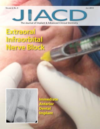
- 37. Negm Background: The application of infraorbital nerve block is often to accomplish regional anesthesia of the face. The procedure offers several advantages over local tissue infiltra- tion. A nerve block often achieves anesthe- sia with a smaller amount of medication than is required for local infiltration. In addition, unlike local tissue infiltration, blocks can pro- vide anesthesia without causing tissue distor- tion. In this article I will discuss the structure of anesthesia, approach of the technique, indi- cation, contraindication and others. The aim of this study was to describe a technique for extraoral infraorbital nerve block using several anatomical points for reference, simplifying the procedure and enabling greater success and a more rapid learning curve. Effective pain control in Dentistry may be achieved by local anesthetic techniques. The success of the anesthetic technique for nerve block in anterior region depends on the proximity of the needle tip to the infraorbital foramen at the moment of anesthetic injection into it. Two techniques are available to reach the infraorbital nerve, namely extraoral and intraoral; these tech- niques differ in the place where we should start. Methods: The application of extraoral infraor- bital nerve block in implant for anterior region has a superior effect rather than the intra- oral approach. A nerve block often achieves anesthesia with a smaller amount of medica- tion than is required for local infiltration. In addition, unlike local tissue infiltration, blocks can provide anesthesia without causing tis- sue distortion. And as we mention before that successful infraorbital nerve block provides anesthesia for the area between the lower eyelid and the upper lip including the anterior part of the alveolar ridge at the side of inner- vation so we can perform the implant surgery effectively with more comfort to the patients. You should pay attention while using this tech- nique because the needle is in very close prox- imity to the facial artery. So that, avoid adding vasoconstrictors to the anesthetic agent. Results: The obtained data showed 100% success rate of anesthe- sia injection for all cases we treated. Conclusions: This method is potentially effective for extraoral infraorbital nerve block, especially in Geriatric Dentistry. The Application of Extraoral Infraorbital Nerve Block Compared to the Intraoral Approach Dr. Shady A. M. Negm1 1. Dentist at Ministry of Health, Fellow of Alexandria Oral Implantology Association (A.O.I.A), Fellow of Egyptian Society of Oral Implantology (E.S.O.I) Abstract KEY WORDS: Extraoral infraorbital nerve block, intraoral infraorbital nerve block, local anesthesia The Journal of Implant Advanced Clinical Dentistry • 35
- 38. 36 • Vol. 6, No. 4 • July 2014 INTRODUCTION In general, regional anesthesia is ideal when the area of interest is innervated by a single superficial nerve. The infraorbital nerve sup- plies sensory innervation to the lower eyelid, the side and ala of the nose, and the upper lip. Since the infraorbital nerve provides a consid- erably large area of sensory innervation, it is a prime candidate for a regional nerve block. A successful infraorbital nerve block provides anesthesia for the area between the lower eye- lid and the upper lip including the anterior part of the alveolar ridge at the side of innervation. The cranial nerve V (trigeminal nerve), pro- vides sensory innervation to the face. The sec- ond division, the maxillary nerve (V2), exits the skull from the foramen rotundum. After giving off numerous branches, the maxillary nerve even- tually enters the face through the infraorbital canal, where it ends as the infraorbital nerve. The infraorbital nerve supplies sensory branches to the lower eyelid, the side of the nose, and the upper lip including the anterior part of the alveolar ridge at the side of innervation. Indications for infraorbital nerve blocks include wound closure, pain relief, and anesthesia for debridement. Contraindica- tions for infraorbital nerve block include any allergy or sensitivity to the anesthetic agent, evidence of infection at the infection site, distortion of anatomical landmarks, and unco- operative patients. The disadvantage of the infraorbital nerve block is that it is performed by blind palpation of the infraorbital foramen. Technique for extraoral infraorbital nerve block During the extraoral technique, the needle is in very close proximity to the facial artery. Because of this proximity, avoid adding vasoconstrictors to the anesthetic agent. Use the landmarks to locate the infraorbital foramen. Prepare the skin overlying the infraorbital foramen with povidone iodine (Betadine) and sterile gauze. Using ster- ile technique, insert the needle through the skin, the subcutaneous tissue, and the muscle. Aspi- Figure 1: The chemical structure of local anesthetic agents. Negm
- 39. The Journal of Implant Advanced Clinical Dentistry • 37 rate to ensure the needle is not within a ves- sel. The facial artery and vein are very close to the needle in this position. Inject the anesthetic solution. The infiltrated tissue appears swollen. Firmly massage this area for 10-15 seconds. Complications that may occur from the extraoral infraorbital injection technique include bleed- ing, hematoma, allergic or systemic reaction to the anesthetic agent, infection, unintentional injection into artery or vein, failure to anesthe- tize, nerve damage, and swelling of the eye lid. CASE PRESENTATION These two Egyptian female patients came to my clinic seeking for implant to restore the miss- ing teeth in anterior segment of the alveolar ridge. I decided to use the extraoral infraorbital nerve block rather than infiltration intraorally. The technique as I mention before is pain- less and less traumatic. The anesthesia was very effective and I complete the surgery with- out any problems. The following figures show- ing the application of the anesthesia: Figure [2] showing the first patient with right extraoral infraorbital injection; Figure [3] showing the first patient with left extraoral infraorbital injec- tion; Figure [4] showing the second patient with left extraoral infraorbital injection; Fig- ure [5] showing the upper anterior region of first patient in where surgery was performed during the implant surgical procedure; Fig- ure [6] showing the upper left anterior region of second patient in where surgery was per- formed during the implant surgical procedure. DISCUSSION The majority of local anesthetic agents share the same basic chemical structure, which con- sists of an aromatic ring, linked to an interme- diate chain (The intermediate chain between the aromatic and hydrophilic segments is either an ester or an amide), linked to a hydrophilic amine segment as seen in Figure 1. The chemi- cal structure of this intermediate group classi- fies the agent into the amide group or the ester group. This structural difference determines Figure 2: The first patient with right extraoral infraorbital injection. Figure 3: The first patient with left extraoral infraorbital injection. Negm
- 40. 38 • Vol. 6, No. 4 • July 2014 Negm Figure 4: The second patient with left extraoral infraorbital injection. Figure 5: The upper anterior region of first patient in where surgery was performed during the implant surgical procedure. Figure 6: The upper left anterior region of second patient in where surgery was performed during the implant surgical procedure. the pathway by which the agent is metabolized and its allergic potential. The toxicities associ- ated with local anesthetics are cardiovascular and neurologic. Methemoglobinemia can occur with use of these agents, though it is rarely clinically significant. An infraorbital nerve block requires 1-3 mL of the chosen anesthetic agent. Lidocaine (Xylocaine) is the most commonly used agent. The onset of action for lidocaine is approximately 4-6 minutes. The duration of effect is approximately 75 minutes, which is suf- ficient enough to complete the implant surgery without needs of giving anesthesia again. ● Correspondence: Dr. Shady A. M. Negm 15 Elahwazy st., Elwardian Alexandria, Egypt +201002944111 smngm@msn.com Disclosure The author declare that he has no financial or personal relationship(s) that may have inappropriately influenced him in writing this article References 1. Gray H, Lewis WH. The trigeminal nerve. Gray’s Anatomy of the Human Body. Bartleby.com. Accessed May 16, 2008. 2. Amsterdam JT, Kilgore KP. Regional anesthesia of the head and neck. In: Roberts JR, Hedges JR, eds.Clinical Procedures in Emergency Medicine. 4th ed. Philadelphia, Pa: WB Saunders Company; 2004:552-66. 3. Crystal CS, Blankenship RB. Local anesthetics and peripheral nerve blocks in the emergency department.Emerg Med Clin North Am. May 2005;23(2):477-502. 4. Trott AT. Wounds and Lacerations: Emergency Care and Closure. 2nd ed. St. Louis, Mo: Mosby; 1997. Negm