Transtornos de desarrollo cortical 2016
•Descargar como PPSX, PDF•
3 recomendaciones•921 vistas

MASTERCLASS: NEUROLOGÍA HOSPITAL NACIONAL DANIEL ALCIDES CARRIÓN

Recomendados
Más contenido relacionado
La actualidad más candente
La actualidad más candente (19)
Similar a Transtornos de desarrollo cortical 2016
Similar a Transtornos de desarrollo cortical 2016 (20)
Último
Último (20)
Transtornos de desarrollo cortical 2016
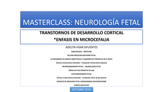
- 1. MASTERCLASS: NEUROLOGÍA FETAL TRANSTORNOS DE DESARROLLO CORTICAL *ENFASIS EN MICROCEFALIA ADELITA HÍJAR SIFUENTES GINECÓLOGA – OBSTETRA FELLOW MEDICINA MATERNO FETAL ULTRASONIDO EN GINECO OBSTETRICIA Y DIAGNÓSTICO PRENATAL DE III NIVEL (Clínica Universitaria Colombia – Fundación Universitaria Dexeus) NEUROSONOGRAFÍA FETAL – NEUROLOGÍA FETAL (Maternal Fetal Medicine Group) ECOCARDIOGRAFÍA FETAL (Clínica Universitaria Colombia – Fundación Clinic de Barcelona) SERVICIO DE MEDICINA FETAL HONADOMANI SAN BARTOLOMÉ CLÍNICA SAN FELIPE OCTUBRE 2016
- 2. TRANSTORNOS DEL DESARROLLO CORTICAL • El desarrollo de la corteza cerebral fetal y la formación de giros y sulcos es un proceso ordenado que se superpone con hitos bien definidos. • Este proceso incluye: • Proliferación de neuronas y células gliales • Migración desde la zona periventricular • Organización de neuronas
- 3. TRANSTORNOS DEL DESARROLLO CORTICAL • Injurias durante este proceso produce un amplio rango de malformaciones corticales que podrían devenir en déficit neurológico y/o convulsiones. • Las malformaciones del desarrollo cortical podrían ser debido a: • Factores genéticos • Lesiones prenatales • Trauma • Infecciones • Teratógenos
- 5. • The cerebral cortex is a modular structure: modules of neurons are induced in a neuroepithelial sheet and subsequently differentiate, migrate and organize into a functioning cerebral cortex. • Neuronal induction is regulated by interplay between intrinsic genetic mechanisms and extrinsic information relayed to cortex by thalamocortical input and other, largely unknown, factors. • Although details of the neural cell proliferation differ among mammalian species.
- 6. HISTOLOGÍA: CORTEZA CEREBRAL FETAL
- 9. Group I: malformations secondary to abnormal neuronal and glial proliferation or apoptosis • Groups I.A and III.D: microcephaly • Group I.B: megalencephalies • Group I.C: cortical dysgeneses with abnormal cell proliferation Group II: malformations due to abnormal neuronal migration A. Lissencephaly/subcortical band heterotopia. B. Cobblestone complex/ congenital muscular dystrophy syndromes. C. Heterotopia: subependymal, subcortical, marginal. Group III: malformations secondary to abnormal postmigrational development A. Polymicrogyria and schizencephaly. B. Cortical dysplasia without balloon cells. C. Microdysgenesis.
- 10. SIGNOS ECOGRÁFICOS DE MDC
- 11. TRANSTORNOS DE PROLIFERACIÓN: MICROCEFALIA • Antenatally is even more difficult, because affected fetuses have usually normal cranial measurements in early gestation, that only decline in late gestation or after birth. • The incidence is estimated to be 1.6 per 1000 singlebirth deliveries. Only 14 % of all microcephalic infants diagnosed by the first year of age had been detected at birth. • The available literature suggests that the risk of mental retardation with an head circumference between – 2 and – 3 SDs is in the range of 10-30%, rising to 50-60% for measurements below – 3 SDs.
- 15. G.PILU
- 16. Daniel-Spiegel et al—Establishment of Fetal Biometric Charts Using Quantile Regression Analysis. J Ultrasound Med 2013; 32:23–33 | 0278-4297
- 18. 24ss
- 19. TRANSTORNOS DE PROLIFERACIÓN: MACROCEFALIA • Head circumference above the 98th percentile or more than 2 standard deviations above the mean. • Macrocephaly is relatively frequent in children and adults and is a common cause for genetic consultation. • The diagnosis assumes that other obvious causes of head enlargement, such as hydrocephalus and cerebral tumors, have been excluded. Usually, macrocephaly is familial and benign and frequently it manifests only late in pregnancy or after delivery.
- 20. TRANSTORNOS DE PROLIFERACIÓN: MACROCEFALIA
- 21. 28ss
- 23. TRANSTORNOS DE PROLIFERACIÓN: HEMIMEGALENCEFALIA • Sobrecrecimiento de un hemisferio cerebral Aislado (50%) o asociado a síndromes neurocutáneos. • Clínica: Convulsiones Hemiparesia Retraso mental Hemihipertrofia en resto del cuerpo
- 24. Unilateral hypertrophy of the affected hemisphere, often associated with ipsilateral ventricular dilatation and midline shift. May be hemihypertrophy of the cerebellum, brainstem and body. Thought to occur as a result of an insult to the germinal matrix between 8 and 16 post menstrual weeks of gestation. May find focal increased echogenicity of the affected hemisphere. No abnormal karyotypic abnormality has been described.
- 27. TRASNTORNOS DE MIGRACIÓN: LISENCEFALIA Lissencephaly, type I is characterized by agyria with or without pachygyria, a wide cortical mantle and minimal or no hydrocephalus. Four layer cortex. · Miller-Dieker syndrome (17p13 deletion) has lissencephaly combined with dysmorphic facial features and other possible associated anomalies. · Norman-Roberts syndrome · Isolated type I lissencephaly Lissencephaly, type II. Vascular bundles and fibroglial tissue are present in the cortex and subarachnoid space. Lissencephaly, type II typically has hydrocephalus and additional serious central nervous system defects. It is usually part of a syndrome. · HARD+/-E syndrome, an acronym for Hydrocephalus, Agyri, Retinal dysplasia, Encephalocele (Walker-Warburg syndrome) · COMS (Cerebro-oculomuscular syndrome) · Other subtypes of type II lissencephaly are possible Lissencephaly is found in Neu-Laxova syndrome, lethal autosomal recessively inherited disorder consisting of growth retardation, microcephaly, lissencephaly, corpus callosum agenesis, intracranial calcifications, cerebellar hypoplasia, facial dysmorphism, microophthalmia, exophthalmus, cataracts, absent eyelids, hydrops, ichthyosis, contractures of extremities and syndactyly.
- 28. 27 ss • Malformación severa de corteza cerebral, que resulta de falla en migración neuronal durante el 3° y 4° mes de gestación. • Clínica: Retraso psicomotor severo Retraso del desarrollo Convulsiones Retraso en crecimiento • Este defecto acarrea un mal pronóstico del RN, con RM y alta tasa de recurrencia, si está ligado a defectos genéticos.
- 30. TRANSTORNOS DE MIGRACIÓN: HETEROTOPIA (CORTICAL – PERIVENTRICULAR)
- 33. 27ss
- 34. 26ss
Notas del editor
- Figure 2 Patient 15. (a) Midsagittal transvaginal ultrasound image at 28 weeks showing thick and echogenic corpus callosum (arrows), and large cavum septi pellucidi (CSP) and third ventricle (3V). Inset (b) Normal corpus callosum at the same gestational age. (c) Midsagittal magnetic resonance image taken 2 days later showing the abnormal callosal genu and splenium (arrows). Note that the body of the corpus callosum is not well depicted in this image (small arrow). The serrated pattern of the parietal cortex (arrowheads) is indicative of the presence of polymicrogyria.
- Figure 3 Patient 5, hemimegalencephaly. (a) Axial transabdominal ultrasound image at 28 weeks showing asymmetrical brain hemispheres with unilateral ventriculomegaly, and abnormal cortex and Sylvian fissure (arrow). (b) Sagittal plane obtained during the same examination showing frontal bossing and thick corpus callosum (arrows). (c) Fetal and (d) neonatal magnetic resonance images showing the abnormal cortex of both hemispheres and thick frontal cortex with polymicrogyria (arrows).
- A, Axial SS-FSE T2-weighted image in a fetus at gestational week 27 demonstrates multiple abnormal infoldings of the developing cortex (white arrow) for expected gestational age, consistent with polymicrogyria. Areas of cystic encephalomalacia with hemorrhage (black arrow) are also seen. B, Low signal intensity consistent with intraventricular hemorrhage is also seen layering in the temporal horns bilaterally (arrowhead). Fetus was referred for ventriculomegaly and choroid plexus cysts detected on prenatal sonogram.