
Recomendados
Más contenido relacionado
Similar a Upper GI tumors - surgery 1.30.02 PM.pptx
Similar a Upper GI tumors - surgery 1.30.02 PM.pptx (20)
Último
Último (20)
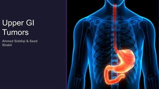
Upper GI tumors - surgery 1.30.02 PM.pptx
- 2. Parts of the upper GI tract
- 4. Case Presentation A 65yr old male presented in OPD with chief complains of progressive dysphagia, weight loss and occasional chest pain. Patient is a known case of GERD since 2 years, along with a 40-pack year smoking hx and heavy alcohol consumption. He is a retired construction worker, exposed to carcinogens, and has a sedentary/unhealthy lifestyle. On examination, patient is pale appearing and has lost over 10kgs over the last few months. Upper GI endoscopy reveals a circumferential ulcerated mass in mid-esophagus & CT scan reveals esophageal wall thickening
- 8. Treatment
- 9. Esophageal Carcinomas It is the 6th most common cancer in the world Mostly presents in mid to late adulthood Has a poor prognosis, only 5-10% of those diagnosed will survive beyond 5 years
- 10. n
- 12. Sign & Symptoms
- 13. ENDOSCOPY – 1st line BIOPSY – for histology & cytology is crucial for accurate diagnosis BARIUM SWALLOW CT/MRI BRONCHOSCOPY LAPROSCOPY ENDOSCOPIC US and/or PET SCAN Investigations
- 14. • Esophageal CA is almost always diagnosed by endoscopic biopsy • Endoscopy should be performed in every patient with dysphagia, even if the barium study is suggestive of a motility disorder • Contrast-enhanced CT scan of the chest and abdomen, and PET scan should be done to assess for distant metastasis • If there is no evidence of distant metastatic disease, endoscopic US should be performed to assess T-stage and regional lymph nodes involvement
- 15. Newer modalities of evaluation • Flow cytometry • P53 immunohistochemistry • Optical coherence tomography • Spectroscopy
- 16. Mid-esophageal proliferative SCC Adenocarcinom a of the lower esophagus Mid-esophageal SCC Adenocarcinom a of the cardia
- 17. NICE Guidelines for oesophagogastric cancer - NG83 • Offer PET-CT for esophageal and gastro-esophageal junctional tumors that are suitable for radical treatment • Do not use endoscopic ultrasound only to distinguish between T2 and T3 tumors • Only offer endoscopic ultrasound when it will help guide ongoing management • Only consider staging laparoscopy when it will help guide ongoing management • Offer HER2 testing in metastatic esophagogastric adenocarcinoma • Offer endoscopic resection for staging in people with suspected stage 1 esophageal adenocarcinoma
- 18. TNM Staging Tis High grade dysplasia T1 Tumor invading lamina propria or submucosa T2 Tumor invading muscularis propria T3 Tumor invading beyond muscularis propria T4 Tumor invading adjacent structures Tx Primary tumor cannot be assessed N0 No regional lymph node metastases N1 Regional lymph node metastases Nx Lymph nodes cannot be assessed M0 No distant metastases M1(a) Celiac node involved (distal tumors) Supraclavicular node involved (proximal tumors) M1(b) Celiac or supraclavicular node involved if not remote from tumor site (i.e. not 1a) All distant metastases Mx Distant metastases cannot be assessed.
- 20. Treatment
- 21. Treatment • Stage 1: Esophagectomy with gastric pull-up • Stage IIa: Esophagectomy with gastric pull-up • Stage IIb: Neoadjuvant chemoradiation, restaging, and esophagectomy with gastric pull-up • Stage III: Neoadjuvant chemoradiation, restaging, and esophagectomy with gastric pull-up • Stage IV: Chemoradiation and esophageal stent or palliative therapy
- 25. Palliative Care
- 26. Palliative Procedures 1. External or intraluminal radiotherapy (brachytherapy) 2. Chemotherapy 3. Intubation tube 4. Endoscopic therapy • Self-expanding metal stents • Endoscopic laser • Endoscopic photodynamic therapy 5. Paliative surgeries
- 27. • Fan et al., conducted a meta-analysis of 27 phase-II/III clinical trials, incorporating a total of 815 patients, with resectable stage I-IV esophageal CA. • The main objective of this meta-analysis was to systematically assess and provide the most updated and comprehensive evidence regarding the safety & efficacy of neoadjuvant immunotherapy combined with chemotherapy, in patients with resectable esophageal CA. • The primary outcomes were the pathological complete response (pCR) rate and the major pathological response (MPR) rate. Secondary outcomes were treatment-related severe adverse events. • pCR: 31.4% (95% CI: 27.6-35.3) & MPR: 48.9% (95% CI: 42.0-55.9). • Severe adverse events: 26.9% (95% CI: 16.7-38.3) • The study demonstrated promising clinical & safety outcomes with neoadjuvant therapy combined with chemo in patients with resectable esophageal CA. Supports the prospective wide application of this treatment option.
- 29. Types of gastro oesophageal junction cancer • Type 1 Type 1 GOJ cancer spreads down into the gastro oesophageal junction from above. So, the cancer cells are in the lower part of the oesophagus and the gastro oesophageal junction. The cancer’s centre is between 1 and 5 cm above the junction. • Type 2 Type 2 GOJ cancers develop at the actual gastro oesophageal junction. The cancer's centre is between 1 cm above and 2 cm below the junction. • Type 3 Type 3 GOJ cancer spreads up into the gastro oesophageal junction from below. So there are cancer cells in the top of the stomach and the gastro oesophageal junction. The cancer’s centre is between 2 and 5 cm below the junction.
- 31. Risk Factors
- 32. Risk Factors There are different risks factors for each type of GOJ cancer. • Type 1 Type 1 GOJ cancers are similar to oesophageal cancers. Barrett's oesophagus increases your risk of type 1 GOJ cancer. This is a condition where the cells lining your oesophagus have become abnormal. This can happen due to long term acid indigestion (acid reflux). • Type 2 We don't have such a good understanding of what causes type 2 cancers. Type 2 characteristics are somewhere between stomach and oesophageal cancer cells. • Type 3 Type 3 GOJ cancers are similar to stomach cancers. They are linked to infection with Helicobacter pylori (H.pylori). H. pylori is a bacteria that lives in the mucous of the lining of the stomach.
- 34. GEJ Cancer Types
- 35. Lymphatic spread
- 36. Diagnosis
- 37. Endoscopic picture A view through the endoscope (Fig A) shows an ulcerated mass in the distal esophagus at the gastroesophageal (GE) junction, which appears malignant. It is arising from abnormal, pink mucosa (Barrett’s mucosa)
- 38. Radiographic imaging Fig A: Radiographs from a double-contrast upper gastrointestinal series show an eccentric filling defect in the anterolateral portion of the distal esophagus Fig B: There is irregularity of the distal esophageal wall, a finding consistent with severe erosive changes, probably from reflux. Fig C: Axial CT of the upper abdomen (without the administration of intravenous contrast material shows thickening of the right anterolateral wall of the distal esophagus (arrow), corresponding in location to the mass seen on the upper gastrointestinal study. Fig D: There is an enlarged (1.0 cm) lymph node in the gastrohepatic ligament
- 39. Fig: Adenocarcinoma at the gastroesophageal junction with central depression shown by asterisk.
- 40. Treatment
- 41. Surgery
- 42. Esophagectomy
- 43. Gastrectomy
- 44. Chemotherapy
- 45. Radiotherapy
- 46. Targeted therapy
- 47. Prognosis
- 48. Research