
Recomendados
Más contenido relacionado
La actualidad más candente
La actualidad más candente (20)
Similar a APOPTOSIS by dr Alfarah.pptx
Similar a APOPTOSIS by dr Alfarah.pptx (20)
Último
Último (20)
APOPTOSIS by dr Alfarah.pptx
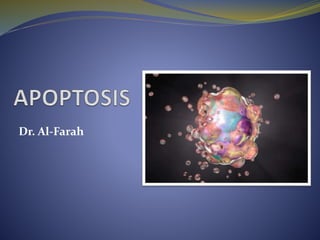
- 1. Dr. Al-Farah
- 2. Learning objectives Discuss causes, morphological and biochemical changes, clinic-pathologic correlations in Apoptosis. Summarize the pathways of apoptosis.
- 4. APOPTOSIS Programmed, enzyme-mediated cell death Apoptosis is a type of cell death that is induced by a tightly regulated suicide program in which cells destined to die activate intrinsic enzymes that degrade the cells’ genomic DNA and nuclear and cytoplasmic proteins.
- 6. APOPTOSIS 1) Cell is broken into apoptotic bodies (morphologic appearance of membrane-bound fragments derived from cells, and named after the Greek designation for “falling off.”). 2) In which membrane remain intact. 3) It become tasty for phagocytosis. 4) Cell death does not illicit inflammatory reaction.
- 7. APOPTOSIS: Purpose/Causes Apoptosis occurs in two broad contexts: As a normal physiologic processes, and As a pathophysiologic processes of cell loss in many different diseases.
- 8. Apoptosis in Physiologic Situations Death by apoptosis is a normal phenomenon that serves to eliminate cells that are no longer needed, or as a mechanism to maintain a constant number of various cell populations in tissues. In the average adult between 50 and 70 billion cells die each day by apoptosis.
- 9. Apoptosis in Physiologic Situations 1) Removal of supernumerary cells (in excess of the required number) during development. EXAMPLE: Apoptosis during embryogenesis i.e., implantation, angiogenesis, organogenesis, developmental involution and metamorphosis. 2) Involution of hormone dependent tissue upon hormone withdrawal EXAMPLE: menstruation, regression of lactating breast after weaning, prostatic atrophy after castration. 3) Cell turnover in proliferating cells EXAMPLE: - Immature lymphocytes in bone marrow and thymus that fail to express useful antigen receptor. - Epithelial cells in intestinal crypts to maintain constant number. - B Lymphocytes in germinal center.
- 10. 4) Elimination of potentially harmful self-reactive lymphocytes EXAMPLE: Induction of Tolerance in Thymus or periphery 5) Death of host cells that have served their useful purpose EXAMPLE: Neutrophils in an acute inflammatory response, and lymphocytes at the end of an immune response. Apoptosis in Physiologic Situations
- 11. Apoptosis in pathologic conditions Apoptosis eliminates cells that are injured beyond repair without eliciting a host reaction
- 12. Apoptosis in pathologic conditions 1) DNA damage: If repair mechanism for damaged DNA cannot cope with the injury, the cell’s intrinsic mechanism induces apoptosis. EXAMPLE: Radiation and cytotoxic anticancer drugs can damage DNA 2) Accumultaion of misfolded protein: Cell death triggered by improperly folded intracellular proteins and the subsequent endoplasmic reticulum (ER) stress response.
- 13. Apoptosis in pathologic conditions 3) Cell death in certain infections: EXAMPLE: a) In viral infections: loss of infected cells by apoptosis induced by virus (adenovirus and HIV). b) By host immune response: In viral hepatitis through cytotoxic cells. c) By cytotoxic lymphocytes: In tumor and cellular rejection of transplant same T- cell mediated (cytotoxic T- cell ). 4) Pathologic atrophy in parenchyma of organ after duct obstruction EXAMPLE: Duct obstruction by stone in pancreas, parotid gland and
- 14. Apoptosis: Morphologic changes 1) Cell shrinkage: Cell size is reduced, the cytoplasm is dense (organelles tightly packed) and eosinophilic. 2) Chromatin condensation: Most characteristic feature. Aggregate peripherally under the nuclear membrane into dense masses of various shapes and sizes. Nucleus may breakup into two or more fragments.
- 16. 3) Formation of cytoplasmic bleb and apoptotic bodies. 4) Phagocytosis of apoptotic bodies by macrophages. Plasma membrane remain intact during apoptosis until last stage. High speed and efficiency. Does not illicit inflammation. Apoptosis: Morphologic changes
- 17. APOPTOSIS: Formation of cytoplasmic bleb
- 18. Summary of Morphological events Cells shrink and become detached from adjoining cells Cytoskeleton collapses. Mitochondria remain intact. Plasma membrane develops bubbles (blebs) on surface. Nucleus and chromatin condense. Chromatin aggregates at periphery of nucleus Nuclear envelope disintegrates DNA fragmentation Budding off and breakage into small membrane wrapped fragments - apoptotic Bodies
- 19. The three main players Regulating gene - p53 gene Protein family - Bcl proteins Family of enzymes - Caspases Common intracellular machinery for apoptosis
- 20. Apoptosis: p53 gene and p53 protein • p53 is tumour suppressor gene • Active gene product p53 produced in response to DNA and cell damage • Prevents cell completing cell cycle • If damage is minor - allows repair • If major - induces apoptosis • Complex mechanisms
- 21. Apoptosis: BCL gene family Large family of proteins There are more than 20 members of the BCL family, which can be divided into three groups based on their function and the BCL2 homology (BH) domains they possess. 1. Anti-apoptotic 2. Pro-apoptotic 3. Sensors (Regulated apoptosis initiators)
- 23. Apoptosis: BCL family Anti-apoptotic Cell survival stimuli induce the expression of anti-apoptotic BCL proteins. BCL2, BCL-XL, and MCL1 are the principal members. Possess four BH domains (called BH1-4). BCL2 was the first apoptosis-related gene that was recognized to play a role in tumorigenesis, and indeed, BCL-2 is overexpressed in a variety of cancers, contributing to cancer cell survival through direct inhibition of apoptosis. Its product is an integral membrane protein (called Bcl-2) located in the membranes of the endoplasmic reticulum (ER), nuclear envelope, and in the outer membrane of the mitochondria. BCL-2 proteins maintain mitochondrial membrane integrity and prevent leakage of mitochondrial proteins that can trigger apoptosis (e.g., cytochrome c).
- 24. BAX and BAK – are the principal members. Possess the first three BH domains (BH1-3) Activated by damage to DNA, misfolded proteins, viral infections, and other injurious events. On activation, BAX and/or BAK oligomerize within the outer mitochondrial membrane and enhance its permeability through forming channels in the mitochondrial membrane that cause leakage of cytochrome c into the cytosol. Apoptosis: BCL family Pro-apoptotic
- 25. BAD, BIM, BID, Puma, and Noxa – are the principal members. Possess only one BH domain, the third of the four BH domains, and hence called BH3-only proteins. Sensors of cellular stress and damage; when upregulated and activated, they can initiate apoptosis. Apoptosis: BCL family Sensor genes
- 27. Apoptosis: Caspases Caspases term is derived from cysteine-dependent aspartate-specific proteases (enzymes). Caspases exist in inactive form , they must undergo cleavage to become active. 10 members – divided into two groups: 1. Inflammatory caspases: 1, 4 and 5 No role in Apoptosis 2. Initiator caspases : 2, 8, 9 and 10 Provide a link between cell signalling and apoptotic execution by activating executioner caspases 3. Executioner caspases: 3, 6, 7 Activated caspases has proteolytic activity, resulting in morphological and biochemical markers of apoptosis. Targets of Executioner caspases: nuclear lamins, DNA (endonucleases), cytoskeleton, cell-cell adhesion molecules.
- 28. Apoptosis: Caspases Granzyme B released by cytotoxic T lymphocytes which is known to activate caspase-3 and -7. Death receptors (like FAS, TRAIL receptors and TNF receptor) which can activate caspase-8 and -10; and Apoptosome, regulated by cytochrome c and the Bcl-2 family, which activates caspase-9.
- 30. Regulation of apoptosis Factors controlling apoptosis thus include substances outside the cell and internal metabolic pathways: Inhibitors include growth factors, extracellular cell matrix, sex steroids, some viral proteins. Inducers include growth factor withdrawal, loss of matrix attachment, glucocorticoids, some viruses, free radicals, ionising radiation, DNA damage, ligand-binding at ‘death receptors’.
- 31. Apoptosis results from the activation of enzymes called Caspases. The process of apoptosis may be divided into an 1. Initiation phase Initiation occur through two distinct pathways (stimuli) which involve different sets of protein. Control and integration of proapoptotic and antiapoptotic signals. Selected caspases become catalytically active and unleash a cascade of other caspases 2. Execution phase During which the terminal caspases trigger cellular fragmentation. 3. Removal of Dead Cells (Phagocytosis) Apoptosis: Mechanism
- 32. Apoptotic transduction pathways There are two distinct pathways that converge on caspase activation: Mitochondrial or intrinsic pathway Death activator or extrinsic pathway The two pathways of apoptosis differ in their induction and regulation, and both culminate in the activation of caspases.
- 34. Apoptosis: Mechanism Initiation phase: Mitochondrial pathway Death Signals Growth factor withdrawal DNA damage (by radiation, UV lights, X-rays, Chemotherapeutic drugs, toxins, free radicals) Protein misfolding (ER stress)
- 35. Apoptosis: Mitochondrial or intrinsic pathway Mitochondrial pathway – is the major mechanism of apoptosis Resulted from increased mitochondrial permeability and release of pro apoptotic molecules into the cytoplasm. Proteins in mitochondria as Cytochrome C (Essential of life). Protein in cytoplasm (released into cytoplasm) – initiate suicide program of apoptosis. Release of mitochondrial protein controlled by – balance between the proapoptotic and antiapoptic members of BCL2 Family In order to understand the cascade of events, an understanding of the BCL family of genes is important.
- 36. Apoptosis: Mitochondrial or intrinsic pathway When a cell is deprived of critical survival signals or their DNA is damaged or misfolded proteins, sensors of damage or stress are activated. These sensors proteins are also BCL proteins i.e., include BIM, BID & BAD. This sensor proteins in-turn activate the pro-apototic factors: BAK & BAX. Activation of BAX and BAK genes produces protein channels in the mitochondrial membrane that cause leakage of cytochrome C into the cytosol. Once Cytochrome C released into the cytoplasm it binds with APAF (apoptotic protease activating factor) to form an apoptosome. Apoptosome formed causes the activation of an initiator caspase (Caspase-9), which in turn activates effector caspases (proteases, endonucleases) that mediate the execution phase.
- 39. Apoptosis: Mechanism Death activator or extrinsic pathway This pathway is initiated by engagement of plasma membrane death receptors. Death receptors are members of the tumor necrosis factor (TNF) receptor family that contain a cytoplasmic domain (called death domain) involved in protein protein interactions. Best known death receptors are Type 1 TNF receptor (TNFR1) activated by Tumour necrosis factor alpha (TNF-α) Fas (CD95) activated by Fas ligand (CD95) TNF-α is an important cytokine that is involved in systemic inflammation, autoimmune disease, and wasting (cachexia) in cancer. It is primarily produced by macrophages; however, it can also be produced by T cells, mast cells, endothelial cells, cardiac cells, and neurons, which explains its multiple disease associations. FasL is expressed on T cells that recognize self antigens and on some Cytotoxic lymphocytes that kill virus-infected and tumor cells. o Apoptosis can also be induced by cytotoxic T-lymphocytes using the enzyme granzyme.
- 40. Apoptosis: Mechanism Death activator or extrinsic pathway Binding of ligand to its specific death receptor is a pro-apoptotic signal. Signals linked to execution phase through an integration stage.
- 41. Apoptosis: Mechanism Death activator or extrinsic pathway FAS ligand bind to FAS receptor Form cross link between three or more FAS receptor (Death inducing signal complex) cytoplasmic death domains form a binding site for an adaptor protein called FADD (Fas-associated death domain) FADD binds inactive caspase-8 (or caspase- 10) in turn recruit and activate caspase-8 Active caspase-8 initiates the executioner caspase sequence which mediate the execution phase of apoptosis
- 42. Apoptosis: Mechanisms Execution Phase Here the 2 initiating pathways converge to a cascade of caspase activation which mediates the final phase of apoptosis. The intrinsic mitochondrial pathway activates the initiator caspase-9, whereas The extrinsic death receptor pathway activates caspase-8 and caspase-10. Active forms of these caspases trigger the rapid and sequential activation of the executioner caspases, such as caspase-3 and caspase-6, which then act on many cellular components.
- 43. Apoptosis: Mechanisms of Apoptosis Execution Phase Activated effector caspases lead to ... Digestion of cytoskeleton proteins Nucleus and chromatin degradation Plasma membrane changes Executioner caspases cause inactivation of DNA repair enzymes, activation of DNAse that degrade DNA and nucleoproteins. Caspases also degrade components of the nuclear matrix and cytoskeleton, leading to fragmentation of cells.
- 44. Plasma Membrane alteration and recognition by phagocytes: Normally, Enzyme system keeps phospholipids (phosphotidylserine) on inner surface During apoptosis Phosphotidylserine “flips” out and is expressed on the outer layer of the membrane. Macrophage receptors recognise and bind Phosphotidylserine followed by engulfment of apoptotic bodies. This process of apoptotic cell phagocytosis is called Efferocytosis Cells that are dying by apoptosis also secrete soluble factors that recruit phagocytes. Apoptosis: Mechanisms of Apoptosis
- 47. Apoptosis: Mechanisms of Apoptosis Lets see the video
- 49. Disorders Associated with Dysregulated Apoptosis Dysregulated apoptosis (“too little or too much”) has been postulated to explain aspects of a wide range of diseases. 1. Disorders associated with defective apoptosis and increased cell survival. Malignancy (mutation of TP53 leading accumulation of mutations) Autoimmune disorders (failure to eliminate potentially harmful cells, such as lymphocytes that can react against self antigens, and failure to eliminate dead cells, a potential source of self antigen) Congenital anomalies Failure of apoptosis in these four sites is a factor in the development of syndactyly (webbed fingers), cleft palate, spina bifida, and bladder diverticulum (pouch) or fistula (open connection) from the bladder to the umbilical skin, respectively.
- 50. • BAX mutation in tumor cells • p53 mutation FLIP resembles initiator caspases, but lacks proteolytic domain, compete with caspase 8 and caspase 10 for binding site in the Death inducing signal complex Inhibitor of apoptosis (IAP) proteins block the inappropriate activation of caspases, including executioners like caspase-3, and keep cells alive. Mainly act on the intrinsic pathway Defective apoptosis and increased cell survival.
- 51. Disorders Associated with Dysregulated Apoptosis 2. Disorders associated with increased apoptosis and excessive cell death. o Neurodegenerative diseases (over-activation of some caspases such as caspase-3) o Ischemic injury and o Death of virus-infected cells in many viral infections
- 52. Review question 1 The apoptosis is classified as a) Programmed cell death b) Non-programmed cell death c) Accidental cell death d) Mitotic cell death
- 53. Review question 2 In which of the following situations would cells die by necrosis, not apoptosis? a) Removal of virus infected cells. b) Removal of developing neurons which fail to make profitable connections with other cells. c) Removal of heart muscle cells damaged by oxygen depletion following cardiac infarction. d) Removal of cells with damaged DNA which cannot be repaired.
- 54. Review question 3 Apoptosis can’t kill which of the following? a) Cell infected with viruses b) Cell with DNA damage c) Cancer cells d) Immune cells
- 55. Identify?
- 56. Review question 5 Apoptotic bodies can be recognized with the presence of these on the surface (a) phosphatidyl tyrosine (b) phosphatidylinositol (c) phosphatidylcholine (d) phosphatidylserine
- 57. Review question 6 Which of the following are killed by the extrinsic apoptosis pathway? a) Virus infected cells. b) Cells with damaged DNA. c) Developing nerve cells which fail to make profitable connections. d) Irradiated cells
- 58. Thank you