Osteoporosis
•Descargar como PPTX, PDF•
4 recomendaciones•1,022 vistas

Osteoporosis management in physiotherapy

Recomendados
Más contenido relacionado
La actualidad más candente
La actualidad más candente (20)
Similar a Osteoporosis
Similar a Osteoporosis (20)
Último
Último (20)
Osteoporosis
- 1. Osteoporosis
- 2. The bones in our skeleton are made of a thick outer shell and a strong inner mesh filled with collagen (protein), calcium salts and other minerals. The inside looks like honeycomb, with blood vessels and bone marrow in the spaces between bone. BONE •Normal bone on left •Osteoporotic bone on right
- 3. Literally translates as “porous bones” Osteoporosis occurs when the holes between bone become bigger, making it fragile and liable to break easily Osteoporosis - Definition “A progressive systematic skeletal disease characterized by low bone mass and micro-architectural deterioration of bone tissue, with a consequent increase in bone fragility and susceptibility to fracture”
- 4. WHO Definition;- Bone Mineral Density ≥2.5 SD’s below the mean seen in young normal subjects True Definition: Bone with lower density and higher fracture risk
- 6. Often called the “silent disease” Bone loss occurs without symptoms › First sign may be a fracture due to weakened bones › A sudden strain or bump can break a bone
- 7. Risk Factors • Chronic liver disease • Excessive secretion of cortisol (Cushing's syndrome) • Radiographic evidence of osteopenia or vertebral deformity • Previous fracture not caused by a major accident • Cancer • Significant loss of height or an abnormal bend in the upper spine (thoracic kyphosis) Risk factors that have the potential to be modified include: • Cigarette smoking • Excessive alcohol intake • Inactivity • Low body weight • Poor general health • Prolonged immobilization Risk factors that cannot be modified include: • Caucasian race • Advanced age • Female sex • Premature menopause (<45 years) • Prolonged time (>1 year) without a menstrual period Conditions associated with osteoporosis: • Anorexia nervosa • Malabsorption syndromes • Excessive secretion of parathyroid hormone • Excessive secretion of thyroid hormone • Post-transplantation • Chronic renal disease
- 8. Osteoporosis – Primary Causes •Osteoporosis results from an unhealthy imbalance between two normal activities of bone: bone resorption and bone formation. •These activities rely on two major types of cells: osteoclasts for bone resorption and osteoblasts for bone formation. The combined processes of bone resorption and bone formation allow the healthy skeleton to be maintained continually by the removal of old bone and its replacement with new bone. •These combined processes are referred to as bone remodeling or bone turnover. During the first 20-25 years of life, these processes are balanced.
- 9. Osteoporosis – Primary Causes •Following a period of balanced bone resorption and bone formation, the destruction of bone begins to exceed the formation of bone; this imbalance leads to a net loss of bone, and the beginnings of osteoporosis. •The risk of fracture increases from 1.5 to 3-fold for every 10% decrease in bone mass. •Bone mineral density (BMD), a measure of bone mass divided by bone area, increases with age until peak bone density is achieved. Bone mineral density is correlated highly with bone strength and is therefore used to quantitatively screen and diagnose patients.
- 10. “If somebody had told me sooner what I know now about osteoporosis, none of this might be happening to me!”
- 11. The most common breaks in weak bones are in the wrist, spine and hip.
- 12. After mid-30’s, you begin to slowly lose bone mass. Women lose bone mass faster after menopause. Men lose bone mass too.
- 13. Classification of Osteoporosis Primary osteoporosis in the elderly can be classified as type I or II Postmenopausal osteoporosis (type I) •Caused by lack of estrogen •Causes PTH to overstimulate osteoclasts •Excessive loss of trabecular bone •Associated with vertebral and Colles' (distal radius) fractures. Age-associated osteoporosis (type II) •Bone loss due to increased bone turnover •Malabsorption •Mineral and vitamin deficiency •Associated with vertebral and hip fractures.
- 14. may be due to many causes. Distinguishing secondary osteoporosis is important in patients of all ages, because many of the causes are treatable or have an important effect on prognosis
- 15. Pathogenesis • Diminished bone mass can result from: – failure to reach an optimal peak bone mass in early adulthood – increased bone resorption – decreased bone formation after peak bone mass has been achieved • All three of these factors probably play a role in most elderly persons. Low bone mass, rapid bone loss, and increased fracture risk correlate with high rates of bone turnover (i.e. resorption and formation). • In osteoporosis, the rate of formation is inadequate to offset the rate of resorption and maintain the structural integrity of the skeleton
- 16. (sometimes called juvenile osteoporosis) can be caused by certain medical conditions, genetic disorders, some medications, or lifestyle factors such as poor diet and lack of exercise. In rare cases, the condition has no cause and is known as idiopathic juvenile osteoporosis.
- 17. Osteoporosis in children may not cause any obvious problems to the child. However, signs and symptoms of osteoporosis can include: Pain in the lower back, hips or feet Spinal deformities such as hunching in the upper back (kyphosis) A chronic limp.
- 18. Medical conditions: hyperthyroidism, Cushing’s syndrome , cystic fibrosis etc Medications: anticonvulsant medications, some types of cancer treatments etc Lifestyle: excessive exercise that leads to disruption of the menstrual cycle. Children who are bedridden or have prolonged periods of immobility are at increased risk of juvenile osteoporosis.
- 19. Secondary osteoporosis refers to osteoporosis that develops as a result of another medical condition. This is by far the most common kind of juvenile osteoporosis. Some of the diseases that can lead to osteoporosis in children include: Juvenile arthritis Diabetes Cystic fibrosis Leukemia Celiac disease Osteogenesis imperfecta ("brittle bone disease") Homocystinuria (a genetic metabolic disorder) Hyperthyroidism Hyperparathyroidism Cushing's syndrome Malabsorption syndromes Anorexia nervosa Kidney disease
- 20. Means that there is no known cause of the disease. This type of juvenile osteoporosis is much less common Generally, idiopathic juvenile osteoporosis tends to resolve by itself, and most children will experience a complete recovery of bone tissue. However, disability may extend into adulthood. The reason for this is unknown.
- 21. The signs and symptoms of juvenile osteoporosis include: Pain in the lower back, hips, knees, ankles, and feet Difficulty walking Fractures in legs, ankles, or feet
- 22. The Surgeon General recommends five simple steps to bone health and osteoporosis prevention …
- 23. Get your daily recommended amounts of calcium and vitamin D.
- 24. Be physically active everyday Improve strength and balance. Even simple activities such as walking, stair climbing and dancing can strengthen bones. •Strength training, including light weights, 2 – 3 times per week •At least 30 minutes of physical activity per day •Any activity that puts stress on bones keeps them strong
- 26. Talk to your doctor about bone health. Be sure to discuss!!! •Your risk factors •Your medication •Calcium & vitamin D intake •Do you need a bone density scan
- 27. Have a bone density test and take medication when appropriate.
- 28. In the USA, the estimated prevalence of osteopenia is 15 million in women and 3 million in men. The estimated prevalence of osteoporosis is 8 million in women and 2 million in men. Although, osteoporosis affects >10 million individuals in the United States, only 10 to 20% are diagnosed and treated Osteoporosis - Prevalence •Osteopenia and osteoporosis are major public health problems, resulting in substantial morbidity and estimated health costs of >$14 billion annually. Estimated global prevalence
- 29. Osteoporosis has been termed a silent disease because, until a fracture occurs, symptoms are absent. Chief clinical manifestations are vertebral and hip fractures Rate of fracture increases exponentially with increasing magnitude of T-scores Increased risk of fracture
- 30. About 300,000 hip fractures occur each year in the United States Hip fractures are associated with a high incidence of deep vein thrombosis and pulmonary embolism (20 to 50%) and a mortality rate between 5 and 20% during the few months after surgery. Increased risk of fracture Increase in risk of hip fractures with decreased bone density
- 31. Increased risk of fracture • About 500,000 vertebral crush fractures per year in the United States • Vertebral fractures rarely require hospitalization but are associated with long-term morbidity and a slight increase in mortality. Multiple fractures lead to height loss (often of several inches), kyphosis, and secondary pain and discomfort related to altered biomechanics of the back.
- 32. Aging vs. Osteoporosis • Bone resorption rates appear to be maintained or even to increase with age • Bone formation rates tend to decrease. • Loss of template due to complete resorption of trabecular elements or to endosteal removal of cortical bone produces irreversible bone loss. • Age-related micro damage and death of osteocytes may also increase skeletal fragility • HOWEVER, Osteoporosis is NOT an inevitable consequence of aging; many persons maintain good bone mass and structural integrity into their 80s and 90s.
- 33. Osteoporosis – Vertebral Body Changes Normal vertebral bodies on right Osteoporosis – compression fracture. Trabecular architecture is classic
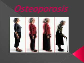
- 35. Osteoporosis – Dorsal Khyphosis Dorsal kyphosis with exaggerated lordosis (dowager's hump) may result from multiple compression fractures. The hump caused by spine fractures is disfiguring. This is the feature of osteoporosis that is the worst thing for most patients. In severe cases, the ribs can touch the pelvic bones.
- 36. Osteoporosis – Other Fractures • Osteoporotic fractures commonly affect the hip because the elderly tend to fall sideways or backwards, landing on this joint. Younger, more agile persons tend to fall forward, landing on the outstretched wrist, thus fracturing the distal radius
- 37. Osteoporosis – Diagnosis Without a fracture or bone density screening there is no way to diagnose the presence of osteoporosis. The goal is to get as much information about compounding risk factors: • A complete history of menstrual function, hormonal history in both genders. • Neurologic deficits and drugs that might increase the risk of falls should be analyzed. • One of the most important predictors of osteoporotic fractures is a history of a fracture after age 40 due to minimal or moderate trauma. In such persons, the fracture risk may be increased several fold. • The physical examination is often unremarkable. Spinal deformity and tenderness over the lower back should be sought.
- 38. Osteoporosis – Screening X-ray findings are generally insufficient for the screening of primary osteoporosis: • A normal x-ray of bone cannot reliably measure bone density but is useful to identify spinal factures, explains back pain, height loss or kyphosis. • X-rays may detect osteopenia only when bone loss is > 30%. Bone densitometry is the only method for diagnosing or confirming osteoporosis in the absence of a fracture • The National Osteoporosis Foundation recommends that bone densitometry be performed routinely in all women > 65, particularly in those who have one or more risk factors. • Densitometry can also be used for monitoring the response to therapy.
- 39. Screening - DEXA Dual energy x-ray absorptiometry (DEXA) • DEXA measures areal density (ie, g/cm2) rather than true volumetric density. • The test is non-invasive and involves no special preparation. • Radiation exposure is minimal, and the procedure is rapid. This is the most popular and accurate test to date and the test only takes about 20 to 40 minutes, with a 5 mrem dose of radiation (a full dental x-ray is 300 mrem).
- 40. Screening - DEXA •Can be used to measure bone mineral density in the spine, hip, wrist, or total body. •However, the standard apparatus is expensive and not portable. Small DEXA machines that can measure the forearm, finger, or heel are less expensive and are portable.
- 41. Screening - DEXA DEXA of the proximal femur in a young woman, age 37, with unsuspected femoral-neck osteopenia (T score, -1.6). DEXA of the lumbar spine in a young woman, age 37, with unsuspected lumbar spine osteopenia (T = -1.8)
- 42. Screening- Ultrasound Densitometry Ultrasound densitometry can assess the density and structure of the skeleton and appears to predict fracture risk in the elderly. The apparatus is relatively inexpensive, portable, and uses no radiation but can be used only in peripheral sites (eg, the heel), where bone is relatively superficial. Ultrasound devices measure the speed of sound (SOS), as well as specific changes in sound waves (broadband attenuation or BUA) as they pass through bone. QUS measurements provide information on fracture risk by providing an indication of bone density and possibly also information on the quality of the bone. Ultrasound devices do not expose the patient to ionizing radiation.
- 43. Diagnosis may include: Physical examination Medical history Medical histories of family members to find out if a genetic disorder is the cause A bone scan – dual energy x-ray absorptiometry (DXA) to test bone density Blood tests.
- 44. Osteoporosis – Treatment & Prevention Treatment of the patient with osteoporosis frequently involves management of acute fractures as well as treatment of the underlying disease Patients should be thoroughly educated to reduce the likelihood of any risk factors associated with bone loss and falling A large body of data indicates that optimal calcium intake reduces bone loss and suppresses bone turnover Routine to recommend supplemental vitamin D Exercise in young individuals increases the likelihood that they will attain the maximal genetically determined peak bone mass. Meta-analyses of studies performed in postmenopausal women indicate that weight-bearing exercise prevents bone loss but does not appear to result in substantial bone gain Osteoporosis does not directly cause death. However, an excess mortality of 10 to 20% occurs in patients with established osteoporosis, particularly those with hip fractures. Prevention of osteoporotic fractures is critical to avoid a worldwide, costly epidemic. Prevention programs should be developed for patients at risk and for patients with diagnosed osteoporosis.
- 45. Osteoporosis – Treatment & Prevention Antiresorptive therapy: Persons with low bone mass and multiple risk factors, particularly those who have already had an osteoporotic fracture, should be considered for antiresorptive therapy. Antiresorptive drugs include estrogens, bisphosphonates, selective estrogen receptor modulators, and calcitonin. Estrogen can prevent menopausal bone loss in most women. Estrogen replacement therapy (ERT) is the treatment of choice for postmenopausal women, particularly those who had an early menopause, and for women who have had a hysterectomy. ERT is particularly effective during the first few years after menopause when bone loss is most rapid. Epidemiologic studies and the few prospective clinical trials of estrogen suggest that ERT or HRT decreases the risk of osteoporotic fractures by 30 to 50%. Because other antiresorptive drugs may have an additive effect when given with estrogen, combination therapy should be considered in patients who have very low bone density, continue to lose bone, or incur a fracture while taking ERT or HRT.
- 46. Osteoporosis – Treatment & Prevention Bisphosphonates are potent antiresorptive drugs that directly inhibit osteoclast activity. For women who cannot tolerate estrogen or have contraindications (e.g., preexisting breast cancer, risk factors for breast cancer), bisphosphonates are considered the next choice; these drugs increase bone mass and decrease the risk of fractures, particularly in patients taking glucocorticoids. Bisphosphonates, particularly alendronate, have also decreased the incidence of vertebral and nonvertebral fractures by >= 50% in large cohorts of postmenopausal women. Alendronate is used to prevent (5 mg/day) and treat (10 mg/day) osteoporosis.
- 47. Osteoporosis – Treatment & Prevention Selective estrogen receptor modulators (SERMs) have been developed that are antiestrogenic and have antiresorptive effects on bone. Calcitonin has been used for many years in the prevention and treatment of osteoporosis. Other therapies: Anabolic therapies are under study; none is approved for osteoporosis. Intermittent injections of parathyroid hormone and fluoride stimulate bone formation and inhibit bone resorption, but their safety and efficacy remain to be established. Thiazides can decrease urinary calcium excretion and slow bone loss.
- 48. . Under supervision by a physical therapist, do repetitive motions with small weights, resistance bands or weight machines in order to strengthen the muscles in your back and other parts of your body. Strength-training will increase the strength of the postural muscles and help you to avoid compression fractures, and stooping, along your spinal column. In healthy bodies, strength-training can increase bone density. In osteoporosis patients, exercises with weight or resistance help you to maintain your current bone mass and avoid future loss of bone density.
- 50. . This should include weight-bearing, strength-training and balance exercise almost every day. Regular, careful physical activity should be taken as seriously as taking an osteoporosis medication.