SlideShare una empresa de Scribd logo
1 de 165
ARTERIAL SUPPLY OF HEAD AND
NECK.
Guided by
Dr. Rudresh K B
Reader
Dept of Oral and Maxillofacial Surgery
Presented by -
Dr. Avinash Rathore
Post Graduate
Dept of Oral and Maxillofacial Surgery
CONTENTS-
• EMBRYOLOGY
• HISTOLOGY
• COMMON CAROTID ARTERY
• EXTERNAL CAROTID ARTERY
• INTERNAL CAROTID ARTERY
• SUBCLAVIAN ARTERY
• LIGATIONS OF ARTERIES
• REFRENCES
DEVELOPMENT
DEVELOPMENT
Aortic arches
The aortic arches are a series of six paired embryological vascular structures which give rise
to several major arteries.
Aortic arches are short vessels connecting ventral and dorsal aortae on each side, they run
within branchial (pharyngeal) arches are based gradually in the 4th and 5th week, in 6 pairs
in total.
The first, second and fifth pairs soon disappear.
The 1st aortic arch – disappears (a small portion persists and forms a piece of the maxillary
artery)
The 2nd aortic arch – disappears (small portion of this arch contributes to the hyoid and
stapedial arteries)
The 3rd aortic arch - has the same development on the right and left side it gives rise to the
initial portion of the internal carotid artery
The external carotid is derived from
the cranial portion of the ventral aorta
The common carotid corresponds to a
portion of the ventral aorta between
exits of the third and fourth arches
The 4th aortic arch - has ultimate fate different on the right and left side
On the left - it forms a part of the arch of the aorta between left common carotid and left
subclavian artery
On the right - it forms the proximal segment of the right subclavian artery
The 5th aortic arch - is transient and soon obliterates
• The 6th aortic arch - pulmonary arch - gives off a branch on each side that
grows toward the developing lung bud
• The proximal part of the sixth right arch persists as the proximal part of the
right pulmonary artery while the distal section degenerates
The sixth left arch gives off the left pulmonary artery and forms the ductus
arteriosus; this duct remains pervious during the whole of fetal life, but then
closes within the first few days after birth due to increased O2concentration
• The outermost layer is known as
the tunica externa also known
as tunica adventitia and is
composed of connective
tissue made up ofcollagen fibers.
• Inside this layer is the tunica
media, or media, which is made up
of smooth muscle cells and elastic
tissue (also called connective tissue
proper).
• The innermost layer, which is in
direct contact with the flow of
blood is the tunica intima,
commonly called theintima. This
layer is made up of
mainly endothelial cells. The hollow
internal cavity in which the blood
flows is called the lumen.
Arteries form part of the circulatory
system. They carry blood that is
oxygenated after it has been
pumped from the heart. Arteries
also aid the heart in pumping
blood. Arteries carry oxygenated
blood away from the heart to the
tissues, except
for pulmonary arteries,which carry
blood to the lungs for oxygenation.
The Structure of Blood
VesselsA Comparison of a Artery and a Vein-
Figure 13-1
• MAJOR ARTERIES
OF HEAD AND
NECK
1. COMMON
CAROTID ARTERIES
2. EXTERNAL
CAROTID ARTERIES
3. INTERNAL
CAROTID ARTERIES
 ADDITIONAL
ARTERIES
- BRANCHES OF
SUBCLAVIAN
ARTERY
COMMON CAROTID ARTERY
15
Cervical part of common carotid artery
 Carotid arteries are generally symmetric and approximately of same
size unlike vertebral arteries.
 In 75% individuals ,CCA bifurcates at the level of C3-C4,roughly at
the upper border of thyroid cartilage.
 In children,the carotid bifurcates one vertebral level higher.
 Variation in the level of bifurcation
Highest seen in –C1 to C2
Lowest seen in –T1 to T2
Common carotid artery
16
CAROTID BODY(CHEMORECEPTORS)
 It is normally 2.5x 5mm to 4 x
7mmflattened structure on the
median and deep side of the upper
end of the common carotid artery.
 Blood supply-from small vesssels
usually from ECA,termed as GLOMIC
ARTERY OR ASCENDING
PHARYNGEAL ARTERY.
 NERVE SUPPLY-9TH 10TH 12TH CN
17
CAROTID SINUS (BARORECEPTORS)
 At the bifurcation of
common carotid artery
 Responsive to changes in the
arterial blood pressure.It
acts as a
baroreceptor(pressure
receptor) and regulates
blood pressure.
 BLOOD SUPPLY-ICA
 NERVE SUPPLY-carotid sinus
nerve or nerve of hering.
RELATION OF COMMON CAROTID
ARTERY
LATERAL-IJV
ANTEROLATERALLY
SKIN,FASCIA,SCM,STERNOHYOID,STERNOTH
YROID,SUP BELLY OF OMOHYOID
POSTEROLATERALLY-
VAGUS NERVE
MEDIAL
PHARYNX, LARYNX, TRACHEA
,OESOPHAGUS
LOBE OF THYROID GLAND
POSTERIOR
Transverse process of C3 C4
PREVERTBRAL FASCIA
SYMPATHETIC TRUNK
APPLIED ANATOMY
Carotid sinus syndrome
• Loss of consciousness due to simple head
movements.
• Hypersensitivity of the carotid sinus due to an
unknown etiology.
• Sudden slight pressure changes, such as that
occasioned by movement of the head, may result in
stimulation of the carotid sinus.
• Impulses transmitted by the sinus reduce blood
pressure and slow the pumping action of the heart.
Thus decreasing blood supply to the brain and
resulting in sudden loss of consciousness.
While supporting the mandible care should be taken
not to apply pressure on the carotid sinus.
21
External carotid artery (Facial carotid)
External carotid artery
• Generally,it lies anterior to the internal
carotid artery.
• It is the chief artery of supply to
structures in the front of the neck and in
the face.
SURFACE MARKING
• ECA is marked by joining
the following two points.
-A) point on the anterior
border of the
sternocleidomastoid
muscle at the level of the
upper border of the
thyroid cartilage.
-B) second point on the
posterior border of the
neck of the mandible.
The artery is slightly convex
forwards in its lower half
and slightly concave
forwards in its upper half.
B
A
COURSE
• ECA begins in the carotid
triangle at the level of
upper border of thyroid
cartilage opposite the disc
between the third and
fourth cervical vertebrae.
• In the carotid triangle,it lies
under cover of the anterior
border of the
sternocleidomastoid muscle
• As the artery ascends,it
passes deep to the post.
Belly of digastric and
stylohyoid muscle and
terminates behind the neck
of the mandible by dividing
into the maxillary and
superficial temporal
arteries.
Has slightly curved course,so that it is anteromedial
to ICA in it lower part,and anterolateral to the ICA
in its upper part.
28
External carotid artery
Superficial
temporal
Maxillary
Facial
Lingual
Superior thyroid
Posterior
auricular
29
Superior thyroid artery
30
Origin and course:
Arises from ECA immediately above the
bifurcation of CCA
Curves anteriorly and downwards to
enter Thyroid gland
31
Branches
Anterior Posterior
Superior thyroid artery
32
RELATION WITH EXTERNAL LARYNGEAL NERVE
APPLIED ANATOMY
• The arch of superior thyroid artery is characteristic –
diagnostic landmark
• The artery and external laryngeal nerve are close to
each other higher up, but diverge slightly near the
gland.
- So, ligature of superior thyroid artery in thyroid
surgery should be made close to the gland in order
to avoid injury of the external laryngeal nerve.
-Damage to the external laryngeal nerve causes some
weakness of phonation due to loss of tightening
effect of the cricothyroid on the vocal cord.
• Intra-arterial infusion chemotherapy for laryngeal
and hypopharyngeal cancers.
Lingual Artery
Origin-
 Lingual Artery arises from the ECA
opposite the tip of greater cornu of the
hyoid bone
Course-
 First part of artery lies in the carotid
triangle
 Second part of artery lies deep to
the hyoglossus muscle which separates
it from the hypoglossal nerve
 Third Part or deep part : runs
upwards along the anterior margin of
the hyoglossus
Branches of Lingual Artery
 Suprahyoid Br
 Dorsal Lingual Br
 Deep Lingual Artery
 Sublingual Artery
APPLIED ANATOMY
• In surgical removal of tongue , first part of
artery is ligated before it gives any branches to
the tongue or tonsil.
LIGATION OF LINGUALARTERY :
Incision – circling the lower pole of
submandibular gland.
- Skin, platysma, deep fascia incised,
submandibular gland exposed , lifted, tendon of
digastric visible.
- Free border of mylohyoid muscle seen,
hypoglossal nerve identified. Digastric tendon
pulled downwards –enlarges the digastric
triangle, hyoglossus muscle visible.
- Muscle divided bluntly, in the gap of its
vertical fibers lingual artery found & ligated.
SUBLINGUALARTERY
Injury occurs in premolar
& molar region, when
sharp instrument or rotating
disks slips off a lower
molar & injure the floor of
mouth.
-May present problems to
the surgeon attempting to
ligate its source because it
may arise from the
submental branch of the
facial artery rather than
from the lingual artery.
For Implants, CBCT to
localise the vascular
canal,injuries to arteries in
vascular canal can cause
sublingual haematoma
leading to blockage of
airway.
FACIAL ARTERY
Facial Artery
Facial artery is the chief artery of the face
Origin :
 Arises from the ECA just above the greater
cornu of the hyoid bone
 It has two parts, first cervical part in the neck
and facial part.
 It enters the face by winding around the base
of the mandible
 At the anteroinferior angle of the masseter
muscle, it can be palpated here and is called as
an “anaesthetist’s artery”
•SURFACE MARKINGS-ANTERO INFERIOR BORDER OF MASSETER
• 1.25 CM LATERAL TO ANGLE OF MOUTH
• MEDIAN ANGLE OF EYE
•TORTUOUS COURSE
•PULSATIONS FELT AT- LOWER BORDER OF MANDIBLE
ORAL CAVITY
Branches of Cervical part
1. Ascending palatine artery- it supplies to root of tongue & tonsil.
2. Tonsillar artery
3. Submental artery- it is a large artery which accompanies the
mylohyoid nerve, and supplies the submental triangle and sub
lingual salivary gland.
4. Glandular branches that supplies submandibular salivary gland
and submental lymph nodes.
ASCENDING
PALATINE ARTERY
• ORIGIN FROM HIGHEST POINT OF
FACIAL ARTERY
• COURSES CRANIALLY ALONG THE
SUPERIOR CONSTRICTOR OF
PHARYNX TO REACH SOFT PALATE
• SMALL BRANCH TO PHARYNGEAL
MUSCLES
• TONSILLAR BRANCH SUPPLYING
PALATINE TONSIL(MAY ARISE
DIRECTLY FROM FACIAL ARTERY)
TERMIAL BRANCHES SUPPLY SOFT
PALATE
TONSILLAR A RTERY:
Passes between the styloglossus and
medial pterygoid muscles and
pierces the superior pharyngeal
constrictor muscle to supply the
palatine tonsil and the posterior
tongue.
SUBMENTAL ARTERY
• RUNS HORIZONTALLY BELOW
THE INFERIOR BORDER OF
MANDIBLE
• TRAVERSES HORIZONTALLY TO
REACH MYLOHYOID MUSCLE
• SUPPLIES SUBMANDIBULAR
NODES,MYLOHYOID AND
SURROUNDING MUSCLE
• ANASTAMOSE WITH
SUBLINGUAL AND INFERIOR
LABIAL ARTERY
Branches of facial part
1. Superior labial- supplies to
upper lip & antero-inferior
part of nasal septum.
2. Inferior labial- supplies to
lower lip.
3. Lateral nasal- to the ala &
dorsum of nose.
4. Angular – supplies the
lacrimal sac and orbicularis
oculi.
FACIAL PART:
INFERIOR LABIAL
ARTERY: Originates near
the corner of the mouth,
passes deep to the depressor
anguli oris muscle, and
pierces the orbicularis oris
muscle.
-The artery courses
superficial to that muscle,
supplying it as well as the
substance of the lower lip.
-It forms an anastomosis
with its counterpart of the
other side and with branches
of the mental and submental
arteries.
SUPERIOR LABIALARTERY:
Arises just above the inferior labial artery. It passes
superficial to the orbicularis oris muscle in the
upper lip to serve that muscle as well as the
substance of the upper lip.
- It sends a small twig, the SEPTAL BRANCH to
supply anteroinferior part of the nasal septum and
another one, the ALAR BRANCH, into the wing of
the nose.
-The terminus of the vessel will anastomose with its
counterpart of the opposite side.
LATERAL NASALARTERY: Small
branch arising at and passing into the
wing and bridge of the nose.
-This supplies ala and dorsum of the
nose. This vessel will anastomose with
various other arteries in its vicinity.
ANGULAR ARTERY: Is the terminal
continuation of the facial artery,
supplying the tissues in the vicinity of
the medial corner of the eye and
anastomosing with dorsal nasal branch
of the ophthalmic artery.
VARIATIONS
APPLIED ANATOMY
• Facial Artery
Compression:
Applying pressure to the
facial artery as it passes
over the inferior border of
the mandible just anterior
to the angle will diminish
blood flow to that side.
o Can be injured –during
operative procedures on
lower premolars &
molars, if instrument
enters the cheek at inferior
vestibular fornix., also
while attempt to open a
buccal abscess or
mucocoele.
• In mand. 1st molar region care
must be taken not to injure the
facial artery while extending the
vertical incision down the
vestibule during surgical
extraction of mandibular
impaction.
• So it is recommended that start
vertical incision from the
vestibule in upward direction.
• While excising the
submandibular gland,the facial
artery should be ligated at two
points and should be secured
before dividing it, otherwise it
may retract through
stylomandibular ligament causing
serious bleeding.
MEDIAL BRANCH
Ascending Pharyngeal Artery
 A small branch arises from
medial side of ECA
 Long, slender vessel, deeply
seated in the neck
 COURSE: Ascends vertically
between the internal carotid and
the side of the pharynx, to the
under surface of the base of the
skull, lying on the Longus
capitis.
BRANCHES
• PHARYNGEAL
BRANCHES
• PALATINE BRANCH
• PREVERTEBRAL
BRANCH
• INFERIOR
TYMPANIC ARTERY
• MENINGEAL
BRANCHES
POSTERIOR BRANCHES
Posterior Auricular Artery
 Small and arises above the
posterior belly of digastric
 It runs upwards and backwards
deep to the parotid gland,
crosses the base of the
mastoid process and ascends
behind the auricle.
 Stylomastoid branch
OCCIPITAL ARTERY
ORIGIN:Arises in carotid
triangle from posterior
aspect of ECA ,opposite
the origin of facial artery.
-It is crossed at its origin by
hypoglossal nerve.
COURSE: Passes backwards
and upwards along & under
cover of lower border of
post. Belly of digastric ,
crossing carotid sheath,
hypoglossal & accessory
nerves.
Then it runs deep to the
mastoid process and
muscles attached to it
i.e.,sternocleidomastiod,
digastric etc.
Then crosses the rectus
capitus
lateralis,superior
oblique,and
semispinalis capitus
muscle at the apex of
the posterior triangle.
Finally it pierces the
trapezius muscle and
ascends in a tortuous
course in the superficial
fascia of the scalp.
Its terminal portion
comes to lie along the
greater occipital nerve.
BRANCHES
IN THE CAROTID TRIANGLE
• STERNOMASTOID BRANCHES – Two
in no.,upper branch accompanies the
accessory nerve and lower branch arises
near the origin of the occipital artery.
Supplies sternomastoid m.
IN THE POSTERIOR TRIANGLE and
SCALP REGION:
• AURICULAR BRANCH: Passes
superficial to the mastoid process to reach
and supply the back of the auricle.
• MASTOID BRANCH:–
Enters cranial cavity through
mastoid foramen, supplies mastoid
air cells in the dura and diploe.
• MENINGEAL BRANCH –
Ascends with the internal
jugular vein and enters the skull
through jugular foramen &
condylar canal, supplies dura of
posterior cranial fossa.
• MUSCULAR BRANCH-
Supply the Digastric,
Stylohyoideus, Splenius, and
Longissimus capitis.
DESCENDING BRANCH :
• The largest branch of the occipital, descends on
the back of the neck, and divides into a
superficial and deep portion.
-The superficial portion runs beneath the
Splenius, giving off branches which pierce that
muscle to supply the Trapezius and anastomose
with the ascending branch of the transverse
cervical artery.
-The deep portion runs down between the
Semispinales capitis and colli, and anastomoses
with the vertebral and with the a. profunda
cervicalis, a branch of the costocervical trunk.
• The terminal branches of
the occipital
artery(occipital branches)
are distributed to the back
of the head: they are very
tortuous, and lie between
the integument and
Occipitalis, anastomosing
with the artery of the
opposite side and with the
posterior auricular and
temporal arteries, and
supplying the Occipitalis,
the integument, and
pericranium
APPLIED ANATOMY
Superficial branch anastomosis with
ascending branch of transverse cervical
artery. Deep branch of descending br of
occipital artery anastomosis with deep
cervical artery.
TERMINAL
BRANCHES
 Larger of the two terminal
branches
 Arises behind the neck of the
mandible, and is embedded
in the substance of the
parotid gland
 It supplies the deep
structures of the face
Maxillary Artery
Branches
1st part (mandibular) :
Lies medial to mandible, it runs along the lower border of
lateral pterygoid muscle
 Deep auricular artery
 Ant.tympanic artery
 Middle meningeal artery
 Accessory meningeal artery
 Inferior alveolar artery
Branches of first and its supply
Branches
1.Deep auricular
2.Anterior tympanic
3.Middle meningeal
4.Accessory meningeal
5.Inferior alveolar
Foramen transmitting
Foramen in the floor of
external acoustic meatus
Petrotympanic fissure
Foramen spinosum
Foramen ovale
Mandibular foramen
Distribution
External acoustic
meatus,outer surface of
tympanic membrane
Inner surface of tympanic
membrane
5th and 7th nerve, middle
ear, tensor tympani
Meninges, Structures in
the infra temporal fossa
Lower teeth and mylohyoid
muscle
 Largest artery that supplies
the dura
 It ascends to the foramen
spinosum through which it
enters the cranium
 Divides into two branches,
anterior and posterior.
 It supplies the dura mater
(the outermost meninges)
and the calvaria.
Middle Meningeal Artery
APPLIED ANATOMY
• FRONTAL BRANCH – Extradural
hemorrhage -hematoma presses on the motor
area – hemiplegia of opposite side
APPROACH- hole in the skull over pterion – 4
cm above mid point of zygomatic arch.(approx
2 finger breadth above zygomatic arch)
• PARIETAL OR POSTERIOR BRANCH -
contralateral deafness
APPROACH- hole is made 4cm above and
4cm behind the external acoustic meatus.
Inferior alveolar artery
 Runs downward & forward
medial to ramus of mandible
to reach mandibular
foramina.
 Before entering mandibular
foramina gives off lingual and
mylohyoid arteries.
 In canal gives branches to
mandibular teeth .
 After coming out of canal
supply chin via mental artery.
BRANCHES
BEFORE ENTERING
MANDIBULAR CANAL:
• Lingual branch to the tongue.
• Mylohyiod branch to the mylohyiod
muscle.
WITHIN THE MANDIBULAR
CANAL:
Branches to the mandible
Branches to the roots of each teeth upto
midline(dental branches)
Incisor branch anastomoses with the
branch from opposite side.
AFTER EMERGING FROM
MENTAL FORAMEN:
mental branch escapes with the nerve
at the mental foramen, supplies the
chin, and anastomoses with the
submental and inferior labial arteries
2nd part (pterygoid part) :
 Artery runs forward &upward superficial to the lower head
of the lateral pterygoid muscle
B. Second part
Branches
1.Deep temporal
2.Pterygoid
3.Masseteric
4.Buccal
Distribution
Temporalis
Lateral and
medial
pterygoid
Masseter
Buccinator
3rd part (pterygopalatine):
 Terminal portion of the artery
passes between the two heads
of the lateral pterygoid muscle
THIRD PART
Branches
1.Post superior alveolar
2.Infraorbital
3.Greater palatine
4.Pharyngeal
4.Artery of pterygoid canal
5.Sphenopalatine(terminal
part)
Foramina
Alveolar canals in the body of
maxilla
Infraorbital fissure
Greater palatine canal
Pharyngeal canal
Pterygoid canal
Sphenopalatine foramen
Distribution
Upper molar and premolar
teeth ; maxillary sinus
Lower orbital muscles,
lacrimal sac ,max sinus
Soft palate, tonsil, palatine
glands and mucosa,upper
gums
Root of nose , pharynx,
auditory tube,sphenoidal
sinus
Auditory tube, upper
pharynx, middle ear
Lateral and medial wall of
nose and air sinuses.
APPLIED ANATOMY
• Site of hematoma during PSA block.
• Produces largest and most esthetically
unappealing hematoma.
• Blood effuses until extravascular pressure
exceeds intravascular pressure or clotting
occurs.
• Infratemporal fossa into which bleeding occurs
accommodates large amount of blood.
• Prevented by aspirating before giving LA in the
site.
• Digital pressure can be applied medial and
superior to the maxillary tuberosity.
Applied anatomy
• In case of abscess
from palatal root of
first molar,incision
should be made in a
antero-posterior
direction parallel to
the artery.
• During lefort I osteotomy:
• Greater palatine artery is easily injured during
osteotomy of the medial or lateral maxillary
sinus walls, pterygomaxillary dysjunction or
during down fracturing of maxilla.
• The average distance from the piriform rim to
the descending palatine artery is 35.4 mm, range
is 31 to 42 mm.
• The average length of the greater palatine canal
above the nasal floor is 10mm, range is 6 to 15
mm.
• The average distance between the
pterygomaxillary fissure and the greater palatine
foramen is 6.6mm.
GUIDELINES TO AVOID INJURY:
• Osteotomy of lateral wall of
maxillary sinus should extend
just beyond the second molar.
• Osteotomy of medial wall of
maxillary sinus should usually
extend 30mm posterior to the
piriform rim in females,in males
it can be carried back to 35mm -
--O’ RYAN
• Because the descending palatine
artery travels in an anterior-
inferior direction as it enters the
greater palatine canal ,injury can
be prevented by closely adapting
the cutting edge of the
osteotome or the saw to the
pterygomaxillary fissure.
APPLIED ANATOMY OF
MAXILLARY ARTERY
• Surgeries involving
condyle-Avoid injury to
maxillary artery as it lies
medial to condyle.
• Ankylotic mass of TMJ
may encircle the artery.So
it is advisable to remove
ankylotic mass in pieces
rather than in toto.
• Trismus involving lateral
pterygoid comprises blood
supply to the nose.
• During Le fort I
osteotomy procedure-
Pterygopalatine
portion of maxillary
artery may be injured
during fracturing the
pterygiod plates if
Tessier’s osteotome is
directed backwards.
-It should be directed
downwards and
medially.
• Can be used as arterial donor in repair
of ICA dissections and aneurysms, due
to close proximity of the artery to the
cranial base.
• Control of epistaxis---If epistaxis is not
controlled after nasal packing,it can be
controlled by ligating IMA via
endonasal,transantral or intraoral
approach.
LITTLE’S AREA or
KIESSELBACH’S
PLEXUS
-Near the anteroinferior part
or vestibule of the septum.
-Contains anastomoses
between
• Superior labial branch of
facial artery
• Branch of sphenopalatine
artery
• Anterior ethmoidal artery
• Greater palatine artery
This is common site of
bleeding from nose or
epistaxis.
 Smaller of the two terminal
branches
 It begins in the substance of
the parotid gland, behind the
neck of the mandible
 Divides into two branches, a
frontal and a parietal
Superficial Temporal Artery
Parietal branch
Frontal branch
Middle temporal artery
Transverse facial artery
 Transverse facial branch
 Anterior auricular branch
 Frontal branch
 Parietal branch
 Zygomatico- orbital branch
Branches
Transverse Facial Artery:
ORIGIN:From STA before it leaves parotid gland.
COURSE: Running forward through the
substance of the gland, it passes transversely
across the side of the face, between the parotid
duct and the lower border of the zygomatic arch.
This vessel rests on the Masseter, and is
accompanied by one or two branches of the
facial nerve.
SUPPLIES: The parotid gland and duct, the
Masseter, and the integument, and anastomose
with the external maxillary, masseteric,
buccinator, and infraorbital arteries.
Middle Temporal Artery: Arises immediately above
the zygomatic arch, and, perforating the temporal
fascia, gives branches to the Temporalis,
anastomosing with the deep temporal branches of
the internal maxillary artery.
- It occasionally gives off a zygomaticoorbital
branch, which runs along the upper border of the
zygomatic arch, between the two layers of the
temporal fascia, to the lateral angle of the orbit.
-This branch, which may arise directly from the
superficial temporal artery, supplies the Orbicularis
oculi, and anastomoses with the lacrimal and
palpebral branches of the ophthalmic artery.
• Anterior
Auricular
Branches :
Distributed to the
anterior portion
of the auricle, the
lobule, and part
of the external
meatus,
anastomosing
with the posterior
auricular.
Frontal Branch :
Runs tortuously upward and
forward to the forehead,
supplying the muscles,
integument, and
pericranium in this region,
and anastomosing with the
supraorbital and frontal
arteries.
Parietal Branch:
Larger than the frontal, curves
upward and backward on the
side of the head, lying
superficial to the temporal
fascia, and anastomosing with
its fellow of the opposite side,
and with the posterior
auricular and occipital
arteries.
APPLIED ANATOMY
• Control of temporal
haemorrhage.
• Anaesthetist’s artery
• Placement of incisions in
craniotomy
• In reduction of zygomatic arch
fractures – Gilli’s approach
-A 2cm incision is placed in the
temporal region at an angle 45
degree to the zygomatic arch,
between two branches of the
superficial temporal artery and
parallel to the anterior branch.
INTERNAL CAROTID ARTERY
• Origin-
• It is one of the terminal
branch of common carotid
artery originates along with
external carotid artery at the
upper border of thyroid
cartilage at the disc of third
and fourth cervical vertebra.
• It supplies two of the four major arteries
supplying blood to the brain.
• CCA CAROTID CANAL(petrous part of
temporal bone) MIDDLE CRANIAL
FOSSA(dorsum sellae of sphenoid bone)
Supplies the hypophsis cerebri,orbit,and most
of the supratentorial part of the brain.
BRANCHES and SEGMENTS
• 1996 –bouthillier divided ICA into 7 anatomical
segments viz.
• C1-Cervical part in the neck
• C2-Petrous part in the petrous temporal bone
• C3-lacerum
• C4-Cavernous part in the cavernous sinus
• C5-clinoid
• C6-opthalmic
• C7-communicating
CEREBRAL PORTION
Cervical part
• It ascends vertically in the neck from its origin to the base of
skull to reach the lower end of the carotid canal. This part is
enclosed in carotid sheath along with internal jugular and
vagus nerve. No branches arises from the internal carotid
artery in the neck.
• Its initial part shows slight dilatation, carotid sinus. Which acts
as a baroreceptor.
PETROUS PART• Within the petrous part of the
temporal bone,in the carotid
canal runs upward forward &
medially at rt. Angle.
Branches-
1) Caroticotympanic- enter
middle ear & anastomose
with ant. & post. Tympanic
branches
2) Artery of the Pterygoid Canal-
anastomose with greater
palatine artery
CAVERNOUS PART
Within the Cavernous Sinus
• Branches
1) Artery to trigeminal
ganglion
2) Superior & inferior
Hypophyseal artery
CEREBRAL PART
• Lies at the base of the brain
after emerging from the
cavernous sinus
Branches
1.Ophthalmic.
2.Anterior Cerebral.
3.Middle Cerebral.
4.Posterior Communicating.
5. Ant. choroidal
On angiogram internal
carotid show ‘S’ shaped
figure ( carotid siphon )
Opthalmic artery
• Arises medial to anterior clinoid
process near optic canal
• In orbit lie inferolateral to optic
nerve
Branches:
1. Central artery of retina
2. Large lacrimal artery
3. Dorsal nasal artery
• Supratrochlear
• Supraorbital
• Posterior ethmoidal
• Anterior ethmoidal
• Palpebral branch
• Recurrent meningeal a.
• Muscular a.
SUBCLAVIAN ARTERY
• MAIN ARTERY OF
UPPER LIMB
ORIGIN-
1.RIGHT-
BRACHIOCEPHALIC
ARTERY
2.LEFT -ARCH OF
AORTAE
COURSE
1.MEDIAL
2.POSTERIOR
3.LATERAL
Circle of Willis
The circle of Willis is a circulatory
anastomosis that supplies blood to
the brain and surrounding structures.
It is named after Thomas Willis (1621–
1675), an English physician
Anterior cerebral artery (left and right)
Anterior communicating artery
Internal carotid artery (left and right)
Posterior cerebral artery (left and right)
Posterior communicating artery (left and
right)
Basilar artery
IMPORTANCE: The arrangement of the
brain's arteries into the circle of Willis
creates collaterals in the cerebral
circulation. If one part of the circle becomes
blocked or narrowed (stenosed) or one of
the arteries supplying the circle is blocked
or narrowed, blood flow from the
other blood vessels can often preserve the
cerebral perfusion well enough to avoid the
symptoms of ischemia.
Anastomoses ICA ECA
Dorsal Nasal Artery and
Angular Artery
Dorsal Nasal Artery
(branch of the
Ophthalmic artery)
Angular Artery (branch of
the Facial Artery)
Supraorbital Artery and Frontal
Artery
Supraorbital Artery
(branch of the
Ophthalmic)
Frontal Artery (terminal
branch of the Superficial
Temporal Artery)
Zygomatico Artery and
Transverse facial artery
Zygomatico (branch
Lacrimal Artery)
Transverse Facial Artery
(branch of Superficial
Temporal Artery)
Branches of the Posterior
Ethmoidal Artery and branches
of the Sphenopalatine Artery
Posterior Ethmoidal
Artery
Sphenopalatine
Artery(branch of the
Internal Maxillary)
Cavernous branches and
Middle Meningeal artery
Cavernous branches
from the cavernous
portion of the ICA
Middle Meningeal Artery
(branch of the Internal
Maxillary)
CARATIDO-JUGULAR REFLEX
Common carotid artery-
It can be compressed against the carotid
tubercle, the anterior tubercle of the
transverse process of vertebra C6 which
lies at the level of cricoid cartilage.
Carotidynia is a syndrome characterized
by unilateral (one-sided) tenderness of
the carotid artery, near the bifurcation.
Carotid Sinus
 Present at the termination of CCA. (or
beginning of ICA.)
 Tunica media is thin, tunica adventia is
thick
 Acts as BARORECEPTOR/PRESSURE
RECEPTOR.
• Carotid sinus hypersensitivity (CSH) is an exaggerated
response to carotid sinus baroreceptor stimulation. It results
in dizziness or syncope from transient diminished cerebral
perfusion.
• For these individuals, even mild stimulation to the neck
results in marked bradycardia and a drop in blood pressure.
Carotid Siphon of Angiogram
 Siphon region is the most common site for atherosclerotic
plaque formation in carotid artery
 Carotid body situated behind the bifurcation of CCA
 Act as a chemoreceptor & respond to change in the O2, CO2 and pH
content of the blood
 Carotid body paragangliomas are vascular lesions, and this is
reflected in their imaging appearance. These lesions splay apart the
internal (ICA) and external carotid arteries (ECA), and as it enlarges, it
will encase, but not narrow the ICA and ECA.
Head Neck Path.Dec 2009; 3(4): 303–306.
Carotid Body
• Definition
• Why ligate?
• Procedure
Individual artery ligation
-External Carotid Artery
-Lingual
-Sublingual
-Facial
-Maxillary
-Sphenopalatine
-Greater Palatine
-Ant./Post. Ethamoidal
-Internal Carotid Artery
• LIGATION Means act of binding or
tying of blood vessels with sutures or wires is
called Ligation…
First ligation was done by AMBROSE
PARE in amputation procedure.
WHY WE LIGATE VESSELS???
1.AFTER AMPUTATION TO ARREST THE
BLOOD FLOW.
2.IN WOUNDS OF ARTERIES WHERE
HAEMMORRHAGE CAN’T BE CONTROLLED.
3.IN SECONDARY HAMMORAGHE WHERE
THEY CAN’T BE CONTROLLED BY OTHER
MEANS.
4.IN LOCAL HYPERTROPHIES TO ARREST THE
NUTRITIONAL SUPPLY TO THAT AREA.
• IN CASE OF ANEURYSMS
• IN CASE OF MALIGNANT TUMOUR TO STOP THE
BLOOD FLOW
• IN ACUTE INFLAMMATION WHERE NEITHER
RESECTION NOR AMPUTATION IS POSSIBLE
• IN VARIOUS OPERATIVE PROCEDURES WHEN WE
ENCOUTER VESSELS TO REDUCE BLOOD FLOW TO
THAT REGION .
PROCEDURE FOR LIGATION
1. EXPOSE THE SHEATH OF VESSEL
2. ISOLATE THE VESSEL
3. PLACE THE LIGATURE
LIGATION OF EXTERNAL
CAROTID
ARTERY
EXPOSED AT TWO SITES
1. IN THE CAROTID TRIANGLE -AT ITS ORIGIN FROM THE
COMMON CAROTID ( ABOVE THE ORIGIN OF SUPERIOR
THYROID ARTERY)
2. IN THE RETROMANIBULAR FOSSA HERE WE LIGATE IT
BEHIND THE ANGLE OF LOWER JAW ( DEALS WITH THE
HAEMORRHAGE FROM ONE OF THE BRANCHES OF
MAXILLARY ARTERY)
• CONTINUE DOWNWARDS / TO
THE ANTERIOR BORDER UP TO
THE LEVEL OF CRICOID
CARTILAGE
• AFTER PENETRATING SKIN,
PLATYSMA SUPERFICIAL SHEATH
OF STERNOCLEDIOMASTOID IS
INCISED
EXPOSURE OF GREAT VESSEL
• WITH BLUNT DISSECTION
ANTERIOR BORDER IS EXPOSED,
MUSCLE IS RETRACTED AND DEEP
LAYER IS SEEN
• IN THIS PART Internal Juglar Vein
IS EXPOSED
IDENTIFICATION OF EXTERNAL CAROTID ARTERY
• THE JUGULAR VEIN IS MOBILIZED BY OPENING
THE CAROTID SHEATH & FREE THE JUGULAR
VEIN.
• RETRACT POSTERIORLY VEIN TO VISUALIZE
ARTERY
EXPOSURE OF THE CAROTID BULB AND EXTERNAL
CAROTID ARTERY
• AS THE DISSECTION
PROCEED
POSTERIORLY THE
CAROTID BULB IS
IDENTIFIED AND
BIFURCATION IS
SEEN
• MANIPULATION OF
BULB AT THIS STAGE
LEAD TO
ARRYTHEMIA AND
ANAESTHESIST
SHOULD BE
INFORMED
• LIGATION
EXTERNAL CAROTID ARTERY IS IDENTIFIED
& LIGATED ABOVE THE SUPERIOR THYROID
ARTERY
• CLOSURE OF WOUND
A VACCUM DRAIN IS PLACED AND WOUND
IS SUTURED IN LAYERS
COMPLICATIONS
• HAMEORRHAGE DUE TO IJV OR ECA( profuse bleeding)
• DAMAGE TO VAGUS NERVE (posteriomedially)
• LIGATION OF ICA( contra lateral hemiplegia & blindness
on the same side)
• HEMATOMA FORMATION
• INFECTION
IN RETROMANDIBULAR
FOSSA
• INCISION
• STARTS THE TIP OF
MASTOID PROCESS AND
CIRCLING THE
MANDIBULAR ANGLE,
CONTINUING
FORWARD BELOW THE
MANDIBLE FOR ABOUT
ONE INCH
• INCISION SHOULD BE
AT EQUAL DISTANCE
FROM THE POSTERIOR
AND INFERIOR BORDER
OF MANDIBLE
EXPOSURE
• AFTER THE BLUNT
DISSECTION OF SKIN, SOME
POST. FIBERS OF PLATYSMA,
RETROMANDIBULAR VEIN OR
EJV IS LOCATED, CUT & TIED
• BRANCHES OF GREATER
AURICULAR NERVE IS CUT &
TIED TO PERMIT THE
MOBILIZATION OF CERVICAL
LOBE OF PAROTID GLAND
• ATTACHMENT OF PAROTID
WITH STERNOMASTOID AT
ANTERIOR BORDER IS
SEVERED & GLAND IS
RETRACTED ANTERIORLY &
UPWARDS
IDENTIFICATION
• UNDERNEATH THE PAROTID GLAND & POST. BELLY OF DIGASTRIC,
SMALL THIN PART OF STYLOHYOID MUSCLE IS VISIBLE
• ABOVE THIS- STYLOID PROCESS & STYLOMANDIBULAR LIGAMENT
IS PALPATED
• NOW MOVING THE JAW FORWARD ENTRANCE TO
RETROMANDIBULAR FOSSA IS WIDENED & PULSE OF ECA IS FELT,
ISOLATE & LIGATE IT
LIGATION OF LINGUAL
ARTERY
LIGATION OF LINGUAL ARTERY
INCISION
• INCISION GIVEN BELOW
THE LOWER BORDER OF
MANDIBLE AFTER
PALPATING THE
SUBMANDIBULAR
GLAND.
• THE POSTERIOR PART OF
INCISION SHOULD BE
TOWARDS THE TIP OF
MASTOID PROCESS AND
ANTERIOR SHOULD
POINT TOWARDS THE
CHIN
EXPOSURE OF ARTERY
• AFTER BLUNT
DISSECTION
SUBMANDIBULAR
GLAND IS EXPOSED
• POST BELLY OF
DIGASTRICS
IDENTIFIED,
MYLOHYOID MUSCLE
REACHED,
HYPOGLOSSAL NERVE
AND ACCOMPANYING
VEIN IDENTIFIED
LIGATION OF ARTERY
• DIGASTRICS TENDON
PULLED DOWNWARD ,
HYOGLOSSUS MUSCLE
DISSECTED AND LINGUAL
ARTERY IS FOUND AND
LIGATED
• FIBERS OF HYOGLOSSUS
MUSCLE SHOWS VERTICAL
COURSE (THIN & FINE)
WHILE THAT OF MYLOHYOID
SHOWS OBLIQUE COURSE
(THICK)
LIGATION OF SUBLINGUAL ARTERY
INDICATIONS
• INJURY IS OBSERVED
WHEN SHARP INSUMENTS
OR ROTATING DISC ARE
SKIPPED ON FLOOR OF THE
MOUTH
• IN VARIOUS SURICAL
PROCEDURE LIKE RANULA
AND TUMOURS OF
SALIVARY GLANDS
• DIFFICULT TO LIGATE
SUBLINGUAL ARTREY MAY BE A BRANCH OF
1. LINGUAL ARTERY
2. SUBMENTAL ARTERY
• INCISION
• IN THE SUBLINGUAL GROOVE
• STRUCTURES IN CLOSE
ASSOCIATION
• SUBLINGUAL GLAND(MED.
&INF.)
• SUBMANDIBULAR DUCT
• LINGUAL NERVE(MED.& INF.)
• HYPOGLOSSAL NERVE AND
SUBLINGUAL VEIN
LIGATION OF FACIAL
ARTERY
LIGATION OF FACIAL ARTERY
INCISION
½ INCH BELOW & PARALLEL TO THE LOWER BORDER OF
MANDIBLE
EXPOSURE
THE SKIN, PLATYSMA MUSCLE AND DEEP FACIA ARE CUT,
SOFT TISSUE IS BLUNTLY CUT AND RETRACTED
LIGATION
PULSE OF FACIAL ARTERY IS FELT & ARTERY IS
ISOLATED AND LIGATED.
• FACIAL ARTERY CROSSES THE LEVEL OF INFERIOR
VESTIBULAR FORNIX IN THE REGION OF 1ST
MANDIBULAR MOLAR.
• DURING BUCCAL SPACE INFECTION THE ARTERY
IS DISLOCATED
• AVOID DEEP INCISION, INCISION SHOULD BE
DOWNWARDS & INWARDS INSTEAD OF
STRAIGHT UPWARDS.
• Incision - at least half inch below the
border of mandible & parallel to it.
Skin,platysma and deep cervical fascia cut
Artery is accompanied by facial vein & crossed superficially by marginal
mandibular branch of facial nerve
Pulse of facial artery felt. Artery- isolated, tied & cut
LIGATION OF
MAXILLARY ARTERY
LIGATION OF MAXILLARY ARTERY
LIGATION CAUSES DECREASE IN INTRA VASCULAR
PRESSURE GRADIENT, RESULTING IN HOMEOSTASIS
APPROACHES:
CAN BE DONE BY
1. TRANSANTRAL APPROACH &
2. INTRAORAL APPROACH
TRANSANTRAL APPROACH
BY CALD WELL LUC APPROACH
PROCEDURE
A LATERALLY BASED U SHAPE MUCOSAL
INCISION IS CREATED
POSTERIOR WALL OF MAXILLARY SINUS IS
IDENTIFIED
POSTERIOR MAXILLARY WALL IS REMOVED
EXPOSURE & LIGATION
• AREA IS ENLARGED,ARTERY IS IDENTIFIED
&LIGATED
SUCCESS RATE
• 87% SUCCESS RATE
INTRA ORAL APPROACH
• THIS PROCEDURE IS GIVEN IN 1984 BY
MACERI & MAKILSKI
• LIGATE INFRATEMPORAL PORTION OF
MAXILLARY ARTERY
INDICATION:
- IN CHILDREN AS AN ALTERNATE TO
EMBOLIZATION & EXTERNAL ARTERY
LIGATION FOR REMOVAL OF VASCULAR
TUMOR
- TO CONTROL BLEEDING IN VARIOUS
MAXILLECTOMY PROCEDURES WHERE
CALD WELL LUC IS CONTRAINDICATED
PROCEDURE:
- BY EXPOSING THE POSTERIOR PORTION OF
MAXILLA THROUGH A POST.
GINGIVOBUCCAL INCISION
- A FINGER IS INSERTED INTO THE DEPTH OF
WOUND TO PALPATE THE MAXILLARY
ARTERY
- THE NERVE HOOK IS USED FOR LIGATION
LIGATION OF
SPHENOPALANTINE
ARTERY
LIGATION OF SPEHNOPALATINE ARTERY
• CAN BE DONE BY TWO METHODS
1. TRANSANTRAL LIGATION
2. ENDOSCOPIC LIGATION
TRANSANTRAL APPROACH
• DESCRIBED BY SIMPSON et al. IN 1982
• APPROACH CALD WELL LUC
• AVOID ENTRANCE TO PTERYGOPALATINE
FOSSA
• MEDIAL, POSTERIOR & INFERIOR WALL IS
REMOVED
• SPHENOPALATINE & VIDIAN NERVE IS
DISSECTED & LIGATION OF ARETRY IS
DONE
ENDOSCOPIC LIGATION FOR SPHENOPALANTINE
ARTERY
• DESCRIBED BY WHITE (MODIFICATION OF
SIMPSONS TECH)
• APPROACH THROUGH
1. MEATAL ANTROSTOMY &
2. CANINE FOSSA
• NOT USING WIDELY AS COSTLY
• ADVANTAGES
1. REDUCE PATIENT DISCOMFORT AND
2. DURATION OF HOSPITALIZATION
LIGATION OF GREATER
PALANTINE ARTERY
LIGATION OF GREATER PALATINE
ARTERY
ENDANGERED DURING MINOR SURGERY PROCEDURES AND DURING
DENTAL TREATMENT
• INCISION
--FROM THE LINGUAL ROOT OF FIRST MOLAR IN AN ANTERIO
POSTERIOR LINE IT SHOULD BE AS NEAR TO THE FREE
MARGINS OF THE GINGIVA AS POSSIBLE
--THE KNIFE EDGES SHOULD BE DIRECTED OUTWARDS AND
UPWARDS , NOT STRAIGHT UPWARDS
ANTERIOR AND
POSTERIOR
ETHMOIDAL ARTERY
LIGATION OF ANTERIOR AND POSTERIOR ETHMOIDAL
ARTERY
INDICATION
1. WHEN LOCAL HAMEORRHAGE CAN’T BE
CONTROLLD BY OTHER MEASURES
2. TO DECREASE BLOOD FLOW TO UPPER NASAL
VAULT FROM THE INTERNAL CAROTID SYSTEM
GENERALLY PERFORMED IN CONJUGATION WITH
MAXILLARY ARTERY OR ECA
FIRST DESCRIBED BY :
KIRCHNER et al. IN 1961
INCISION
A CIRCUMLINEAR
INCISION IS NORMALLY
MADE BETWEEEN THE
INNER CANTHUS OF EYE
AND MIDDLE OF
NOSE(LYNCH INCISION)
• PROCEDURE
• THE PERIOSTEUM IS INCISED
AND ELEVATED
• THE FRONTOETHMOIDAL
SUTURE LINE IS FOLLOWED IN A
POSTERIOR DIRECTION ABOUT
14-22mm TO THE ANTERIOR
ETHMOIDAL ARTERY AND ITS
FORAMEN
• THE POSTERIOR ARTERY IS LIES
AT FURTHER AT VARIABLE
DISTANCE
• THE OPTIC NERVE LIES 4-7mm
POSTERIOR TO POSTERIOR
ETHMOIDAL FORAMEN
LIGATION OF INTERNAL
CAROTID ARTERY
LIGATION OF ICA
GENERALLY IT IS NOT DONE AS THE CHANCES
OF BRAIN DAMAGE (CONTRALATERAL SIDE
HEMIPLEGIA) ARE THERE, BUT IN SOME
SELECTIVE CASES WE HAVE TO LIGATE THE ICA AS
IN CASES OF ICA ANEURYSMS AND HEAD
INJURIES.
WHY EVEN AFTER LIGATION OF CAROTID ARTERY
BLEEDING PERSISTS????
• COLLATERAL CIRCULATION OF COMMON CAROTID OCCURS
AS FOLLOWS:
1 OCCIPITAL ANASTOMOSIS---
B/W THE TRANSVERSE CERVICAL & DEEP CERVICAL
BRANCHES OF SUBCLAVIAN ARTERY AND OCCIPITAL ARTERY
2 ANASTOMOSIS IN & AROUND THYROID GLAND B/W
SUPERIOR THYROID BRANCH & INFERIOR THYROID BRANCH
3 ANASTOMOSIS B/W MIDDLE LINE B/W THE BRANCHES OF
EXTERNAL CAROTID ARTERIES OF BOTH SIDES
REFERENCES:
• SICHER’S ORAL ANATOMY- 8TH EDITION
• LORE AND MEDINA-6TH EDITION
• PRINCIPLES OF SURGERY BY EDWARD WARN
HEAD AND NECK SURGERY- OTOLARYNGOLOGY BY
BYRON. J. BAILEY- 2ND EDITION
• Human Anatomy by B.D. Chaurasia, 6th Edition,Vol 3.
• Grey’s Anatomy
• Netter atlas
• JOURNAL OF MAXILLOFACIAL AND ORAL SURGERY-
LOCATION OF DESCENDING PALATINE ARTERY DURING
LEFORT I OSTEOTOMY
• Int. JOURNAL OF Oral Maxillofac. Surg. 2017; 46: 845–850
http://dx.doi.org/10.1016/j.ijom.2017.03.005,

Más contenido relacionado

La actualidad más candente

Arterial supply of head and neck
Arterial supply of head and neckArterial supply of head and neck
Arterial supply of head and neckDr. Samarth Johari
 
VENOUS DRAINAGE OF HEAD, FACE, NECK AND BRAIN
VENOUS DRAINAGE OF HEAD, FACE, NECK AND BRAINVENOUS DRAINAGE OF HEAD, FACE, NECK AND BRAIN
VENOUS DRAINAGE OF HEAD, FACE, NECK AND BRAINDrVishal2
 
Blood supply of face
Blood supply of faceBlood supply of face
Blood supply of face1423262214
 
Thyroid, facial and lingual artery -- Anatomy and anastomosis
Thyroid, facial and lingual artery -- Anatomy and anastomosisThyroid, facial and lingual artery -- Anatomy and anastomosis
Thyroid, facial and lingual artery -- Anatomy and anastomosisSarbesh Tiwari
 
Segments of internal carotid artery
Segments of internal carotid arterySegments of internal carotid artery
Segments of internal carotid arteryANDREA TITUS
 
Anatomy of Submandibular Gland
Anatomy of Submandibular GlandAnatomy of Submandibular Gland
Anatomy of Submandibular GlandFuad Ridha Mahabot
 
Ligation of arteries in maxillofacial region
Ligation of arteries in maxillofacial regionLigation of arteries in maxillofacial region
Ligation of arteries in maxillofacial regionIndian dental academy
 
Arteries of head and neck; subclavian vessels
Arteries of head and neck; subclavian vesselsArteries of head and neck; subclavian vessels
Arteries of head and neck; subclavian vesselsDr. Mohammad Mahmoud
 
Deep cervical fascia (fascia colli)
Deep cervical fascia (fascia colli)Deep cervical fascia (fascia colli)
Deep cervical fascia (fascia colli)surajitkundu
 
Submandibular salivary gland
Submandibular salivary glandSubmandibular salivary gland
Submandibular salivary glandChitransha03
 
Facial artery dr gosai
Facial artery dr gosaiFacial artery dr gosai
Facial artery dr gosaiDr.B.B. Gosai
 
Anterior triangle of the neck
Anterior triangle of the neckAnterior triangle of the neck
Anterior triangle of the neckMoamer Gabsa
 

La actualidad más candente (20)

Arterial supply of head and neck
Arterial supply of head and neckArterial supply of head and neck
Arterial supply of head and neck
 
Arteries of the head and neck
Arteries of the head and neckArteries of the head and neck
Arteries of the head and neck
 
VENOUS DRAINAGE OF HEAD, FACE, NECK AND BRAIN
VENOUS DRAINAGE OF HEAD, FACE, NECK AND BRAINVENOUS DRAINAGE OF HEAD, FACE, NECK AND BRAIN
VENOUS DRAINAGE OF HEAD, FACE, NECK AND BRAIN
 
Maxillary artery
Maxillary arteryMaxillary artery
Maxillary artery
 
Facial artery
Facial arteryFacial artery
Facial artery
 
Blood supply of face
Blood supply of faceBlood supply of face
Blood supply of face
 
Thyroid, facial and lingual artery -- Anatomy and anastomosis
Thyroid, facial and lingual artery -- Anatomy and anastomosisThyroid, facial and lingual artery -- Anatomy and anastomosis
Thyroid, facial and lingual artery -- Anatomy and anastomosis
 
Segments of internal carotid artery
Segments of internal carotid arterySegments of internal carotid artery
Segments of internal carotid artery
 
External carotid artery
External carotid arteryExternal carotid artery
External carotid artery
 
Anatomy of Submandibular Gland
Anatomy of Submandibular GlandAnatomy of Submandibular Gland
Anatomy of Submandibular Gland
 
Ligation of arteries in maxillofacial region
Ligation of arteries in maxillofacial regionLigation of arteries in maxillofacial region
Ligation of arteries in maxillofacial region
 
Arteries of head and neck; subclavian vessels
Arteries of head and neck; subclavian vesselsArteries of head and neck; subclavian vessels
Arteries of head and neck; subclavian vessels
 
Anatomy of scalp
Anatomy of scalp Anatomy of scalp
Anatomy of scalp
 
Mandibular nerve dental surgery
Mandibular nerve dental surgeryMandibular nerve dental surgery
Mandibular nerve dental surgery
 
Deep cervical fascia (fascia colli)
Deep cervical fascia (fascia colli)Deep cervical fascia (fascia colli)
Deep cervical fascia (fascia colli)
 
Mandibular nerve
Mandibular nerveMandibular nerve
Mandibular nerve
 
Submandibular salivary gland
Submandibular salivary glandSubmandibular salivary gland
Submandibular salivary gland
 
Facial artery dr gosai
Facial artery dr gosaiFacial artery dr gosai
Facial artery dr gosai
 
Anterior triangle of the neck
Anterior triangle of the neckAnterior triangle of the neck
Anterior triangle of the neck
 
Hard and soft palate
Hard and soft palateHard and soft palate
Hard and soft palate
 

Similar a Arterial supply of head and neck

Blood supply of head and neck.pdf
Blood supply of head and neck.pdfBlood supply of head and neck.pdf
Blood supply of head and neck.pdfParulGarg894243
 
Arterial supply of the head and neck
Arterial supply of the head and neckArterial supply of the head and neck
Arterial supply of the head and neckMahima Shanker
 
Arterial supply of head and neck
Arterial supply of head and neckArterial supply of head and neck
Arterial supply of head and neckshwetasabu1
 
Anatomy of eca.pptx anatomy physiology and pharmacology
Anatomy of eca.pptx anatomy physiology and pharmacologyAnatomy of eca.pptx anatomy physiology and pharmacology
Anatomy of eca.pptx anatomy physiology and pharmacologyDivuuJain
 
External carotid artery by dr.meher
External carotid artery by dr.meherExternal carotid artery by dr.meher
External carotid artery by dr.mehermehermoinkhan
 
arterial supply of head and neck
arterial supply of head and neckarterial supply of head and neck
arterial supply of head and neckshalini sampreethi
 
Blood vessels of head and neck
Blood vessels of head and neckBlood vessels of head and neck
Blood vessels of head and neckrineekhanna
 
Arterial supply of brain
Arterial supply of brainArterial supply of brain
Arterial supply of brainAhmed Mohamed
 
Arterial supply of face
Arterial  supply of faceArterial  supply of face
Arterial supply of faceDr. Aparna Ray
 
Blood supply of brain and spinal cord.pdf
Blood supply of brain and spinal cord.pdfBlood supply of brain and spinal cord.pdf
Blood supply of brain and spinal cord.pdfOmpriyaS
 
Blood supply of head and neck
Blood supply of head and neck Blood supply of head and neck
Blood supply of head and neck Pratik Warade
 
Arterial supply of head and neck
Arterial supply of head and neckArterial supply of head and neck
Arterial supply of head and neckDr Bhavik Miyani
 
Anatomy of thorax /certified fixed orthodontic courses by Indian dental academy
Anatomy of thorax /certified fixed orthodontic courses by Indian dental academy Anatomy of thorax /certified fixed orthodontic courses by Indian dental academy
Anatomy of thorax /certified fixed orthodontic courses by Indian dental academy Indian dental academy
 
Anatomy of thorax onlilne dental courses /certified fixed orthodontic courses...
Anatomy of thorax onlilne dental courses /certified fixed orthodontic courses...Anatomy of thorax onlilne dental courses /certified fixed orthodontic courses...
Anatomy of thorax onlilne dental courses /certified fixed orthodontic courses...Indian dental academy
 
Blood supply of face
Blood supply of faceBlood supply of face
Blood supply of faceRam Yadav
 
Blood and venous supply to head and neck
Blood and venous supply to head and neckBlood and venous supply to head and neck
Blood and venous supply to head and neckchaithrashree16
 
Venous Supply of head, neck and face ish.pptx
Venous Supply of head, neck and face ish.pptxVenous Supply of head, neck and face ish.pptx
Venous Supply of head, neck and face ish.pptxishwaryar19
 

Similar a Arterial supply of head and neck (20)

Blood supply of head and neck.pdf
Blood supply of head and neck.pdfBlood supply of head and neck.pdf
Blood supply of head and neck.pdf
 
Arterial supply of the head and neck
Arterial supply of the head and neckArterial supply of the head and neck
Arterial supply of the head and neck
 
Arterial supply of head and neck
Arterial supply of head and neckArterial supply of head and neck
Arterial supply of head and neck
 
Anatomy of eca.pptx anatomy physiology and pharmacology
Anatomy of eca.pptx anatomy physiology and pharmacologyAnatomy of eca.pptx anatomy physiology and pharmacology
Anatomy of eca.pptx anatomy physiology and pharmacology
 
External carotid artery by dr.meher
External carotid artery by dr.meherExternal carotid artery by dr.meher
External carotid artery by dr.meher
 
arterial supply of head and neck
arterial supply of head and neckarterial supply of head and neck
arterial supply of head and neck
 
Blood vessels of head and neck
Blood vessels of head and neckBlood vessels of head and neck
Blood vessels of head and neck
 
Arterial supply of brain
Arterial supply of brainArterial supply of brain
Arterial supply of brain
 
ECA & ICA.pptx
ECA & ICA.pptxECA & ICA.pptx
ECA & ICA.pptx
 
Arterial supply of face
Arterial  supply of faceArterial  supply of face
Arterial supply of face
 
Blood supply of brain and spinal cord.pdf
Blood supply of brain and spinal cord.pdfBlood supply of brain and spinal cord.pdf
Blood supply of brain and spinal cord.pdf
 
Blood supply of head and neck
Blood supply of head and neck Blood supply of head and neck
Blood supply of head and neck
 
Arterial supply of head and neck
Arterial supply of head and neckArterial supply of head and neck
Arterial supply of head and neck
 
Anatomy of thorax /certified fixed orthodontic courses by Indian dental academy
Anatomy of thorax /certified fixed orthodontic courses by Indian dental academy Anatomy of thorax /certified fixed orthodontic courses by Indian dental academy
Anatomy of thorax /certified fixed orthodontic courses by Indian dental academy
 
Anatomy of thorax onlilne dental courses /certified fixed orthodontic courses...
Anatomy of thorax onlilne dental courses /certified fixed orthodontic courses...Anatomy of thorax onlilne dental courses /certified fixed orthodontic courses...
Anatomy of thorax onlilne dental courses /certified fixed orthodontic courses...
 
Blood supply of face
Blood supply of faceBlood supply of face
Blood supply of face
 
ARTERIAL SUPPLY.pptx
ARTERIAL SUPPLY.pptxARTERIAL SUPPLY.pptx
ARTERIAL SUPPLY.pptx
 
Blood and venous supply to head and neck
Blood and venous supply to head and neckBlood and venous supply to head and neck
Blood and venous supply to head and neck
 
The pericardium
The pericardiumThe pericardium
The pericardium
 
Venous Supply of head, neck and face ish.pptx
Venous Supply of head, neck and face ish.pptxVenous Supply of head, neck and face ish.pptx
Venous Supply of head, neck and face ish.pptx
 

Último

Call Girls Hosur Just Call 9630942363 Top Class Call Girl Service Available
Call Girls Hosur Just Call 9630942363 Top Class Call Girl Service AvailableCall Girls Hosur Just Call 9630942363 Top Class Call Girl Service Available
Call Girls Hosur Just Call 9630942363 Top Class Call Girl Service AvailableGENUINE ESCORT AGENCY
 
Trichy Call Girls Book Now 9630942363 Top Class Trichy Escort Service Available
Trichy Call Girls Book Now 9630942363 Top Class Trichy Escort Service AvailableTrichy Call Girls Book Now 9630942363 Top Class Trichy Escort Service Available
Trichy Call Girls Book Now 9630942363 Top Class Trichy Escort Service AvailableGENUINE ESCORT AGENCY
 
VIP Hyderabad Call Girls Bahadurpally 7877925207 ₹5000 To 25K With AC Room 💚😋
VIP Hyderabad Call Girls Bahadurpally 7877925207 ₹5000 To 25K With AC Room 💚😋VIP Hyderabad Call Girls Bahadurpally 7877925207 ₹5000 To 25K With AC Room 💚😋
VIP Hyderabad Call Girls Bahadurpally 7877925207 ₹5000 To 25K With AC Room 💚😋TANUJA PANDEY
 
Models Call Girls In Hyderabad 9630942363 Hyderabad Call Girl & Hyderabad Esc...
Models Call Girls In Hyderabad 9630942363 Hyderabad Call Girl & Hyderabad Esc...Models Call Girls In Hyderabad 9630942363 Hyderabad Call Girl & Hyderabad Esc...
Models Call Girls In Hyderabad 9630942363 Hyderabad Call Girl & Hyderabad Esc...GENUINE ESCORT AGENCY
 
Most Beautiful Call Girl in Bangalore Contact on Whatsapp
Most Beautiful Call Girl in Bangalore Contact on WhatsappMost Beautiful Call Girl in Bangalore Contact on Whatsapp
Most Beautiful Call Girl in Bangalore Contact on WhatsappInaaya Sharma
 
Call Girls Rishikesh Just Call 9667172968 Top Class Call Girl Service Available
Call Girls Rishikesh Just Call 9667172968 Top Class Call Girl Service AvailableCall Girls Rishikesh Just Call 9667172968 Top Class Call Girl Service Available
Call Girls Rishikesh Just Call 9667172968 Top Class Call Girl Service Availableperfect solution
 
9630942363 Genuine Call Girls In Ahmedabad Gujarat Call Girls Service
9630942363 Genuine Call Girls In Ahmedabad Gujarat Call Girls Service9630942363 Genuine Call Girls In Ahmedabad Gujarat Call Girls Service
9630942363 Genuine Call Girls In Ahmedabad Gujarat Call Girls ServiceGENUINE ESCORT AGENCY
 
Call Girl In Pune 👉 Just CALL ME: 9352988975 💋 Call Out Call Both With High p...
Call Girl In Pune 👉 Just CALL ME: 9352988975 💋 Call Out Call Both With High p...Call Girl In Pune 👉 Just CALL ME: 9352988975 💋 Call Out Call Both With High p...
Call Girl In Pune 👉 Just CALL ME: 9352988975 💋 Call Out Call Both With High p...chetankumar9855
 
Call Girl in Indore 8827247818 {LowPrice} ❤️ (ahana) Indore Call Girls * UPA...
Call Girl in Indore 8827247818 {LowPrice} ❤️ (ahana) Indore Call Girls  * UPA...Call Girl in Indore 8827247818 {LowPrice} ❤️ (ahana) Indore Call Girls  * UPA...
Call Girl in Indore 8827247818 {LowPrice} ❤️ (ahana) Indore Call Girls * UPA...mahaiklolahd
 
All Time Service Available Call Girls Marine Drive 📳 9820252231 For 18+ VIP C...
All Time Service Available Call Girls Marine Drive 📳 9820252231 For 18+ VIP C...All Time Service Available Call Girls Marine Drive 📳 9820252231 For 18+ VIP C...
All Time Service Available Call Girls Marine Drive 📳 9820252231 For 18+ VIP C...Arohi Goyal
 
Manyata Tech Park ( Call Girls ) Bangalore ✔ 6297143586 ✔ Hot Model With Sexy...
Manyata Tech Park ( Call Girls ) Bangalore ✔ 6297143586 ✔ Hot Model With Sexy...Manyata Tech Park ( Call Girls ) Bangalore ✔ 6297143586 ✔ Hot Model With Sexy...
Manyata Tech Park ( Call Girls ) Bangalore ✔ 6297143586 ✔ Hot Model With Sexy...vidya singh
 
Best Rate (Guwahati ) Call Girls Guwahati ⟟ 8617370543 ⟟ High Class Call Girl...
Best Rate (Guwahati ) Call Girls Guwahati ⟟ 8617370543 ⟟ High Class Call Girl...Best Rate (Guwahati ) Call Girls Guwahati ⟟ 8617370543 ⟟ High Class Call Girl...
Best Rate (Guwahati ) Call Girls Guwahati ⟟ 8617370543 ⟟ High Class Call Girl...Dipal Arora
 
Andheri East ) Call Girls in Mumbai Phone No 9004268417 Elite Escort Service ...
Andheri East ) Call Girls in Mumbai Phone No 9004268417 Elite Escort Service ...Andheri East ) Call Girls in Mumbai Phone No 9004268417 Elite Escort Service ...
Andheri East ) Call Girls in Mumbai Phone No 9004268417 Elite Escort Service ...Anamika Rawat
 
Model Call Girls In Chennai WhatsApp Booking 7427069034 call girl service 24 ...
Model Call Girls In Chennai WhatsApp Booking 7427069034 call girl service 24 ...Model Call Girls In Chennai WhatsApp Booking 7427069034 call girl service 24 ...
Model Call Girls In Chennai WhatsApp Booking 7427069034 call girl service 24 ...hotbabesbook
 
Saket * Call Girls in Delhi - Phone 9711199012 Escorts Service at 6k to 50k a...
Saket * Call Girls in Delhi - Phone 9711199012 Escorts Service at 6k to 50k a...Saket * Call Girls in Delhi - Phone 9711199012 Escorts Service at 6k to 50k a...
Saket * Call Girls in Delhi - Phone 9711199012 Escorts Service at 6k to 50k a...BhumiSaxena1
 
Top Rated Hyderabad Call Girls Erragadda ⟟ 9332606886 ⟟ Call Me For Genuine ...
Top Rated  Hyderabad Call Girls Erragadda ⟟ 9332606886 ⟟ Call Me For Genuine ...Top Rated  Hyderabad Call Girls Erragadda ⟟ 9332606886 ⟟ Call Me For Genuine ...
Top Rated Hyderabad Call Girls Erragadda ⟟ 9332606886 ⟟ Call Me For Genuine ...chandars293
 
Coimbatore Call Girls in Coimbatore 7427069034 genuine Escort Service Girl 10...
Coimbatore Call Girls in Coimbatore 7427069034 genuine Escort Service Girl 10...Coimbatore Call Girls in Coimbatore 7427069034 genuine Escort Service Girl 10...
Coimbatore Call Girls in Coimbatore 7427069034 genuine Escort Service Girl 10...chennailover
 
Russian Call Girls Service Jaipur {8445551418} ❤️PALLAVI VIP Jaipur Call Gir...
Russian Call Girls Service  Jaipur {8445551418} ❤️PALLAVI VIP Jaipur Call Gir...Russian Call Girls Service  Jaipur {8445551418} ❤️PALLAVI VIP Jaipur Call Gir...
Russian Call Girls Service Jaipur {8445551418} ❤️PALLAVI VIP Jaipur Call Gir...parulsinha
 
Call Girls Raipur Just Call 9630942363 Top Class Call Girl Service Available
Call Girls Raipur Just Call 9630942363 Top Class Call Girl Service AvailableCall Girls Raipur Just Call 9630942363 Top Class Call Girl Service Available
Call Girls Raipur Just Call 9630942363 Top Class Call Girl Service AvailableGENUINE ESCORT AGENCY
 
Russian Call Girls Lucknow Just Call 👉👉7877925207 Top Class Call Girl Service...
Russian Call Girls Lucknow Just Call 👉👉7877925207 Top Class Call Girl Service...Russian Call Girls Lucknow Just Call 👉👉7877925207 Top Class Call Girl Service...
Russian Call Girls Lucknow Just Call 👉👉7877925207 Top Class Call Girl Service...adilkhan87451
 

Último (20)

Call Girls Hosur Just Call 9630942363 Top Class Call Girl Service Available
Call Girls Hosur Just Call 9630942363 Top Class Call Girl Service AvailableCall Girls Hosur Just Call 9630942363 Top Class Call Girl Service Available
Call Girls Hosur Just Call 9630942363 Top Class Call Girl Service Available
 
Trichy Call Girls Book Now 9630942363 Top Class Trichy Escort Service Available
Trichy Call Girls Book Now 9630942363 Top Class Trichy Escort Service AvailableTrichy Call Girls Book Now 9630942363 Top Class Trichy Escort Service Available
Trichy Call Girls Book Now 9630942363 Top Class Trichy Escort Service Available
 
VIP Hyderabad Call Girls Bahadurpally 7877925207 ₹5000 To 25K With AC Room 💚😋
VIP Hyderabad Call Girls Bahadurpally 7877925207 ₹5000 To 25K With AC Room 💚😋VIP Hyderabad Call Girls Bahadurpally 7877925207 ₹5000 To 25K With AC Room 💚😋
VIP Hyderabad Call Girls Bahadurpally 7877925207 ₹5000 To 25K With AC Room 💚😋
 
Models Call Girls In Hyderabad 9630942363 Hyderabad Call Girl & Hyderabad Esc...
Models Call Girls In Hyderabad 9630942363 Hyderabad Call Girl & Hyderabad Esc...Models Call Girls In Hyderabad 9630942363 Hyderabad Call Girl & Hyderabad Esc...
Models Call Girls In Hyderabad 9630942363 Hyderabad Call Girl & Hyderabad Esc...
 
Most Beautiful Call Girl in Bangalore Contact on Whatsapp
Most Beautiful Call Girl in Bangalore Contact on WhatsappMost Beautiful Call Girl in Bangalore Contact on Whatsapp
Most Beautiful Call Girl in Bangalore Contact on Whatsapp
 
Call Girls Rishikesh Just Call 9667172968 Top Class Call Girl Service Available
Call Girls Rishikesh Just Call 9667172968 Top Class Call Girl Service AvailableCall Girls Rishikesh Just Call 9667172968 Top Class Call Girl Service Available
Call Girls Rishikesh Just Call 9667172968 Top Class Call Girl Service Available
 
9630942363 Genuine Call Girls In Ahmedabad Gujarat Call Girls Service
9630942363 Genuine Call Girls In Ahmedabad Gujarat Call Girls Service9630942363 Genuine Call Girls In Ahmedabad Gujarat Call Girls Service
9630942363 Genuine Call Girls In Ahmedabad Gujarat Call Girls Service
 
Call Girl In Pune 👉 Just CALL ME: 9352988975 💋 Call Out Call Both With High p...
Call Girl In Pune 👉 Just CALL ME: 9352988975 💋 Call Out Call Both With High p...Call Girl In Pune 👉 Just CALL ME: 9352988975 💋 Call Out Call Both With High p...
Call Girl In Pune 👉 Just CALL ME: 9352988975 💋 Call Out Call Both With High p...
 
Call Girl in Indore 8827247818 {LowPrice} ❤️ (ahana) Indore Call Girls * UPA...
Call Girl in Indore 8827247818 {LowPrice} ❤️ (ahana) Indore Call Girls  * UPA...Call Girl in Indore 8827247818 {LowPrice} ❤️ (ahana) Indore Call Girls  * UPA...
Call Girl in Indore 8827247818 {LowPrice} ❤️ (ahana) Indore Call Girls * UPA...
 
All Time Service Available Call Girls Marine Drive 📳 9820252231 For 18+ VIP C...
All Time Service Available Call Girls Marine Drive 📳 9820252231 For 18+ VIP C...All Time Service Available Call Girls Marine Drive 📳 9820252231 For 18+ VIP C...
All Time Service Available Call Girls Marine Drive 📳 9820252231 For 18+ VIP C...
 
Manyata Tech Park ( Call Girls ) Bangalore ✔ 6297143586 ✔ Hot Model With Sexy...
Manyata Tech Park ( Call Girls ) Bangalore ✔ 6297143586 ✔ Hot Model With Sexy...Manyata Tech Park ( Call Girls ) Bangalore ✔ 6297143586 ✔ Hot Model With Sexy...
Manyata Tech Park ( Call Girls ) Bangalore ✔ 6297143586 ✔ Hot Model With Sexy...
 
Best Rate (Guwahati ) Call Girls Guwahati ⟟ 8617370543 ⟟ High Class Call Girl...
Best Rate (Guwahati ) Call Girls Guwahati ⟟ 8617370543 ⟟ High Class Call Girl...Best Rate (Guwahati ) Call Girls Guwahati ⟟ 8617370543 ⟟ High Class Call Girl...
Best Rate (Guwahati ) Call Girls Guwahati ⟟ 8617370543 ⟟ High Class Call Girl...
 
Andheri East ) Call Girls in Mumbai Phone No 9004268417 Elite Escort Service ...
Andheri East ) Call Girls in Mumbai Phone No 9004268417 Elite Escort Service ...Andheri East ) Call Girls in Mumbai Phone No 9004268417 Elite Escort Service ...
Andheri East ) Call Girls in Mumbai Phone No 9004268417 Elite Escort Service ...
 
Model Call Girls In Chennai WhatsApp Booking 7427069034 call girl service 24 ...
Model Call Girls In Chennai WhatsApp Booking 7427069034 call girl service 24 ...Model Call Girls In Chennai WhatsApp Booking 7427069034 call girl service 24 ...
Model Call Girls In Chennai WhatsApp Booking 7427069034 call girl service 24 ...
 
Saket * Call Girls in Delhi - Phone 9711199012 Escorts Service at 6k to 50k a...
Saket * Call Girls in Delhi - Phone 9711199012 Escorts Service at 6k to 50k a...Saket * Call Girls in Delhi - Phone 9711199012 Escorts Service at 6k to 50k a...
Saket * Call Girls in Delhi - Phone 9711199012 Escorts Service at 6k to 50k a...
 
Top Rated Hyderabad Call Girls Erragadda ⟟ 9332606886 ⟟ Call Me For Genuine ...
Top Rated  Hyderabad Call Girls Erragadda ⟟ 9332606886 ⟟ Call Me For Genuine ...Top Rated  Hyderabad Call Girls Erragadda ⟟ 9332606886 ⟟ Call Me For Genuine ...
Top Rated Hyderabad Call Girls Erragadda ⟟ 9332606886 ⟟ Call Me For Genuine ...
 
Coimbatore Call Girls in Coimbatore 7427069034 genuine Escort Service Girl 10...
Coimbatore Call Girls in Coimbatore 7427069034 genuine Escort Service Girl 10...Coimbatore Call Girls in Coimbatore 7427069034 genuine Escort Service Girl 10...
Coimbatore Call Girls in Coimbatore 7427069034 genuine Escort Service Girl 10...
 
Russian Call Girls Service Jaipur {8445551418} ❤️PALLAVI VIP Jaipur Call Gir...
Russian Call Girls Service  Jaipur {8445551418} ❤️PALLAVI VIP Jaipur Call Gir...Russian Call Girls Service  Jaipur {8445551418} ❤️PALLAVI VIP Jaipur Call Gir...
Russian Call Girls Service Jaipur {8445551418} ❤️PALLAVI VIP Jaipur Call Gir...
 
Call Girls Raipur Just Call 9630942363 Top Class Call Girl Service Available
Call Girls Raipur Just Call 9630942363 Top Class Call Girl Service AvailableCall Girls Raipur Just Call 9630942363 Top Class Call Girl Service Available
Call Girls Raipur Just Call 9630942363 Top Class Call Girl Service Available
 
Russian Call Girls Lucknow Just Call 👉👉7877925207 Top Class Call Girl Service...
Russian Call Girls Lucknow Just Call 👉👉7877925207 Top Class Call Girl Service...Russian Call Girls Lucknow Just Call 👉👉7877925207 Top Class Call Girl Service...
Russian Call Girls Lucknow Just Call 👉👉7877925207 Top Class Call Girl Service...
 

Arterial supply of head and neck

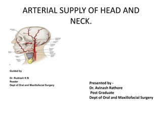
  • 1. ARTERIAL SUPPLY OF HEAD AND NECK. Guided by Dr. Rudresh K B Reader Dept of Oral and Maxillofacial Surgery Presented by - Dr. Avinash Rathore Post Graduate Dept of Oral and Maxillofacial Surgery
  • 2. CONTENTS- • EMBRYOLOGY • HISTOLOGY • COMMON CAROTID ARTERY • EXTERNAL CAROTID ARTERY • INTERNAL CAROTID ARTERY • SUBCLAVIAN ARTERY • LIGATIONS OF ARTERIES • REFRENCES DEVELOPMENT
  • 4. Aortic arches The aortic arches are a series of six paired embryological vascular structures which give rise to several major arteries. Aortic arches are short vessels connecting ventral and dorsal aortae on each side, they run within branchial (pharyngeal) arches are based gradually in the 4th and 5th week, in 6 pairs in total. The first, second and fifth pairs soon disappear.
  • 5. The 1st aortic arch – disappears (a small portion persists and forms a piece of the maxillary artery) The 2nd aortic arch – disappears (small portion of this arch contributes to the hyoid and stapedial arteries) The 3rd aortic arch - has the same development on the right and left side it gives rise to the initial portion of the internal carotid artery The external carotid is derived from the cranial portion of the ventral aorta The common carotid corresponds to a portion of the ventral aorta between exits of the third and fourth arches
  • 6. The 4th aortic arch - has ultimate fate different on the right and left side On the left - it forms a part of the arch of the aorta between left common carotid and left subclavian artery On the right - it forms the proximal segment of the right subclavian artery The 5th aortic arch - is transient and soon obliterates
  • 7. • The 6th aortic arch - pulmonary arch - gives off a branch on each side that grows toward the developing lung bud • The proximal part of the sixth right arch persists as the proximal part of the right pulmonary artery while the distal section degenerates The sixth left arch gives off the left pulmonary artery and forms the ductus arteriosus; this duct remains pervious during the whole of fetal life, but then closes within the first few days after birth due to increased O2concentration
  • 8. • The outermost layer is known as the tunica externa also known as tunica adventitia and is composed of connective tissue made up ofcollagen fibers. • Inside this layer is the tunica media, or media, which is made up of smooth muscle cells and elastic tissue (also called connective tissue proper). • The innermost layer, which is in direct contact with the flow of blood is the tunica intima, commonly called theintima. This layer is made up of mainly endothelial cells. The hollow internal cavity in which the blood flows is called the lumen. Arteries form part of the circulatory system. They carry blood that is oxygenated after it has been pumped from the heart. Arteries also aid the heart in pumping blood. Arteries carry oxygenated blood away from the heart to the tissues, except for pulmonary arteries,which carry blood to the lungs for oxygenation.
  • 9.
  • 10.
  • 11. The Structure of Blood VesselsA Comparison of a Artery and a Vein- Figure 13-1
  • 12.
  • 13. • MAJOR ARTERIES OF HEAD AND NECK 1. COMMON CAROTID ARTERIES 2. EXTERNAL CAROTID ARTERIES 3. INTERNAL CAROTID ARTERIES  ADDITIONAL ARTERIES - BRANCHES OF SUBCLAVIAN ARTERY
  • 15. 15 Cervical part of common carotid artery  Carotid arteries are generally symmetric and approximately of same size unlike vertebral arteries.  In 75% individuals ,CCA bifurcates at the level of C3-C4,roughly at the upper border of thyroid cartilage.  In children,the carotid bifurcates one vertebral level higher.  Variation in the level of bifurcation Highest seen in –C1 to C2 Lowest seen in –T1 to T2 Common carotid artery
  • 16. 16 CAROTID BODY(CHEMORECEPTORS)  It is normally 2.5x 5mm to 4 x 7mmflattened structure on the median and deep side of the upper end of the common carotid artery.  Blood supply-from small vesssels usually from ECA,termed as GLOMIC ARTERY OR ASCENDING PHARYNGEAL ARTERY.  NERVE SUPPLY-9TH 10TH 12TH CN
  • 17. 17 CAROTID SINUS (BARORECEPTORS)  At the bifurcation of common carotid artery  Responsive to changes in the arterial blood pressure.It acts as a baroreceptor(pressure receptor) and regulates blood pressure.  BLOOD SUPPLY-ICA  NERVE SUPPLY-carotid sinus nerve or nerve of hering.
  • 18. RELATION OF COMMON CAROTID ARTERY LATERAL-IJV ANTEROLATERALLY SKIN,FASCIA,SCM,STERNOHYOID,STERNOTH YROID,SUP BELLY OF OMOHYOID POSTEROLATERALLY- VAGUS NERVE MEDIAL PHARYNX, LARYNX, TRACHEA ,OESOPHAGUS LOBE OF THYROID GLAND POSTERIOR Transverse process of C3 C4 PREVERTBRAL FASCIA SYMPATHETIC TRUNK
  • 19. APPLIED ANATOMY Carotid sinus syndrome • Loss of consciousness due to simple head movements. • Hypersensitivity of the carotid sinus due to an unknown etiology. • Sudden slight pressure changes, such as that occasioned by movement of the head, may result in stimulation of the carotid sinus. • Impulses transmitted by the sinus reduce blood pressure and slow the pumping action of the heart. Thus decreasing blood supply to the brain and resulting in sudden loss of consciousness. While supporting the mandible care should be taken not to apply pressure on the carotid sinus.
  • 20. 21 External carotid artery (Facial carotid)
  • 21. External carotid artery • Generally,it lies anterior to the internal carotid artery. • It is the chief artery of supply to structures in the front of the neck and in the face.
  • 22. SURFACE MARKING • ECA is marked by joining the following two points. -A) point on the anterior border of the sternocleidomastoid muscle at the level of the upper border of the thyroid cartilage. -B) second point on the posterior border of the neck of the mandible. The artery is slightly convex forwards in its lower half and slightly concave forwards in its upper half. B A
  • 23. COURSE • ECA begins in the carotid triangle at the level of upper border of thyroid cartilage opposite the disc between the third and fourth cervical vertebrae. • In the carotid triangle,it lies under cover of the anterior border of the sternocleidomastoid muscle • As the artery ascends,it passes deep to the post. Belly of digastric and stylohyoid muscle and terminates behind the neck of the mandible by dividing into the maxillary and superficial temporal arteries.
  • 24. Has slightly curved course,so that it is anteromedial to ICA in it lower part,and anterolateral to the ICA in its upper part.
  • 25.
  • 26.
  • 29. 30 Origin and course: Arises from ECA immediately above the bifurcation of CCA Curves anteriorly and downwards to enter Thyroid gland
  • 31. 32
  • 32. RELATION WITH EXTERNAL LARYNGEAL NERVE
  • 33. APPLIED ANATOMY • The arch of superior thyroid artery is characteristic – diagnostic landmark • The artery and external laryngeal nerve are close to each other higher up, but diverge slightly near the gland. - So, ligature of superior thyroid artery in thyroid surgery should be made close to the gland in order to avoid injury of the external laryngeal nerve. -Damage to the external laryngeal nerve causes some weakness of phonation due to loss of tightening effect of the cricothyroid on the vocal cord. • Intra-arterial infusion chemotherapy for laryngeal and hypopharyngeal cancers.
  • 34. Lingual Artery Origin-  Lingual Artery arises from the ECA opposite the tip of greater cornu of the hyoid bone Course-  First part of artery lies in the carotid triangle  Second part of artery lies deep to the hyoglossus muscle which separates it from the hypoglossal nerve  Third Part or deep part : runs upwards along the anterior margin of the hyoglossus
  • 35. Branches of Lingual Artery  Suprahyoid Br  Dorsal Lingual Br  Deep Lingual Artery  Sublingual Artery
  • 36.
  • 37. APPLIED ANATOMY • In surgical removal of tongue , first part of artery is ligated before it gives any branches to the tongue or tonsil. LIGATION OF LINGUALARTERY : Incision – circling the lower pole of submandibular gland. - Skin, platysma, deep fascia incised, submandibular gland exposed , lifted, tendon of digastric visible.
  • 38. - Free border of mylohyoid muscle seen, hypoglossal nerve identified. Digastric tendon pulled downwards –enlarges the digastric triangle, hyoglossus muscle visible. - Muscle divided bluntly, in the gap of its vertical fibers lingual artery found & ligated.
  • 39. SUBLINGUALARTERY Injury occurs in premolar & molar region, when sharp instrument or rotating disks slips off a lower molar & injure the floor of mouth. -May present problems to the surgeon attempting to ligate its source because it may arise from the submental branch of the facial artery rather than from the lingual artery.
  • 40. For Implants, CBCT to localise the vascular canal,injuries to arteries in vascular canal can cause sublingual haematoma leading to blockage of airway.
  • 42. Facial Artery Facial artery is the chief artery of the face Origin :  Arises from the ECA just above the greater cornu of the hyoid bone  It has two parts, first cervical part in the neck and facial part.  It enters the face by winding around the base of the mandible  At the anteroinferior angle of the masseter muscle, it can be palpated here and is called as an “anaesthetist’s artery”
  • 43. •SURFACE MARKINGS-ANTERO INFERIOR BORDER OF MASSETER • 1.25 CM LATERAL TO ANGLE OF MOUTH • MEDIAN ANGLE OF EYE •TORTUOUS COURSE •PULSATIONS FELT AT- LOWER BORDER OF MANDIBLE ORAL CAVITY
  • 44. Branches of Cervical part 1. Ascending palatine artery- it supplies to root of tongue & tonsil. 2. Tonsillar artery 3. Submental artery- it is a large artery which accompanies the mylohyoid nerve, and supplies the submental triangle and sub lingual salivary gland. 4. Glandular branches that supplies submandibular salivary gland and submental lymph nodes.
  • 45. ASCENDING PALATINE ARTERY • ORIGIN FROM HIGHEST POINT OF FACIAL ARTERY • COURSES CRANIALLY ALONG THE SUPERIOR CONSTRICTOR OF PHARYNX TO REACH SOFT PALATE • SMALL BRANCH TO PHARYNGEAL MUSCLES • TONSILLAR BRANCH SUPPLYING PALATINE TONSIL(MAY ARISE DIRECTLY FROM FACIAL ARTERY) TERMIAL BRANCHES SUPPLY SOFT PALATE TONSILLAR A RTERY: Passes between the styloglossus and medial pterygoid muscles and pierces the superior pharyngeal constrictor muscle to supply the palatine tonsil and the posterior tongue.
  • 46. SUBMENTAL ARTERY • RUNS HORIZONTALLY BELOW THE INFERIOR BORDER OF MANDIBLE • TRAVERSES HORIZONTALLY TO REACH MYLOHYOID MUSCLE • SUPPLIES SUBMANDIBULAR NODES,MYLOHYOID AND SURROUNDING MUSCLE • ANASTAMOSE WITH SUBLINGUAL AND INFERIOR LABIAL ARTERY
  • 47. Branches of facial part 1. Superior labial- supplies to upper lip & antero-inferior part of nasal septum. 2. Inferior labial- supplies to lower lip. 3. Lateral nasal- to the ala & dorsum of nose. 4. Angular – supplies the lacrimal sac and orbicularis oculi.
  • 48. FACIAL PART: INFERIOR LABIAL ARTERY: Originates near the corner of the mouth, passes deep to the depressor anguli oris muscle, and pierces the orbicularis oris muscle. -The artery courses superficial to that muscle, supplying it as well as the substance of the lower lip. -It forms an anastomosis with its counterpart of the other side and with branches of the mental and submental arteries.
  • 49. SUPERIOR LABIALARTERY: Arises just above the inferior labial artery. It passes superficial to the orbicularis oris muscle in the upper lip to serve that muscle as well as the substance of the upper lip. - It sends a small twig, the SEPTAL BRANCH to supply anteroinferior part of the nasal septum and another one, the ALAR BRANCH, into the wing of the nose. -The terminus of the vessel will anastomose with its counterpart of the opposite side.
  • 50. LATERAL NASALARTERY: Small branch arising at and passing into the wing and bridge of the nose. -This supplies ala and dorsum of the nose. This vessel will anastomose with various other arteries in its vicinity. ANGULAR ARTERY: Is the terminal continuation of the facial artery, supplying the tissues in the vicinity of the medial corner of the eye and anastomosing with dorsal nasal branch of the ophthalmic artery.
  • 52. APPLIED ANATOMY • Facial Artery Compression: Applying pressure to the facial artery as it passes over the inferior border of the mandible just anterior to the angle will diminish blood flow to that side. o Can be injured –during operative procedures on lower premolars & molars, if instrument enters the cheek at inferior vestibular fornix., also while attempt to open a buccal abscess or mucocoele.
  • 53. • In mand. 1st molar region care must be taken not to injure the facial artery while extending the vertical incision down the vestibule during surgical extraction of mandibular impaction. • So it is recommended that start vertical incision from the vestibule in upward direction. • While excising the submandibular gland,the facial artery should be ligated at two points and should be secured before dividing it, otherwise it may retract through stylomandibular ligament causing serious bleeding.
  • 55. Ascending Pharyngeal Artery  A small branch arises from medial side of ECA  Long, slender vessel, deeply seated in the neck  COURSE: Ascends vertically between the internal carotid and the side of the pharynx, to the under surface of the base of the skull, lying on the Longus capitis.
  • 56. BRANCHES • PHARYNGEAL BRANCHES • PALATINE BRANCH • PREVERTEBRAL BRANCH • INFERIOR TYMPANIC ARTERY • MENINGEAL BRANCHES
  • 58. Posterior Auricular Artery  Small and arises above the posterior belly of digastric  It runs upwards and backwards deep to the parotid gland, crosses the base of the mastoid process and ascends behind the auricle.  Stylomastoid branch
  • 59. OCCIPITAL ARTERY ORIGIN:Arises in carotid triangle from posterior aspect of ECA ,opposite the origin of facial artery. -It is crossed at its origin by hypoglossal nerve. COURSE: Passes backwards and upwards along & under cover of lower border of post. Belly of digastric , crossing carotid sheath, hypoglossal & accessory nerves. Then it runs deep to the mastoid process and muscles attached to it i.e.,sternocleidomastiod, digastric etc.
  • 60. Then crosses the rectus capitus lateralis,superior oblique,and semispinalis capitus muscle at the apex of the posterior triangle. Finally it pierces the trapezius muscle and ascends in a tortuous course in the superficial fascia of the scalp. Its terminal portion comes to lie along the greater occipital nerve.
  • 61. BRANCHES IN THE CAROTID TRIANGLE • STERNOMASTOID BRANCHES – Two in no.,upper branch accompanies the accessory nerve and lower branch arises near the origin of the occipital artery. Supplies sternomastoid m. IN THE POSTERIOR TRIANGLE and SCALP REGION: • AURICULAR BRANCH: Passes superficial to the mastoid process to reach and supply the back of the auricle.
  • 62. • MASTOID BRANCH:– Enters cranial cavity through mastoid foramen, supplies mastoid air cells in the dura and diploe. • MENINGEAL BRANCH – Ascends with the internal jugular vein and enters the skull through jugular foramen & condylar canal, supplies dura of posterior cranial fossa. • MUSCULAR BRANCH- Supply the Digastric, Stylohyoideus, Splenius, and Longissimus capitis.
  • 63. DESCENDING BRANCH : • The largest branch of the occipital, descends on the back of the neck, and divides into a superficial and deep portion. -The superficial portion runs beneath the Splenius, giving off branches which pierce that muscle to supply the Trapezius and anastomose with the ascending branch of the transverse cervical artery. -The deep portion runs down between the Semispinales capitis and colli, and anastomoses with the vertebral and with the a. profunda cervicalis, a branch of the costocervical trunk.
  • 64. • The terminal branches of the occipital artery(occipital branches) are distributed to the back of the head: they are very tortuous, and lie between the integument and Occipitalis, anastomosing with the artery of the opposite side and with the posterior auricular and temporal arteries, and supplying the Occipitalis, the integument, and pericranium
  • 65. APPLIED ANATOMY Superficial branch anastomosis with ascending branch of transverse cervical artery. Deep branch of descending br of occipital artery anastomosis with deep cervical artery.
  • 67.  Larger of the two terminal branches  Arises behind the neck of the mandible, and is embedded in the substance of the parotid gland  It supplies the deep structures of the face Maxillary Artery
  • 68.
  • 69. Branches 1st part (mandibular) : Lies medial to mandible, it runs along the lower border of lateral pterygoid muscle  Deep auricular artery  Ant.tympanic artery  Middle meningeal artery  Accessory meningeal artery  Inferior alveolar artery
  • 70. Branches of first and its supply Branches 1.Deep auricular 2.Anterior tympanic 3.Middle meningeal 4.Accessory meningeal 5.Inferior alveolar Foramen transmitting Foramen in the floor of external acoustic meatus Petrotympanic fissure Foramen spinosum Foramen ovale Mandibular foramen Distribution External acoustic meatus,outer surface of tympanic membrane Inner surface of tympanic membrane 5th and 7th nerve, middle ear, tensor tympani Meninges, Structures in the infra temporal fossa Lower teeth and mylohyoid muscle
  • 71.  Largest artery that supplies the dura  It ascends to the foramen spinosum through which it enters the cranium  Divides into two branches, anterior and posterior.  It supplies the dura mater (the outermost meninges) and the calvaria. Middle Meningeal Artery
  • 72. APPLIED ANATOMY • FRONTAL BRANCH – Extradural hemorrhage -hematoma presses on the motor area – hemiplegia of opposite side APPROACH- hole in the skull over pterion – 4 cm above mid point of zygomatic arch.(approx 2 finger breadth above zygomatic arch) • PARIETAL OR POSTERIOR BRANCH - contralateral deafness APPROACH- hole is made 4cm above and 4cm behind the external acoustic meatus.
  • 73.
  • 74. Inferior alveolar artery  Runs downward & forward medial to ramus of mandible to reach mandibular foramina.  Before entering mandibular foramina gives off lingual and mylohyoid arteries.  In canal gives branches to mandibular teeth .  After coming out of canal supply chin via mental artery.
  • 75. BRANCHES BEFORE ENTERING MANDIBULAR CANAL: • Lingual branch to the tongue. • Mylohyiod branch to the mylohyiod muscle. WITHIN THE MANDIBULAR CANAL: Branches to the mandible Branches to the roots of each teeth upto midline(dental branches) Incisor branch anastomoses with the branch from opposite side. AFTER EMERGING FROM MENTAL FORAMEN: mental branch escapes with the nerve at the mental foramen, supplies the chin, and anastomoses with the submental and inferior labial arteries
  • 76. 2nd part (pterygoid part) :  Artery runs forward &upward superficial to the lower head of the lateral pterygoid muscle
  • 77. B. Second part Branches 1.Deep temporal 2.Pterygoid 3.Masseteric 4.Buccal Distribution Temporalis Lateral and medial pterygoid Masseter Buccinator
  • 78. 3rd part (pterygopalatine):  Terminal portion of the artery passes between the two heads of the lateral pterygoid muscle
  • 79. THIRD PART Branches 1.Post superior alveolar 2.Infraorbital 3.Greater palatine 4.Pharyngeal 4.Artery of pterygoid canal 5.Sphenopalatine(terminal part) Foramina Alveolar canals in the body of maxilla Infraorbital fissure Greater palatine canal Pharyngeal canal Pterygoid canal Sphenopalatine foramen Distribution Upper molar and premolar teeth ; maxillary sinus Lower orbital muscles, lacrimal sac ,max sinus Soft palate, tonsil, palatine glands and mucosa,upper gums Root of nose , pharynx, auditory tube,sphenoidal sinus Auditory tube, upper pharynx, middle ear Lateral and medial wall of nose and air sinuses.
  • 80. APPLIED ANATOMY • Site of hematoma during PSA block. • Produces largest and most esthetically unappealing hematoma. • Blood effuses until extravascular pressure exceeds intravascular pressure or clotting occurs. • Infratemporal fossa into which bleeding occurs accommodates large amount of blood. • Prevented by aspirating before giving LA in the site. • Digital pressure can be applied medial and superior to the maxillary tuberosity.
  • 81. Applied anatomy • In case of abscess from palatal root of first molar,incision should be made in a antero-posterior direction parallel to the artery.
  • 82. • During lefort I osteotomy: • Greater palatine artery is easily injured during osteotomy of the medial or lateral maxillary sinus walls, pterygomaxillary dysjunction or during down fracturing of maxilla. • The average distance from the piriform rim to the descending palatine artery is 35.4 mm, range is 31 to 42 mm. • The average length of the greater palatine canal above the nasal floor is 10mm, range is 6 to 15 mm. • The average distance between the pterygomaxillary fissure and the greater palatine foramen is 6.6mm.
  • 83. GUIDELINES TO AVOID INJURY: • Osteotomy of lateral wall of maxillary sinus should extend just beyond the second molar. • Osteotomy of medial wall of maxillary sinus should usually extend 30mm posterior to the piriform rim in females,in males it can be carried back to 35mm - --O’ RYAN • Because the descending palatine artery travels in an anterior- inferior direction as it enters the greater palatine canal ,injury can be prevented by closely adapting the cutting edge of the osteotome or the saw to the pterygomaxillary fissure.
  • 84. APPLIED ANATOMY OF MAXILLARY ARTERY • Surgeries involving condyle-Avoid injury to maxillary artery as it lies medial to condyle. • Ankylotic mass of TMJ may encircle the artery.So it is advisable to remove ankylotic mass in pieces rather than in toto. • Trismus involving lateral pterygoid comprises blood supply to the nose.
  • 85. • During Le fort I osteotomy procedure- Pterygopalatine portion of maxillary artery may be injured during fracturing the pterygiod plates if Tessier’s osteotome is directed backwards. -It should be directed downwards and medially.
  • 86. • Can be used as arterial donor in repair of ICA dissections and aneurysms, due to close proximity of the artery to the cranial base. • Control of epistaxis---If epistaxis is not controlled after nasal packing,it can be controlled by ligating IMA via endonasal,transantral or intraoral approach.
  • 87. LITTLE’S AREA or KIESSELBACH’S PLEXUS -Near the anteroinferior part or vestibule of the septum. -Contains anastomoses between • Superior labial branch of facial artery • Branch of sphenopalatine artery • Anterior ethmoidal artery • Greater palatine artery This is common site of bleeding from nose or epistaxis.
  • 88.  Smaller of the two terminal branches  It begins in the substance of the parotid gland, behind the neck of the mandible  Divides into two branches, a frontal and a parietal Superficial Temporal Artery
  • 89. Parietal branch Frontal branch Middle temporal artery Transverse facial artery
  • 90.  Transverse facial branch  Anterior auricular branch  Frontal branch  Parietal branch  Zygomatico- orbital branch Branches
  • 91. Transverse Facial Artery: ORIGIN:From STA before it leaves parotid gland. COURSE: Running forward through the substance of the gland, it passes transversely across the side of the face, between the parotid duct and the lower border of the zygomatic arch. This vessel rests on the Masseter, and is accompanied by one or two branches of the facial nerve. SUPPLIES: The parotid gland and duct, the Masseter, and the integument, and anastomose with the external maxillary, masseteric, buccinator, and infraorbital arteries.
  • 92. Middle Temporal Artery: Arises immediately above the zygomatic arch, and, perforating the temporal fascia, gives branches to the Temporalis, anastomosing with the deep temporal branches of the internal maxillary artery. - It occasionally gives off a zygomaticoorbital branch, which runs along the upper border of the zygomatic arch, between the two layers of the temporal fascia, to the lateral angle of the orbit. -This branch, which may arise directly from the superficial temporal artery, supplies the Orbicularis oculi, and anastomoses with the lacrimal and palpebral branches of the ophthalmic artery.
  • 93. • Anterior Auricular Branches : Distributed to the anterior portion of the auricle, the lobule, and part of the external meatus, anastomosing with the posterior auricular.
  • 94. Frontal Branch : Runs tortuously upward and forward to the forehead, supplying the muscles, integument, and pericranium in this region, and anastomosing with the supraorbital and frontal arteries. Parietal Branch: Larger than the frontal, curves upward and backward on the side of the head, lying superficial to the temporal fascia, and anastomosing with its fellow of the opposite side, and with the posterior auricular and occipital arteries.
  • 95. APPLIED ANATOMY • Control of temporal haemorrhage. • Anaesthetist’s artery • Placement of incisions in craniotomy • In reduction of zygomatic arch fractures – Gilli’s approach -A 2cm incision is placed in the temporal region at an angle 45 degree to the zygomatic arch, between two branches of the superficial temporal artery and parallel to the anterior branch.
  • 96. INTERNAL CAROTID ARTERY • Origin- • It is one of the terminal branch of common carotid artery originates along with external carotid artery at the upper border of thyroid cartilage at the disc of third and fourth cervical vertebra.
  • 97.
  • 98. • It supplies two of the four major arteries supplying blood to the brain. • CCA CAROTID CANAL(petrous part of temporal bone) MIDDLE CRANIAL FOSSA(dorsum sellae of sphenoid bone) Supplies the hypophsis cerebri,orbit,and most of the supratentorial part of the brain.
  • 99. BRANCHES and SEGMENTS • 1996 –bouthillier divided ICA into 7 anatomical segments viz. • C1-Cervical part in the neck • C2-Petrous part in the petrous temporal bone • C3-lacerum • C4-Cavernous part in the cavernous sinus • C5-clinoid • C6-opthalmic • C7-communicating CEREBRAL PORTION
  • 100. Cervical part • It ascends vertically in the neck from its origin to the base of skull to reach the lower end of the carotid canal. This part is enclosed in carotid sheath along with internal jugular and vagus nerve. No branches arises from the internal carotid artery in the neck. • Its initial part shows slight dilatation, carotid sinus. Which acts as a baroreceptor.
  • 101. PETROUS PART• Within the petrous part of the temporal bone,in the carotid canal runs upward forward & medially at rt. Angle. Branches- 1) Caroticotympanic- enter middle ear & anastomose with ant. & post. Tympanic branches 2) Artery of the Pterygoid Canal- anastomose with greater palatine artery
  • 102. CAVERNOUS PART Within the Cavernous Sinus • Branches 1) Artery to trigeminal ganglion 2) Superior & inferior Hypophyseal artery
  • 103. CEREBRAL PART • Lies at the base of the brain after emerging from the cavernous sinus Branches 1.Ophthalmic. 2.Anterior Cerebral. 3.Middle Cerebral. 4.Posterior Communicating. 5. Ant. choroidal On angiogram internal carotid show ‘S’ shaped figure ( carotid siphon )
  • 104. Opthalmic artery • Arises medial to anterior clinoid process near optic canal • In orbit lie inferolateral to optic nerve Branches: 1. Central artery of retina 2. Large lacrimal artery 3. Dorsal nasal artery • Supratrochlear • Supraorbital • Posterior ethmoidal • Anterior ethmoidal • Palpebral branch • Recurrent meningeal a. • Muscular a.
  • 105. SUBCLAVIAN ARTERY • MAIN ARTERY OF UPPER LIMB ORIGIN- 1.RIGHT- BRACHIOCEPHALIC ARTERY 2.LEFT -ARCH OF AORTAE COURSE 1.MEDIAL 2.POSTERIOR 3.LATERAL
  • 106.
  • 107. Circle of Willis The circle of Willis is a circulatory anastomosis that supplies blood to the brain and surrounding structures. It is named after Thomas Willis (1621– 1675), an English physician Anterior cerebral artery (left and right) Anterior communicating artery Internal carotid artery (left and right) Posterior cerebral artery (left and right) Posterior communicating artery (left and right) Basilar artery IMPORTANCE: The arrangement of the brain's arteries into the circle of Willis creates collaterals in the cerebral circulation. If one part of the circle becomes blocked or narrowed (stenosed) or one of the arteries supplying the circle is blocked or narrowed, blood flow from the other blood vessels can often preserve the cerebral perfusion well enough to avoid the symptoms of ischemia.
  • 108. Anastomoses ICA ECA Dorsal Nasal Artery and Angular Artery Dorsal Nasal Artery (branch of the Ophthalmic artery) Angular Artery (branch of the Facial Artery) Supraorbital Artery and Frontal Artery Supraorbital Artery (branch of the Ophthalmic) Frontal Artery (terminal branch of the Superficial Temporal Artery) Zygomatico Artery and Transverse facial artery Zygomatico (branch Lacrimal Artery) Transverse Facial Artery (branch of Superficial Temporal Artery) Branches of the Posterior Ethmoidal Artery and branches of the Sphenopalatine Artery Posterior Ethmoidal Artery Sphenopalatine Artery(branch of the Internal Maxillary) Cavernous branches and Middle Meningeal artery Cavernous branches from the cavernous portion of the ICA Middle Meningeal Artery (branch of the Internal Maxillary)
  • 110.
  • 111. Common carotid artery- It can be compressed against the carotid tubercle, the anterior tubercle of the transverse process of vertebra C6 which lies at the level of cricoid cartilage. Carotidynia is a syndrome characterized by unilateral (one-sided) tenderness of the carotid artery, near the bifurcation. Carotid Sinus  Present at the termination of CCA. (or beginning of ICA.)  Tunica media is thin, tunica adventia is thick  Acts as BARORECEPTOR/PRESSURE RECEPTOR.
  • 112. • Carotid sinus hypersensitivity (CSH) is an exaggerated response to carotid sinus baroreceptor stimulation. It results in dizziness or syncope from transient diminished cerebral perfusion. • For these individuals, even mild stimulation to the neck results in marked bradycardia and a drop in blood pressure. Carotid Siphon of Angiogram  Siphon region is the most common site for atherosclerotic plaque formation in carotid artery
  • 113.  Carotid body situated behind the bifurcation of CCA  Act as a chemoreceptor & respond to change in the O2, CO2 and pH content of the blood  Carotid body paragangliomas are vascular lesions, and this is reflected in their imaging appearance. These lesions splay apart the internal (ICA) and external carotid arteries (ECA), and as it enlarges, it will encase, but not narrow the ICA and ECA. Head Neck Path.Dec 2009; 3(4): 303–306. Carotid Body
  • 114. • Definition • Why ligate? • Procedure Individual artery ligation -External Carotid Artery -Lingual -Sublingual -Facial -Maxillary -Sphenopalatine -Greater Palatine -Ant./Post. Ethamoidal -Internal Carotid Artery
  • 115. • LIGATION Means act of binding or tying of blood vessels with sutures or wires is called Ligation… First ligation was done by AMBROSE PARE in amputation procedure.
  • 116. WHY WE LIGATE VESSELS??? 1.AFTER AMPUTATION TO ARREST THE BLOOD FLOW. 2.IN WOUNDS OF ARTERIES WHERE HAEMMORRHAGE CAN’T BE CONTROLLED. 3.IN SECONDARY HAMMORAGHE WHERE THEY CAN’T BE CONTROLLED BY OTHER MEANS. 4.IN LOCAL HYPERTROPHIES TO ARREST THE NUTRITIONAL SUPPLY TO THAT AREA.
  • 117. • IN CASE OF ANEURYSMS • IN CASE OF MALIGNANT TUMOUR TO STOP THE BLOOD FLOW • IN ACUTE INFLAMMATION WHERE NEITHER RESECTION NOR AMPUTATION IS POSSIBLE • IN VARIOUS OPERATIVE PROCEDURES WHEN WE ENCOUTER VESSELS TO REDUCE BLOOD FLOW TO THAT REGION .
  • 118. PROCEDURE FOR LIGATION 1. EXPOSE THE SHEATH OF VESSEL 2. ISOLATE THE VESSEL 3. PLACE THE LIGATURE
  • 120. EXPOSED AT TWO SITES 1. IN THE CAROTID TRIANGLE -AT ITS ORIGIN FROM THE COMMON CAROTID ( ABOVE THE ORIGIN OF SUPERIOR THYROID ARTERY) 2. IN THE RETROMANIBULAR FOSSA HERE WE LIGATE IT BEHIND THE ANGLE OF LOWER JAW ( DEALS WITH THE HAEMORRHAGE FROM ONE OF THE BRANCHES OF MAXILLARY ARTERY)
  • 121. • CONTINUE DOWNWARDS / TO THE ANTERIOR BORDER UP TO THE LEVEL OF CRICOID CARTILAGE • AFTER PENETRATING SKIN, PLATYSMA SUPERFICIAL SHEATH OF STERNOCLEDIOMASTOID IS INCISED EXPOSURE OF GREAT VESSEL • WITH BLUNT DISSECTION ANTERIOR BORDER IS EXPOSED, MUSCLE IS RETRACTED AND DEEP LAYER IS SEEN • IN THIS PART Internal Juglar Vein IS EXPOSED
  • 122. IDENTIFICATION OF EXTERNAL CAROTID ARTERY • THE JUGULAR VEIN IS MOBILIZED BY OPENING THE CAROTID SHEATH & FREE THE JUGULAR VEIN. • RETRACT POSTERIORLY VEIN TO VISUALIZE ARTERY
  • 123. EXPOSURE OF THE CAROTID BULB AND EXTERNAL CAROTID ARTERY • AS THE DISSECTION PROCEED POSTERIORLY THE CAROTID BULB IS IDENTIFIED AND BIFURCATION IS SEEN • MANIPULATION OF BULB AT THIS STAGE LEAD TO ARRYTHEMIA AND ANAESTHESIST SHOULD BE INFORMED
  • 124. • LIGATION EXTERNAL CAROTID ARTERY IS IDENTIFIED & LIGATED ABOVE THE SUPERIOR THYROID ARTERY • CLOSURE OF WOUND A VACCUM DRAIN IS PLACED AND WOUND IS SUTURED IN LAYERS
  • 125. COMPLICATIONS • HAMEORRHAGE DUE TO IJV OR ECA( profuse bleeding) • DAMAGE TO VAGUS NERVE (posteriomedially) • LIGATION OF ICA( contra lateral hemiplegia & blindness on the same side) • HEMATOMA FORMATION • INFECTION
  • 127. • INCISION • STARTS THE TIP OF MASTOID PROCESS AND CIRCLING THE MANDIBULAR ANGLE, CONTINUING FORWARD BELOW THE MANDIBLE FOR ABOUT ONE INCH • INCISION SHOULD BE AT EQUAL DISTANCE FROM THE POSTERIOR AND INFERIOR BORDER OF MANDIBLE
  • 128. EXPOSURE • AFTER THE BLUNT DISSECTION OF SKIN, SOME POST. FIBERS OF PLATYSMA, RETROMANDIBULAR VEIN OR EJV IS LOCATED, CUT & TIED • BRANCHES OF GREATER AURICULAR NERVE IS CUT & TIED TO PERMIT THE MOBILIZATION OF CERVICAL LOBE OF PAROTID GLAND • ATTACHMENT OF PAROTID WITH STERNOMASTOID AT ANTERIOR BORDER IS SEVERED & GLAND IS RETRACTED ANTERIORLY & UPWARDS
  • 129. IDENTIFICATION • UNDERNEATH THE PAROTID GLAND & POST. BELLY OF DIGASTRIC, SMALL THIN PART OF STYLOHYOID MUSCLE IS VISIBLE • ABOVE THIS- STYLOID PROCESS & STYLOMANDIBULAR LIGAMENT IS PALPATED • NOW MOVING THE JAW FORWARD ENTRANCE TO RETROMANDIBULAR FOSSA IS WIDENED & PULSE OF ECA IS FELT, ISOLATE & LIGATE IT
  • 131. LIGATION OF LINGUAL ARTERY INCISION • INCISION GIVEN BELOW THE LOWER BORDER OF MANDIBLE AFTER PALPATING THE SUBMANDIBULAR GLAND. • THE POSTERIOR PART OF INCISION SHOULD BE TOWARDS THE TIP OF MASTOID PROCESS AND ANTERIOR SHOULD POINT TOWARDS THE CHIN
  • 132. EXPOSURE OF ARTERY • AFTER BLUNT DISSECTION SUBMANDIBULAR GLAND IS EXPOSED • POST BELLY OF DIGASTRICS IDENTIFIED, MYLOHYOID MUSCLE REACHED, HYPOGLOSSAL NERVE AND ACCOMPANYING VEIN IDENTIFIED
  • 133. LIGATION OF ARTERY • DIGASTRICS TENDON PULLED DOWNWARD , HYOGLOSSUS MUSCLE DISSECTED AND LINGUAL ARTERY IS FOUND AND LIGATED • FIBERS OF HYOGLOSSUS MUSCLE SHOWS VERTICAL COURSE (THIN & FINE) WHILE THAT OF MYLOHYOID SHOWS OBLIQUE COURSE (THICK)
  • 134. LIGATION OF SUBLINGUAL ARTERY INDICATIONS • INJURY IS OBSERVED WHEN SHARP INSUMENTS OR ROTATING DISC ARE SKIPPED ON FLOOR OF THE MOUTH • IN VARIOUS SURICAL PROCEDURE LIKE RANULA AND TUMOURS OF SALIVARY GLANDS
  • 135. • DIFFICULT TO LIGATE SUBLINGUAL ARTREY MAY BE A BRANCH OF 1. LINGUAL ARTERY 2. SUBMENTAL ARTERY
  • 136. • INCISION • IN THE SUBLINGUAL GROOVE • STRUCTURES IN CLOSE ASSOCIATION • SUBLINGUAL GLAND(MED. &INF.) • SUBMANDIBULAR DUCT • LINGUAL NERVE(MED.& INF.) • HYPOGLOSSAL NERVE AND SUBLINGUAL VEIN
  • 138. LIGATION OF FACIAL ARTERY INCISION ½ INCH BELOW & PARALLEL TO THE LOWER BORDER OF MANDIBLE EXPOSURE THE SKIN, PLATYSMA MUSCLE AND DEEP FACIA ARE CUT, SOFT TISSUE IS BLUNTLY CUT AND RETRACTED
  • 139. LIGATION PULSE OF FACIAL ARTERY IS FELT & ARTERY IS ISOLATED AND LIGATED. • FACIAL ARTERY CROSSES THE LEVEL OF INFERIOR VESTIBULAR FORNIX IN THE REGION OF 1ST MANDIBULAR MOLAR. • DURING BUCCAL SPACE INFECTION THE ARTERY IS DISLOCATED • AVOID DEEP INCISION, INCISION SHOULD BE DOWNWARDS & INWARDS INSTEAD OF STRAIGHT UPWARDS.
  • 140. • Incision - at least half inch below the border of mandible & parallel to it.
  • 141. Skin,platysma and deep cervical fascia cut
  • 142. Skin,platysma and deep cervical fascia cut
  • 143. Artery is accompanied by facial vein & crossed superficially by marginal mandibular branch of facial nerve
  • 144. Pulse of facial artery felt. Artery- isolated, tied & cut
  • 146. LIGATION OF MAXILLARY ARTERY LIGATION CAUSES DECREASE IN INTRA VASCULAR PRESSURE GRADIENT, RESULTING IN HOMEOSTASIS APPROACHES: CAN BE DONE BY 1. TRANSANTRAL APPROACH & 2. INTRAORAL APPROACH
  • 147. TRANSANTRAL APPROACH BY CALD WELL LUC APPROACH PROCEDURE A LATERALLY BASED U SHAPE MUCOSAL INCISION IS CREATED POSTERIOR WALL OF MAXILLARY SINUS IS IDENTIFIED POSTERIOR MAXILLARY WALL IS REMOVED
  • 148. EXPOSURE & LIGATION • AREA IS ENLARGED,ARTERY IS IDENTIFIED &LIGATED SUCCESS RATE • 87% SUCCESS RATE
  • 149. INTRA ORAL APPROACH • THIS PROCEDURE IS GIVEN IN 1984 BY MACERI & MAKILSKI • LIGATE INFRATEMPORAL PORTION OF MAXILLARY ARTERY
  • 150. INDICATION: - IN CHILDREN AS AN ALTERNATE TO EMBOLIZATION & EXTERNAL ARTERY LIGATION FOR REMOVAL OF VASCULAR TUMOR - TO CONTROL BLEEDING IN VARIOUS MAXILLECTOMY PROCEDURES WHERE CALD WELL LUC IS CONTRAINDICATED
  • 151. PROCEDURE: - BY EXPOSING THE POSTERIOR PORTION OF MAXILLA THROUGH A POST. GINGIVOBUCCAL INCISION - A FINGER IS INSERTED INTO THE DEPTH OF WOUND TO PALPATE THE MAXILLARY ARTERY - THE NERVE HOOK IS USED FOR LIGATION
  • 153. LIGATION OF SPEHNOPALATINE ARTERY • CAN BE DONE BY TWO METHODS 1. TRANSANTRAL LIGATION 2. ENDOSCOPIC LIGATION
  • 154. TRANSANTRAL APPROACH • DESCRIBED BY SIMPSON et al. IN 1982 • APPROACH CALD WELL LUC • AVOID ENTRANCE TO PTERYGOPALATINE FOSSA • MEDIAL, POSTERIOR & INFERIOR WALL IS REMOVED • SPHENOPALATINE & VIDIAN NERVE IS DISSECTED & LIGATION OF ARETRY IS DONE
  • 155. ENDOSCOPIC LIGATION FOR SPHENOPALANTINE ARTERY • DESCRIBED BY WHITE (MODIFICATION OF SIMPSONS TECH) • APPROACH THROUGH 1. MEATAL ANTROSTOMY & 2. CANINE FOSSA • NOT USING WIDELY AS COSTLY • ADVANTAGES 1. REDUCE PATIENT DISCOMFORT AND 2. DURATION OF HOSPITALIZATION
  • 157. LIGATION OF GREATER PALATINE ARTERY ENDANGERED DURING MINOR SURGERY PROCEDURES AND DURING DENTAL TREATMENT • INCISION --FROM THE LINGUAL ROOT OF FIRST MOLAR IN AN ANTERIO POSTERIOR LINE IT SHOULD BE AS NEAR TO THE FREE MARGINS OF THE GINGIVA AS POSSIBLE --THE KNIFE EDGES SHOULD BE DIRECTED OUTWARDS AND UPWARDS , NOT STRAIGHT UPWARDS
  • 159. LIGATION OF ANTERIOR AND POSTERIOR ETHMOIDAL ARTERY INDICATION 1. WHEN LOCAL HAMEORRHAGE CAN’T BE CONTROLLD BY OTHER MEASURES 2. TO DECREASE BLOOD FLOW TO UPPER NASAL VAULT FROM THE INTERNAL CAROTID SYSTEM GENERALLY PERFORMED IN CONJUGATION WITH MAXILLARY ARTERY OR ECA
  • 160. FIRST DESCRIBED BY : KIRCHNER et al. IN 1961 INCISION A CIRCUMLINEAR INCISION IS NORMALLY MADE BETWEEEN THE INNER CANTHUS OF EYE AND MIDDLE OF NOSE(LYNCH INCISION)
  • 161. • PROCEDURE • THE PERIOSTEUM IS INCISED AND ELEVATED • THE FRONTOETHMOIDAL SUTURE LINE IS FOLLOWED IN A POSTERIOR DIRECTION ABOUT 14-22mm TO THE ANTERIOR ETHMOIDAL ARTERY AND ITS FORAMEN • THE POSTERIOR ARTERY IS LIES AT FURTHER AT VARIABLE DISTANCE • THE OPTIC NERVE LIES 4-7mm POSTERIOR TO POSTERIOR ETHMOIDAL FORAMEN
  • 163. LIGATION OF ICA GENERALLY IT IS NOT DONE AS THE CHANCES OF BRAIN DAMAGE (CONTRALATERAL SIDE HEMIPLEGIA) ARE THERE, BUT IN SOME SELECTIVE CASES WE HAVE TO LIGATE THE ICA AS IN CASES OF ICA ANEURYSMS AND HEAD INJURIES.
  • 164. WHY EVEN AFTER LIGATION OF CAROTID ARTERY BLEEDING PERSISTS???? • COLLATERAL CIRCULATION OF COMMON CAROTID OCCURS AS FOLLOWS: 1 OCCIPITAL ANASTOMOSIS--- B/W THE TRANSVERSE CERVICAL & DEEP CERVICAL BRANCHES OF SUBCLAVIAN ARTERY AND OCCIPITAL ARTERY 2 ANASTOMOSIS IN & AROUND THYROID GLAND B/W SUPERIOR THYROID BRANCH & INFERIOR THYROID BRANCH 3 ANASTOMOSIS B/W MIDDLE LINE B/W THE BRANCHES OF EXTERNAL CAROTID ARTERIES OF BOTH SIDES
  • 165. REFERENCES: • SICHER’S ORAL ANATOMY- 8TH EDITION • LORE AND MEDINA-6TH EDITION • PRINCIPLES OF SURGERY BY EDWARD WARN HEAD AND NECK SURGERY- OTOLARYNGOLOGY BY BYRON. J. BAILEY- 2ND EDITION • Human Anatomy by B.D. Chaurasia, 6th Edition,Vol 3. • Grey’s Anatomy • Netter atlas • JOURNAL OF MAXILLOFACIAL AND ORAL SURGERY- LOCATION OF DESCENDING PALATINE ARTERY DURING LEFORT I OSTEOTOMY • Int. JOURNAL OF Oral Maxillofac. Surg. 2017; 46: 845–850 http://dx.doi.org/10.1016/j.ijom.2017.03.005,