Life cycle of pinus
•Descargar como PPTX, PDF•
15 recomendaciones•19,960 vistas

This document provides information about the genus Pinus. It discusses the systematic position of Pinus, describing that it is in the division Gymnospermae, class Coniferopsida, order Coniferales, and family Pinaceae. It then discusses the geographical distribution, morphology, internal stem and leaf structure, reproduction, development of microsporangium and female cone, structure of ovule, pollination, embryology, and germination of Pinus. Key details include that Pinus is widely distributed in the Northern hemisphere, has scale and needle-like foliage leaves, and reproduces through monoecious cones that produce microspores and megaspores through meiosis.

Recomendados
Más contenido relacionado
La actualidad más candente
La actualidad más candente (20)
Similar a Life cycle of pinus
Similar a Life cycle of pinus (20)
Último
Último (20)
Life cycle of pinus
- 1. Dr. Avinash Gholave Department of Botany K. V. N. Naik Arts, Commerce And Science College Nashik. agholave@gmail.com
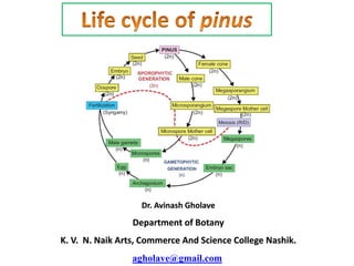
- 2. Introduction: Gymnosperms like all vascular plants have a sporophyte-dominant life cycle. The gametophyte (Gamete-bearing phase) is relatively short lived. Two spore types, microspores and megaspores are typically produced in pollen cones or ovulate cones, respectively. Genus pinus comes under the family pinaceae. Systematic position of Pinus: Division- Gymnospermae Class-Coniferopsida Order-Coniferales Family-Pinaceae Genus-Pinus.
- 3. Geographical distribution of Pinus: The genus Pinus is widely distributed in the Northern hemisphere. There are about 75 species of this genus. About six species have been recorded from different parts of our country. The blue pine, Pinus wallichiana (Syn. P. excelsa) is largely found in North-West Himalayan region at 1,800 metres to 7,000 metres elevation. In India the Pinus is represented by the following species. 1) Pinus gerardiana: Found in Kashmir and Himachal. 2) P. roxburghii: Found in Himalayas, kashmir, Himachal, Punjab and Uttaranchal. 3) P. wallichiana: Found in outer Himalayas, Kashmir, Punjab and Uttaranchal. 4) P. insularis: Found in Khasi hills, Nagaland and Burma. 5) P. merkusi: Found in east India and Burma. 6) P. armandi: Found in Arunachal Pradesh.
- 4. Morphology: Branches of unlimited growth/Long shoot: The main branches have an unlimited growth. They bear only scale Leaves. Branches of limited growth/ Dwarf shoot: Branches of limited growth or dwarf branches are produced in the axil of the scale leaves on the main branches. These are about 1-2cm Long. These are covered by one or two scale leaves. These branches also bear foliage leaves. A dwarf shoot with its foliage leaves is called spur. Leaf: Pinus has two types of Leaves: scale leaves and foliage leaves. Scale Leaves: The scale leaves are small. membranous and brownish in colour. These are protective in function. They are present on the long and dwarf shoots. Foliage Leaves: The foliage leaves are green and needle-like. They are found only on the dwarf shoots forming the spur. Roots: Pinus has a well developed tap root. It remains short and grows on hard ground or rocks. The lateral roots are well developed. These roots spread over a large area. Young roots are infested with a fungus to form mycorrhizae.
- 5. Internal Structure of the Stem: Stem is differentiated internally into epidermis, cortex, vascular tissue and central pith. 1) Epidermis: The surface is covered by an epidermis. It consists of a sing e layer of cells. Outer walls of these cells are highly cutinized. Below the epidermis is a hypodermis which is formed of Layers of lignified cells. 2) Cortex: The cortex is formed of parenchymatous cells. Cortex has a large number of resin canals. Each resin canal is surrounded by a layer of resin secreting glandular epithelial cells.
- 6. 3) Vascular bundle. The vascular bundles are conjoint collateral and open. They form a ring around the pith. In each vascular bundle the xylem is towards the inner side and phloem towards the outerside. A narrow strip of cambium is present between xylem and phloem. Pericycle is present outer to the ring of vascular bundles. A single layered thin walled endodermis is present outside the pericycle. Secondary growth takes place by the activity of cambium ring. There are distinct annual growth rings in the wood. 4) Bark Phellogen originates in the deeper layers of the cortex. It is present in the form of strips. It produces characteristic scaly bark. Fig. T. S. of pinus stem
- 7. Internal Structure of Leaf A transverse section shows following internal parts of the leaf: 1) Epidermis: Leaf s covered by thick walled epidermis. Epidermis s covered by a thick layer of cuticle. Sunken stomata are present below the general surface. Two or three Layered hypodermis is present underneath the epidermis. Hypodermis is composed of sclerenchymatous tissues. This hypodermis is the main strengthening tissue of the leaf. 2) Mesophyll: The mesophyll of the leaf is parenchymatous. It is not differentiated in to palisade and spongy parenchyma. Resin canals are present below the hypodermis. Each resin canal is lined by a layer of small epithelial cells. Each leaf is supplied by two unbranched veins. 3) Endodermis and vascular tissues: Endodermis is present outside the pericycle. Pericycle surrounds the xylem Pericycle is formed of parenchymatous cells. Its cell adjacent to the phloem are called aluminous cells. The cells adjacent to the xylem are called tracheidal cells. These specialized cells form the transfusion tissue. They help in the lateral flow of nutrients.
- 8. Internal Structure of Root: The internal structure of root resembles to that of a dicotyledonous root. In transverse section the root shows a piliferous layer bearing unicellular root hairs. The root hairs are found only in the young roots and root tips. In young roots there is fungal growth of ectophytic mycorrhiza. With the appearance of this fungus the root hairs of the root disappear. Just beneath the piliferous layer there lies a broad cortex which consists of 4 to 5 layers of thin-walled parenchymatous cells. The inner-most layer of the cortex is single- layered endodermis consisting of brown suberized cells containing tannin in them. Just below the endodermis there is multi-layered pericycle containing tannin and starch grains. Lateral roots are developed from the second layer of the pericycle. The outermost layer of the pericycle helps in the formation of the digestive sac which enables the lateral roots to penetrate through the cortex to the outside. In the center of the stele there are two to six Y-shaped xylem bundles alternating with them. The xylem has no true vessels and consists of tracheids. The phloem consists of sieve tubes and phloem parenchyma.
- 9. Reproduction: Pinus is monoecious. Plant develops both male and female strobili on the same plant. The strobili are monosporous. There is no vegetative reproduction in Pinus. Male Cone: The male cones are much smaller. They are produced in clusters near the tip of the long shoots. The male cones are produced in the spring. Each male cone has a central axis. It bears a number of spirally arranged microsporophyll's . Each microsporophyll has saclike microsporangia on the ventral side. Each Microsporangium produces a large number of microspores (pollen grains).The wall of each microspore (pollen grain) consists of inner intine and an outer exine. It has balloon like wings. The wings help in the dispersal of spores by wind.
- 10. Development of Microsporangium (Stamen) 1) A number of hypodermal cells act sporangial initials. The sporangial initials divides to form outer wall initials and the inner archesporial initial. 2) The wall initials divide to form a many layered wall of the sporangium. The archesporial initials also increase in number by the repeated divisions. The peripheral cells of the archesporium form the tapetum. 3.Some of the archesporial cells are transformed into microspore mother cells. The remaining archesporial cells and the tapetal layer provide nourishment to the developing microspore mother cells. 4. The microspore mother cell divides by meiosis to form four microspores or pollen grains. The exine of spore forms wings. The pollen grain divides in to smaller and larger cells. The smaller cell again divides to form two small prothalial cells. The larger cell becomes antheridial cell the sporangium splits and microspores are released from the microsporangia at this stage.
- 12. Female cone: Female cones are produced in the axils of the scale Leaves. The production of female cones is initiated in the winter. These become ready for pollination during the following spring. Each young female cone has a central axis. It bears spirally arranged scales. The scales are of two types. Some are thin membranous and are directly attached to the central axis they are called bract scales. Woody ovuliferous scales are present on the ventral surface of each bract scale. The broader end of the ovuliferous scale has projection called the umbo. Each ovuliferous scale bears two ovules. They are situated side by side on upper side each ovule (megasporangium) has a mass of nucellar tissue. They are surrounded by a single integument. The micropylar end of the ovule is directed towards the central axis. A single megaspore mother cells differentiated in the nucellus near the micropylar end. This megaspore mother cell undergoes meiosis to form four megaspores only the lower most megaspore remains functional the others disintegrate. Functional megaspore (embryo sac) increases in size. It occupies the major part of the nucellus. Pollination takes place at this stage. Images copyright to google
- 13. Structure of Ovule: In Pinus two anatropous ovules are formed on the upper side of the ovuliferous scale. Each ovule consists of a central mass of tissue called 'nucellus' which is surrounded by a covering called 'integument'. The integument arises from the base of the ovule covers nucelluson all sides, except at the top, where it leaves a small passage called 'micropyle. The integument is differentiated into an outer fleshy layer (sarcotesta), middle stony layer (sclerotesta) and the inner fleshy layer (sarcotesta). The micropyle leads to the top of the nucellus. Where nucellus develops a cup like chamber called "pollen chamber is present. In pollen chamber the pollen grains are lodged after the pollination. In Pinus nucellar beak is not formed. Both inner and outer vascular strands are absent. In nucellus, towards the micropylar end a single large cell gets differentiated. It is the archesporial cell. It divides periclinally into an outer parietal cell end inner sporogenous cell or megaspore mother cell. The parietal cell divides and develops into a nourishing layer. Megaspore mother cell undergose meiosis and forms a liner tetrad of megaspore of which the basal one is the functional megaspore. It is called as embryo sac cell. nucellus
- 14. Pollination: Each ovule secretes a mucilaginous drop at the micropylar end. A gap is produced between the ends of the ovuliferous scales. It forms a passage for the entry of pollen grains. Wind carried pollen grains. The mucilage drop entangles the pollen grain. Pollen grain is carried through the micropyle to the surface of the nucellus. Diploid nucleus divides thrice to form eight cells. The lower four cells becomes proembryonal cells. The upper four nuclei are separated by incomplete cell walls. Four proembryonal divides to produce three tiers of cells:
- 15. Embryonal cell; The cells of the lower tier become embryonal cells. The four embryonal cells separate from each other. Each develops into a separate embryo independently. Each embryonal cell forms secondary suspensor cells. The formation of more than one embryo from a single fertilized oosphere is called polyembryony. Only one embryo reaches maturity. The rest are aborted. Suspensor cells: The cells of the middle tier become suspensor cells. suspensor cells elongate very much. It pushes the developing embryos into the prothalial tissue for nutrition. Rosette cells: The cells of the upper most tiers are called the rosette cells. These cells do not take part in the development of the embryo. A fully developed embryo is in the form a short straight axis. Its radicle is present towards the micropylar end. Plumule is present towards the inner side. Plumule is surrounded by the cotyledons.The unutilized prothalial tissue forms the endosperm. The persistant nucellus tissues near the micropylar end form the perisperm. The integument becomes hard testa. Some part of the ovuliferous scale fuses with the developing seed. It makes a large wing for dispersal of seed. The axis of the female cone rapidly increases. It produces gaps in ovuliferous scales. The cone becomes woody for the dispersal of seeds
- 16. Germination of Seed The radicle grows out it splits the testa at the micropylar end. This radicle grows down into the soil and forms the primary root. The hypocotyl elongates to form a loop. Then it becomes straight. It carries with it the plumule and the cotyledons. The testa is also carried up with the cotyledons.