
Recomendados
Más contenido relacionado
La actualidad más candente
La actualidad más candente (20)
Similar a Malaria
Similar a Malaria (20)
Último
Último (20)
Malaria
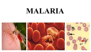
- 1. MALARIA 1
- 2. Introduction: • Linked with poisonous vapors of swamps or stagnant water. • Italian: Mala = Bad ; aria = Air • Term used by Italians to describe cause of intermittent fevers associated with exposure to marsh air. • Other names: Jungle fever, Marsh fever, Swamp fever. • Malaria continues to be most important cause of fever and morbidity in the Tropical world. • “HIV, Malaria and TB are among the most important infectious agents in the world. • Malaria has been eradicated from Europe, Most of North America, USA, Korea and Japan. 2
- 3. Introduction • Malaria is probably one of the oldest diseases known to mankind that has had profound impact on our history. • Malaria is a vector-borne infectious disease caused by single-celled protozoan parasites of the genus Plasmodium. • Malaria is transmitted from person to person by the bite of female mosquitoes. • Malaria remains the world's most devastating human parasitic infection. • Malaria affects over 40% of the world's population. • WHO, estimates that there are 350 - 500 million cases of malaria worldwide, of which 270 - 400 million are Falciparum malaria, the most severe form of the disease. 3
- 5. Malaria Kills more people than AIDS • Malaria kills in one year what AIDS kills in 15 years. • For every death due to HIV/AIDS there are about 50 deaths due to malaria. • To add to the problem is the increasing drug resistance to the established drug. 5
- 6. Ancient History of Malaria • Malaria parasites have been with us since the dawn of time. They probably originated in Africa (along with mankind), and fossils of mosquitoes up to 30 million years old, show that the malaria vector, the malaria mosquito, was present well before the earliest history. • Hippocrates, a physician born in ancient Greece, today regarded as the "Father of Medicine", was the first to describe the manifestations of the disease. 6
- 7. History • 1880 - Charles Louis Alphose Lavern discovered malarial parasite. • 1891 - Polychrome stain- Romanowsky. • 1898 - Italian scientists Amico Bignami, Battista Grassi and Giovanni Bastianelli - Life cycle of parasite. • 1902 – Ronald Ross was awarded Nobel Prize for demonstration of vector Anopheles mosquito while serving in India. • 1976 – Trager and Jensen in vitro cultivation of parasite. 7
- 8. Nature of parasite as Drawn by Lavern 8
- 9. What causes Malaria? • Malaria is caused by a parasite called Plasmodium, which is transmitted via the bites of infected mosquitoes. In the human body, the parasites multiply in the liver, and then infect red blood cells. 9 •Malaria is caused by an infection by one of four single celled Plasmodia species, they are: falciparum, vivax, malariae, and ovale. •The most dangerous of the four is P. falciparum.
- 10. • Phylum: Apicomplexa • Class: Haematozoea • Order: Haemosporida • Genus: Plasmodium 10
- 11. Etiology of Malaria Four Species known to infect Human • Plasmodium vivax – Benign Tertian Malaria • P. ovale - Ovale tertian Malaria • P. malariae – Quartan malaria • P. falciparum – Falciparum malaria or Malignant Tertian malaria. • Falciparum accounts for 90% of deaths due to malaria and vivax is the most widely spread species because it exists in both temperate and tropical climates. • The malaria life cycle is a complex cycle with both sexual and asexual aspects. 11
- 12. Life Cycle: • Intermediate host : Human • Definitive host : Mosquito • Infective stage : Sporozoite • Infective way : mosquito bite skin of human • Parasitic position : liver and red blood cells • Transmitted stage : gametocytes 12
- 13. • Human Cycle: (Intermediate host) – Primary exoerythrocytic / pre- erythrocytic schizogony – Erythrocytic schizogony – Gametogony – Secondary exoerythrocytic or dormant schizogony • Mosquito cycle (Definitive host) – Sporogony 13
- 14. 14
- 15. Primary exoerythrocytic or pre-erythrocytic schizogony: • Sporozoites leave the blood stream within one hour and enter liver parenchyma cells. • Becomes rounded and multiple nuclear division, followed by cytoplasmic division and develop into primary exoerythrocytic schizont. • Size varies according to species, from 24-60 um in diameter. • Contains 2000 – 50,000 merozoites. • Duration: – P. falciparum: 6 days – P. vivax: 8 days – P. ovale: 9 days – P. malariae: 13-16 days • Liver cell ruptures and merozoites released to blood stream. 15
- 16. Erythrocytic schizogony: • Merozoites enter blood stream and invade RBCs. • Trophozoites, Schizonts and Merozoites stages are passed on here. • 6-32 nuclei are produced followed by cytoplasmic division. • RBC ruptures to release merozoites. • Metabolism dependent on digestion of Haemoglobin which transformed to malaria pigment. • Clinical attack of malaria is due to this stage; parasitic multiplication. • Only young ring forms are found in peripheral blood in case of P. falciparum due to the tendency of its developing erythrocytic schizonts to aggregate in capillaries of brain and other internal organs. 16
- 17. 17
- 18. 18
- 19. 19
- 20. 20
- 21. Gametogony: • Some merozoites develop into male and female gametocytes in RBC after erythrocytic schizogony. – Microgametocytes (male) – Macrogametocytes (female) • Only mature gametocytes are found in peripheral blood. • Do not cause any febrile condition. • Hyperparasitaemia, anaemia and antimalarial drugs stimulates Gametogony. • CARRIER - Host carrying gametocytes. • Half life of mature gametocytes in blood stream may be up to 2- 3 days only. • Taken up by mosquito. 21
- 22. Secondary exoerythrocytic or dormant schizogony: • Sporozoites of P. vivax and P. ovale enter into resting (dormant) stage before undergoing asexual multiplication in hepatocytes. • Sporozoites attain rounded shape of 4-6 um in diameter, uninucleate known as Hypnozoite. • These reactivates after weeks or months or years to become secondary exoerythrocytic schizonts and release merozoites. • Infects RBC causing RELAPSE. 22 • This does not occur in P. falciparum & P. malariae. • But RECRUDESCENCE may occur, which is caused may be due to drug resistance and waning of host immunity. • This may occur in all species of Plasmodium.
- 23. Mosquito Cycle: • Sexual cycle starts in human. • Only mature sexual forms are capable of further development in mosquito. • Human blood must contain at least 12 gametocytes/ul to infect mosquito. 23
- 24. CULTURE • Trager and Jensen (1976), successfully cultivated and maintained P. falciparum in vitro in human RBC. • Medium RPMI (Roswell Park Memorial Institute) 1640 was used in a continuous flow system in which human erythrocytes were in a shallow stationary layer covered by a shallow layer of medium. • The medium was made to flow slowly and continuously over the layer of settled red cells, under an atmosphere with 7% carbon dioxide and 1-5% oxygen. • Culture is used for the production of antigen. 24
- 25. Mode of transmission: • Through infected Mosquito. • Trophozoite induced malaria: – Transfusion associated malaria – Congenital malaria – By the use of contaminated syringe particularly in drug addicts. 25
- 26. PATHOGENICITY INCUBATION PERIOD: • Following the infective bite by the Anopheles mosquito a period of time (the "incubation period") goes by before the first symptoms appear. • The incubation period in most cases varies from 7 to 30 days. • The shorter periods are observed most frequently with P. falciparum and the longer ones with P. malariae. – P. falciparum: 12 days – P. vivax & P. ovale: 13-17 days – P. malariae: 28-30 days 26
- 27. 27
- 28. Clinical Features: 1. Periodic bout of fever with chills and rigor (Febrile paroxysm) 2. Anaemia 3. Splenomegaly 4. Pernicious malaria – Cerebral malaria – Algid malaria – Septicaemic malaria 28
- 29. Febrile Paroxysm: • Clinical manifestation is because of Erythrocytic schizogony. • Begins early afternoon. • Cold stage: last for 15 to 60 minutes; intense cold feeling. • Hot stage: Last for 2 to 6 hrs; intense hot feel; fever (40-40.6 C); severe headache, nausea and vomiting. • Profuse Sweating • Periodicity varies according to species: – P. vivax & P. ovale – 48 hrs – P. malariae- 72 hrs – P. falciparum- typical tertian fever is not usual 29
- 30. Anaemia • Occurs after few paroxysms. • Microcytic or normocytic hypochromic anaemia develops. • Causes: – RBC lysis – Splenic removal of both infected and uninfected RBCs – Autoimmune lysis of coated infected and uninfected RBCs. – Decreased incorporation of iron into heme. – RBC fragility. – Decreased RBC production from bone marrow suppression. • P. vivax and P. ovale- infects young RBC • P. malariae- old RBC • P. falciparum- all age type RBC ( may infect upto 50% of RBCs) 30
- 31. Splenomegaly: • Followed after few paroxysms. • Becomes palpable. • Caused due to massive proliferation of macrophages which phagocytose parasitised and non-parasitized RBC. 31
- 32. Pernicious Malaria: • It is a complex of complications of falciparum malaria: • Cerebral malaria: – Severe complication frequently leads to death. – Hyperpyrexia, coma and paralysis. – Capillaries of brain plugged by parasitized RBCs which contains malaria pigment. – Commonly occurs in children of age 3 – 4 yrs. • Algid Malaria: – Resembles surgical shock with cold clammy skin. – Peripheral circulatory failure and profound shock. • Septicemic malaria: – High degree of prostration, continuous fever and involves various organs. 32
- 33. Blackwater fever: • Manifestation of repeated falciparum infection. • Due to inadequate treatment with quinine. • Mechanism yet unknown but Autoimmune mechanism has been suggested. • Parasitized and quininized RBCs during previous infection act as antigen. • Blackwater fever has now become rare due to replacement of quinine with other antimalarial drugs. 33
- 34. Host Immunity: Innate immunity: • Age of RBC • Nature of haemoglobin: – Resistance to all Plasmodium spp.- Thalassemia haemoglobin, & Hb-F – P. falciparum - Hb-S – P. vivax - Hb-E • Enzyme content of RBC: – G6PD deficiency • Presence or absence of certain factor: – Duffy antigen 34
- 35. Acquired immunity: • Antibodies against sporozoites and asexual and sexual blood stages develops which protects by inhibiting invasion to RBCs. • Activation of T- cells, Macrophages & Killer cells. • P. falciparum in other hand, avoid frequent passage through spleen and thus exposure to immune effector mechanisms by cytoadherence to capillary lining. • This does not occur in other species so the difference in disease severity. 35
- 36. Laboratory Diagnosis • Laboratory diagnosis of malaria can be made through microscopic examination of thick and thin blood smears. • Thick blood smears are more sensitive in detecting malaria parasites because the blood is more concentrated allowing for a greater volume of blood to be examined; however, thick smears are more difficult to read. 36
- 37. Blood collected with sterile technique 37
- 38. Making of Thick smear 38
- 39. Microscopy • Malaria parasites can be identified by examining under the microscope a drop of the patient's blood, spread out as a "blood smear" on a microscope slide. • Prior to examination, the specimen is stained (most often with the Giemsa stain) to give to the parasites a distinctive appearance. • This technique remains the gold standard for laboratory confirmation of malaria 39
- 40. 40
- 41. QBC system has evolved as rapid and precise method in Diagnosis • The QBC Malaria method is the simplest and most sensitive method for diagnosing the following diseases. – Malaria – Babesiosis – Trypanosomiasis (Chagas disease, Sleeping Sickness) – Filariasis (Elephantiasis, Loa-Loa) – Relapsing Fever (Borreliosis) • It involves staining of centrifuged and compressed red cell layer with acridine orange and its examination under UV light source. 41
- 42. Principle of QBC System 42
- 43. Rapid Diagnostic Test (RDTs) :Antigen Detection Methods • Various test kits are available to detect antigens derived from malaria parasites. • Such immunologic ("immunochromatographic") tests most often use a dipstick or cassette format, and provide results in 2-15 minutes. • Antigens targeted are: – Histidine-rich protein II (HRP II)- Produced by trophozoites and young gametocytes of P. falciparum. – Parasite lactate dehydrogenase (pLDH): produced by asexual and sexual stages (gametocytes) of all 4 species. – Aldolase (pan-specific)malarial antigen of all species. 43
- 44. 44
- 45. Serology • Serology detects antibodies against malaria parasites, using either indirect immunofluorescence (IFA) or enzyme-linked immunosorbent assay (ELISA). • Serology does not detect current infection but rather measures past experience. 45
- 46. Newer Diagnostic methods Molecular Diagnosis • Parasite nucleic acids are detected using polymerase chain reaction (PCR). This technique is more accurate than microscopy. • However, it is expensive, and requires a specialized laboratory and technician. 46
- 47. Other Laboratory Findings • Normocytic anemia of variable severity. • CBC: Leukopenia, Thrombocytopenia, Eosinophilia – Increased ESR • Liver function tests may be abnormal. • Presence of protein in the Urine of children with P. malariae is suggestive of Quartan nephrosis. • In severe Falciparum malaria with renal damage may cause oliguria and appearance of protein, and red cells in the Urine. 47
- 48. Treatment: • Chloroquine was the drug of choice. • Quinine being the most reliable alternative. • Tetracycline and clindamycin used as an adjunct to quinine therapy. • Mefloquine and halofantrine active against chloroquine resistant strains. • Primaquine used for eliminating exoerythrocytic parasites in liver. • In patient with G6PD deficiency, Primaquine may precipitate haemolysis. 48
- 49. Prophylaxis • Elimination of mosquito breeding sites. – Spraying insecticides, petroleum oils in the breeding sites. – Using larvivorous fish and bacterium, Bacillus thuringiensis var. israelensis, in breeding places. • Protection of individuals. – Wearing long sleeved clothing and trousers after sunset. – Using bed nets impregnated with pyrethroids. – Application of mosquito repellents containing diethyltoluamide to skin. – Early diagnosis and prompt treatment. 49
- 50. Vaccine: • No effective malarial vaccine is yet available. • Vaccines under research: – Anti-sporozoite vaccines: aimed to prevent entry of sporozoites in liver cell by blocking the invasion. – Vaccines against asexual forms of parasite in blood- surface antigen of trophozoites and schizonts have been characterised and cloned. – Antigametocyte vaccine: Antigametocyte antibody might control transmission and fertilization of parasite in vector. 50