2013 Vlincl lehesjoki
•Descargar como PPTX, PDF•
1 recomendación•548 vistas

Basic Research on the Neuropathology of CLN8; Variant Late-Infantile Batten Disease
Recomendados

Recomendados
Más contenido relacionado
La actualidad más candente
La actualidad más candente (20)
Similar a 2013 Vlincl lehesjoki
Similar a 2013 Vlincl lehesjoki (20)
Más de Batten Disease Support and Research Association
Más de Batten Disease Support and Research Association (20)
Último
Último (20)
2013 Vlincl lehesjoki
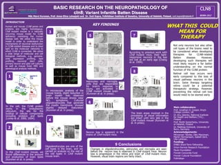
- 1. BASIC RESEARCH ON THE NEUROPATHOLOGY OF cln8; Variant Infantile Batten Disease MSc Mervi Kuronen, Prof. Anna-Elina Lehesjoki and Dr. Outi Kopra, Folkhälsan Institute of Genetics, University of Helsinki, Finland, outi.koprai@helsinki.fi INTRODUCTION Human and mouse CLN8 genes and proteins are highly homologous. Cln8 mutant mouse is a naturally occurring mouse model for CLN8 deficiency mimicking the human disease well. We utilize this mouse model in order to understand the mechanisms of neuronal dysfunction in CLN8-related diseases and to shed light on the molecular networks to which CLN8 is connected. We utilize a wide range of molecular and cell biological methods, e.g. genome- wide expression profiling, lipid profiling, neuronal stem cells, primary neuron and glial cell cultures as well as mouse brain MRI and stereological analysis methods. In the cell, the CLN8 protein works in the endoplasmic reticulum, the site for the buildup of cellular components including proteins and lipids (Lonka et al. 2000, 2004). 2 KEY FINDINGS 7 8 WHAT THIS COULD MEAN FOR THERAPY Neuron loss is apparent in the brains of Cln8 mutant mice. Oligodendrocytes are one of the cell types in the brain, and we carried on analysis on the rest of the cell types in Cln8 mutant mouse brain. According to previous work with Cln8 mutant mice, their light- responsive neurons in the eye are lost at an early age (Chang et al. 1994). The brain areas involved in the processing of visual information are intact until very late in the Cln8 mutant mouse (Kuronen et al. in press). Astrocytes and microglia, cells that are responsible for brain’s response to injury, are activated in CLN8 mutant mouse brain (Kuronen et al. in press). In microscopic analysis of the mouse brains, slight reduction in brain myelin was observed before the mouse shows clear NCL symptoms. Cultured oligodendrocytes that generate the myelin membrane, showed slightly delayed maturation (Kuronen et al. in press). In the Cln8 mutant mouse, we found differences in the amount and production of brain lipids (Kuronen et al. in press) Main collaborators: Prof. Jonathan D. Cooper, King’s College London, UK Dr. Anu Jalanko, National Institute for Health and Welfare (THL) , Finland Prof. Pentti Somerharju, University of Helsinki, Finland Dr. Matthias Eckhardt, University of Bonn, Germany Acknowledgements: Minnamari Talvitie, Otto Manninen, Martin Hermansson, Isabell Zech, Ulla Lahtinen, Funding: EMBO Short-Term Fellowship Orion-Farmos Research Foundation Paulo Foundation Finnish Concordia Fund Finnish Cultural Foundation Folkhälsan Modified from PhD thesis of Liina Lonka, 2004 CTSD 1 3 4 5 astrocytes microglia 6 neurons 9 Conclusions Changes in oligodendrocytes, astrocytes and microglia are seen before the neuron loss is observed in Cln8 mutant mice. Neuron loss is progressive in the retina and brain of Cln8 mutant mice. However, visual brain regions are fairly intact. Not only neurons but also other cell types of the brains need to be considered when developing therapies for CLN8-related Batten disease. However, developing such therapies will most likely require a far better understanding on the normal function of the CLN8 protein. Retinal cell loss occurs very early compared to the loss of visual pathway neurons in the brain. Gene therapy targeting retina could be a potential therapeutic strategy. However, preventing the retinal cell loss would need to be started early.