Juvenile NCL White Matter Abnormalities
•
1 recomendación•90 vistas

Rustic global and widespread local white matter abnormalities in juvenile neuronal ceroid lipofuscinosis

Recomendados
Más contenido relacionado
La actualidad más candente
La actualidad más candente (20)
Similar a Juvenile NCL White Matter Abnormalities
Similar a Juvenile NCL White Matter Abnormalities (20)
Más de Batten Disease Support and Research Association
Más de Batten Disease Support and Research Association (20)
Último
Último (20)
Juvenile NCL White Matter Abnormalities
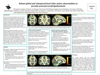
- 1. University or Laboratory Logo Juvenile NCL Robust global and widespread local white matter abnormalities in juvenile neuronal ceroid lipofuscinosis Principal investigator: Professor of radiology Taina Autti, HUS Medical Imaging Center, Radiology, University of Helsinki, Helsinki University Hospital, Finland. Other investigators: Ulrika Roine, Timo Roine, Laura Åberg, Anna Tokola, Marja Balk, Antti Hakkarainen, Minna Mannerkoski and Tuula Lönnqvist Introduction: Juvenile neuronal ceroid lipofuscinosis (CLN3) is a progressive neurodegenerative lysosomal storage disease of the childhood. It manifests with loss of vision, seizures and loss of cognitive and motor functions leading to premature death. Previous magnetic resonance imaging (MRI) studies have reported cerebral and cerebellar atrophy, progressive hippocampal atrophy, thalamic signal intensity alterations and decreased white matter volume in the corona radiata. Diffusion-weighted (DW) magnetic resonance imaging (MRI) is a noninvasive method, which can be used to investigate the white matter microstructure in the brain by measuring the diffusion of water molecules in white matter bundles. For the first time in CLN3, we investigated global and local white matter microstructure with DW-MRI in 14 children with CLN3 (age 9.6±3.4, 71% males) and 14 control subjects (age 11.2±2.3, 50% males). The acquisition was repeated for the children with CLN3 two years later (12 subjects, age 11.4±3.2, 67% males). Discussion: Here, we present the first diffusion weighted magnetic resonance imaging study in juvenile neuronal ceroid lipofuscinosis (CLN3). Using both global and local approaches, we show robust findings involving widespread decreases in FA in CLN3. FA can be affected by dense packing of nerve fibers, intact cell membranes, thickness of the nerve fibers, myelinisation, orientation of the nerve fibers and the amount of crossing fibers. As we also found decreased planarity in CLN3, the differences in FA could not be explained by changes in fiber complexity, i.e. the amount of crossing fibers. Moreover, diffusivity was in general increased in CLN3 compared to control subjects, which is in accordance with a previous study performed in infantile NCL. As conventional MRI is visually normal in CLN3 up to an age of 10, we showed that DW-MRI is more capable of detecting the microstructural changes related to the disease. We found no local or global differences between the two CLN3 time points. In other words, the microstructural changes were already present at the time of the first scan (average age 9.6) and do not progress rapidly in the prepubertal stage (the second scan was acquired at an average age of 11.4). Conclusion: Our results indicate that the widespread microstructural white matter abnormalities are present already in less than 10 year old children with CLN3, even though the conventional MRI is usually visually normal under the age of 10. Furthermore, our results suggest that the microstructural abnormalities do not progress rapidly in the prepubertal stage. Further studies are needed to detect whether these abnormalities are present already at a younger age, and to investigate the specific nature of these differences with more detailed microstructural models. Acknowledgements: Batten Disease Support & Research Association, Paulo Foundation, Finnish Brain Foundation Methods: Investigation of global microstructural white matter abnormalities with two methods 1) Whole-brain tractography: reconstruction of nerve fibers in the brain with an advanced tractography method: constrained spherical deconvolution (CSD) 2) White matter tract skeleton: central voxels of white matter tracts form a white matter tract skeleton Investigation of local microstructural white matter abnormalities 1) Tract-based spatial statistics (TBSS) was used to compare microstructural properties in the white matter skeleton locally between the children with CLN3 and control subjects Microstructural properties: Fractional anisotropy (FA): anisotropy (directedness) of diffusion is generally higher in coherent white matter fiber bundles as the water molecules can diffuse more freely along the white matter fiber than perpendicular to it. Mean diffusivity (MD): average diffusion distance in all directions Axial diffusivity (AD): diffusivity along the orientation of the white matter bundle Radial diffusivity (RD): diffusivity perpendicular to the orientation of the white matter bundle Planarity (CP): Provides knowledge about the fiber complexity, i.e. crossing nerve fibers Global results shown with both the tractography and skeleton approach: • Decreased global FA (p=0.000001) and planarity values in CLN3 • Increased axial, radial and mean diffusivity in CLN3 Similar results in the CLN3 children two years later, but no significant difference between the first and second time point. Local results shown with TBSS: • Widespread local decreases in FA (Fig. 1) for example in the corona radiata and posterior thalamic radiation (both p<0.0002) • Widespread increases in mean, axial and radial diffusivity (p<0.0002) (Fig. 2) Figure 1. Local white matter abnormalities in fractional anisotropy (FA) investigated with tract-based spatial statistics. Decreased FA values in CLN3 compared to controls illustrated in axial (A), coronal (B), and sagittal (C) slices. Figure 2. Local increases in mean diffusivity (MD) investigated with tract-based spatial statistics. Increased MD values in CLN3 compared to controls illustrated in axial (A), coronal (B), and sagittal (C) slices.