
Recomendados
Más contenido relacionado
La actualidad más candente
La actualidad más candente (20)
Similar a Introduction of tissue - Epithelial tissue
Similar a Introduction of tissue - Epithelial tissue (20)
Último
Último (20)
Introduction of tissue - Epithelial tissue
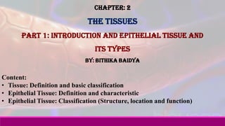
- 1. Chapter: 2 The Tissues Part 1: Introduction and Epithelial tissue and its types By: Bithika Baidya Content: • Tissue: Definition and basic classification • Epithelial Tissue: Definition and characteristic • Epithelial Tissue: Classification (Structure, location and function)
- 2. • Tissue: Definition The word ‘Tissue’comes from an old French verb meaning ‘to weave’ • Definition: ✓ “Tissues are aggregates or groups of cells organized to perform one or more specific functions.” ✓ “Tissues are a group of cell that have similar structure, act together to perform specific function and have same origin.” ✓ “A tissue is a group of cells that usually have a common embryonic origin and function together to carry out specialized activities.”
- 3. • Types of tissues Body tissues can be classified into four basic types according to structure and function: 1. Epithelial tissue covers body surfaces and lines hollow organs, body cavities, and ducts. It also forms glands. (Origin Ectoderm) 2. Connective tissue protects and supports the body and its organs. Various types of connective tissue bind organs together, store energy reserves as fat, and help provide immunity to disease-causing organisms. (Origin Mesoderm) 3. Muscular tissue generates the physical force needed to make body structures move and generates body heat. (origin ) 4. Nervous tissue detects changes in a variety of conditions inside and outside the body and responds by generating action potentials (nerve impulses) that activate muscular contractions and glandular secretions. (Origin Ectoderm)
- 4. • Epithelial Tissue: Definition and characteristic An epithelial tissue (ep-i-THE¯ -le¯-al) or epithelium (plural is epithelia) consists of cells arranged in continuous sheets, in either single or multiple layers. Epithelial tissue forms coverings and linings throughout the body. • Characteristic ✓ The cells are closely packed and are held tightly together by many cell junctions. ✓ There is little intercellular space between adjacent plasma membranes. ✓ It is never covered by another tissue, so it always has a free surface. ✓ There are various surfaces- Apical surface of the epithelium faces the body surface or body cavity and it may contain cilia or microvilli. The lateral surfaces of an epithelial cell face the adjacent cells on either side. The basal surface of an epithelial cell is opposite the apical surface, and the basal surfaces adhere to basement membrane ✓ The basement membrane is a thin extracellular that functions as a point of attachment and support for the overlying epithelial tissue.
- 5. • Types of epithelial tissue • The bases for classifying epithelium tissue is Cell shapes: 1. Squamous cells arranged like floor tiles and are thin. 2. Cuboidal cells are shaped like cubes or hexagon 3. Columnar cells are much taller than they are wide, like columns. Arrangement of layers: 1. Simple:- single layer of cells present 2. Stratified:- multiple layers of cells are present
- 6. Combining the two characteristics (arrangements of layers and cell shapes), the types of covering and lining epithelia are as follows: 1. Simple epithelium a) Simple squamous epithelium b) Simple cuboidal epithelium c) Simple columnar epithelium (nonciliated and ciliated) d) Pseudostratified columnar epithelium (nonciliated and ciliated) 2. Stratified epithelium a) Stratified squamous epithelium b) Stratified cuboidal epithelium c) Stratified columnar epithelium d) Transitional epithelium
- 7. 1. Simple epithelium a) Simple squamous epithelium Description: Single layer of flattened cells with disc-shaped centrally located nucleus and sparse cytoplasm; the simplest of the epithelia. Location: Lines heart, blood vessels, lymphatic vessels, air sacs of lungs, glomerular (Bowman’s) capsule of kidneys, and inner surface of the tympanic membrane (eardrum); forms epithelial layer of serous membranes, such as the peritoneum, pericardium, and pleura. Function: Filtration, diffusion, osmosis, and secretion in serous membranes. Nuclei of squamous Epithelial cells Air sacs of lung tissue Photomicrograph: Simple squamous epithelium forming part of the alveolar (air sac) walls (125x). Lungs
- 8. b) Simple cuboidal epithelium Description: Single layer of cube-shaped cells; centrally located nucleus. Location: Covers surface of ovary, lines anterior surface of capsule of the lens of the eye, forms the pigmented epithelium at the posterior surface of the eye, lines kidney tubules and smaller ducts of many glands, and makes up the secreting portion of some glands such as the thyroid gland and the ducts of some glands such as the pancreas. Function: Secretion and absorption. Simple cuboidal Epithelial cells Kidney tubules Photomicrograph: Simple cuboidal epithelium in kidney tubules (430x).
- 9. b) Simple columnar epithelium Description: Single layer of tall cells with round to oval nuclei; some cells bear cilia; layer may contain mucus-secreting unicellular glands (goblet cells). Location: (Ciliated) Covers surface of ovary, lines anterior surface of capsule of the lens of the eye, forms the pigmented epithelium at the posterior surface of the eye, lines kidney tubules and smaller ducts of many glands, and makes up the secreting portion of some glands such as the thyroid gland and the ducts of some glands such as the pancreas. (Non-ciliated)Lines the gastrointestinal tract (from the stomach to the anus), ducts of many glands, and gallbladder Function: Secretion and absorption Photomicrograph: Simple columnar epithelium of the stomach mucosa (860X). Simple columnar Epithelial cell Basement membrane gastrointestinal tract
- 10. d) Pseudostratified columnar epithelium: Description: Not a true stratified tissue; nuclei of cells are at different levels; all cells are attached to basement membrane, but not all reach the apical surface. Location: Pseudostratified ciliated columnar epithelium lines the airways of most of upper respiratory tract; pseudostratified non-ciliated columnar epithelium lines larger ducts of many glands, epididymis, and part of male urethra. Function: Secretion and movement of mucus by ciliary action. Photomicrograph: pseudostratified columnar epithelium lining the human trachea (570x). Pseudostratified Epithelial layer Trachea cilia
- 11. 2. Stratified epithelium a) Stratified squamous epithelium: Description: Several layers of cells; cuboidal to columnar shape in deep layers; squamous cells form the apical layer and several layers deep to it; cells from the basal layer replace surface cells as they are lost. Location: Keratinized variety forms superficial layer of skin; non-keratinized variety lines wet surfaces, such as lining of the mouth, esophagus, part of larynx, part of pharynx, and vagina, and covers the tongue. Function: Protects underlying tissues in areas subjected to abrasion Photomicrograph: Stratified squamous epithelium lining the esophagus (285x). Stratified squamous epithelium Mouth, part of larynx, pharynx
- 12. b) Stratified cuboidal epithelium (Rare) Description: Two or more layers of cells in which the cells in the apical layer are cube-shaped. Location: Ducts of adult sweat glands, salivary gland, mammary gland and part of male urethra. Function: Protection and limited secretion and absorption. Photomicrograph: Stratified cuboidal epithelium Forming salivary gland duct salivary gland Stratified cuboidal epithelium
- 13. c) Stratified columnar epithelium (Rare) Description: Several layers of irregularly shaped cells; only the apical layer has columnar cells. Location: Lines part of urethra, large excretory ducts of some glands, such as esophageal glands, small areas in anal mucous membrane, and part of the conjunctiva of the eye. Function: Protection and secretion Photomicrograph: Stratified columnar epithelium Lining male urethra Stratified columnar epithelium male urethra
- 14. d) Transitional epithelium Description: Appearance is variable (transitional); shape of cells in apical layer ranges from squamous (when stretched) to cuboidal (when relaxed). Location: Lines urinary bladder and portions of ureters and urethra. Function: Stretches readily and permits distension of urinary organ by contained urine. urinary bladder Photomicrograph: Transitional epithelium lining the urinary bladder, relaxed state (360X); Transitional epithelium
- 15. Questions: 1. Define Tissue 2. Enlist the basic types of tissue found in the body. 3. Define: Epithelium tissue 4. What are the characteristics of epithelial tissues? 5. Enlist the various function of epithelial tissue. 6. What is the function of the basement membrane? 7. Write a note on Simple Epithelial tissue and its types. 8. Write a note on Stratified Epithelial tissue and its types. References: Principles of Anatomy and Physiology - 12th Edition Gerard J. Tortora and Bryan Derrickson Essentials of Anatomy and Physiology (Pictures credit) THANK YOU