Canon Medical Systems VISIONS Magazine - issue 30

VISIONS magazine is a publication of Canon Medical Systems Europe and is offered free of charge to medical and health professionals. The magazine is published twice a year. Registration to access full, previously published, digital editions can be done via the web site: https://eu.medical.canon/visions. Canon Medical Systems stores and uses personal data of the registration to send out the magazine and inform members about new developments. Members can customize preferences or opt-out, after registration, in the online VISIONS profile. VISIONS magazine is covering Canon Medical Systems European region and as such reflects products, technologies and services for this particular area. The mentioned products may not be available in other geographic regions. Please consult your Canon representative sales office in case of any questions. No part of this publication may be reproduced in whole or in part, stored in an automated storage and retrieval system or transmitted in any manner whatsoever without written permission of the publisher. VISIONS Magazine is not responsible for any inaccuracies in this publication. News items and articles are announced firstly, as pre-publication, via the dedicated VISIONS LinkedIn Group: https://www.linkedin.com/groups/3698045. In this group you can actively participate in discussions about the content and future direction of the magazine. Follow us also on SlideShare: http://www.slideshare.net/canonmedicaleu. ©2018 by Toshiba Medical Systems Europe B.V. All rights reserved

Recomendados
Recomendados
Más contenido relacionado
Similar a Canon Medical Systems VISIONS Magazine - issue 30
Similar a Canon Medical Systems VISIONS Magazine - issue 30 (20)
Más de Canon Medical Systems Europe
Más de Canon Medical Systems Europe (13)
Último
Último (20)
Canon Medical Systems VISIONS Magazine - issue 30
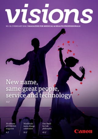
- 1. 30 editions of VISIONS magazine 4 // NO. 30 // FEBRUARY 2018 // MAGAZINE FOR MEDICAL & HEALTH PROFESSIONALS New name, same great people, service and technology 11 // Our Made for Life philosophy 45 // Worldwide name change celebration 10 //
- 2. Two dancers (ballet dancer and 'pop and locking' dancer) expressing the name change (transition) from Toshiba Medical to Canon Medical Systems. (photo: Jaco Terlouw) VISIONS magazine is a publication of Canon Medical Europe and is offered free of charge to medical and health professionals. The magazine is published twice a year. Registration to access full, previously published, digital editions can be done via the web site: https://nl.medical.canon/visions-magazine. Canon Medical stores and uses personal data of the registration to send out the magazine and inform members about new developments. Members can customize preferences or opt-out, after registration, in the online VISIONS profile. VISIONS magazine is covering Canon Medical’s European region and as such reflects products, technologies and services for this particular area. The mentioned products may not be available in other geographic regions. Please consult your Canon Medical representative sales office in case of any questions. No part of this publication may be reproduced in whole or in part, stored in an automated storage and retrieval system or transmitted in any manner whatsoever without written permission of the publisher. The opinions expressed in this publication are solely those of the authors and not necessarily those of Canon Medical. Canon Medical does not guarantee the accuracy or reliability of the information provided herein. News items and articles are announced firstly, as pre-publication, via the dedicated VISIONS LinkedIn Group: https://www.linkedin.com/groups/3698045. In this group you can actively participate in discussions about the content and future direction of the magazine. Aquilion ONE, Aquilion ONE / ViSION Edition, Aquilion PRIME, Aquilion ONE GENESIS Edition, Celesteion, Aquilion Prime SP, Aquilion CXL, Aquilion RXL, Infinix-i 4D CT, Infinix-i Hybrid +, Vantage Galan 3T, FIRST, PURE ViSION Optics, AIDR 3D, Aplio-i series, Aplio i800, are trademarks of Canon Medical Systems Corporation. Secondlife is a trademark of Canon Medical Systems Europe B.V. Publisher Canon Medical Systems Europe B.V. Zilverstraat 1 NL-2718 RP Zoetermeer +31 79 368 92 22 https://eu.medical.canon/ marketing.eu@eu.medical.canon Editor-in-chief Jack Hoogendoorn jack.hoogendoorn@eu.medical.canon Editor Jacqueline de Graaf jacqueline.degraaf@eu.medical.canon Modality coordinators and reviewers CT: Roy Irwan, Roy Verlaan UL: Jeroen Uijttenhout, Marcel Vermooten X-Ray: Rene Degros Design & Layout Boerma Reclame boermareclame.com Printmanagement Printweb Media B.V. printweb.nl Text contributions and editing The Creative Practice thecreativepractice.com Photography Cojan van Toor www.cojanvantoor.nl © 2018 by Canon. All rights reserved. ISSN 1617-2876 Folow us:
- 3. VISIONS 30 // 3 Before you lies perhaps the most unique issue of VISIONS to date. At first glance it might look like any other one but nothing could be further from the truth. This is not only the thirtieth issue of VISIONS, it also carries a brand new logo and company name on the cover. Did you notice? Reason is, as you probably know, that we are being warmly welcomed in the Canon family and thus changed our name to Canon Medical Systems all over the world at the beginning of the year. Both are memorable facts in the history of our company and good news for you. Why? Well, we have kept all that you value so highly: the same great people, reliable products and technologies, unconditional customer focus and unsurpassed service – now endorsed and supported by a strong and recognized worldwide brand of a renowned company that considers health care and research to be of paramount importance. Around eight percent of revenue is invested in R&D every year and globally Canon ranked third among the top patent holders for the sixth straight year. Combine this with the fact that health care is one of the strategic pillars of the company and the message becomes immediately clear. Moreover, we don’t only work hard to stay innovative but we are fundamentally changing how innovation is made. Medical imaging and health care have always been part of the Canon DNA. Did you know that the first president of Canon, Takeshi Mitarai, was an obstetrician? And that Canon developed the first indirect X-ray camera in Japan? A great and respected heritage! Now bolstered with over one hundred years of experience in medical imaging and treatment, we will further innovate solutions, from health care data to precision medicine, and bring products on the market like never seen before. I would like to suggest keeping this issue of VISIONS as it is the first one bearing the Canon logo. Undoubtedly many more will follow to inform you about important business developments, new products and experiences of our customers. In our history of the many medical ‘firsts’ we have developed, this is also a ‘first’ and, who knows, it might become a collector’s item one day. JACK HOOGENDOORN Senior Manager Marketing Canon Medical Systems Europe BV © 2018 CANON MEDICAL SYSTEMS Kind regards, //EDITORIAL
- 4. 4 // VISIONS 30 Celebrating 30editions of innovative technologies and applications in medical imaging.
- 5. VISIONS 30 // 5 Register online for VISIONS Stay up-to-date with the latest innovations in medical imaging Subscribe to have access to full, previously published, digital editions of VISIONS Magazine. Visit our web site at: https://nl.medical.canon/visions-magazine Do you also want to receive a paper copy of VISIONS magazine at home or at work? Please tell us so during registration and we will send you VISIONS magazine twice a year! Registration is easy and absolutely free. Alternatively you can follow us at SlideShare (https://www.slideshare.net/canonmedicaleu) and dedicated VISIONS LinkedIn Group (https://www.linkedin.com/groups/3698045) © 2018 CANON MEDICAL SYSTEMS
- 6. 6 // VISIONS 30 //CONTENTS 10Worldwide name change celebration 12Expanding horizons in Interventional Oncology in a state-of-the-art Angio-CT environment X-RAY 28Unique interventional platform creates new opportunities for minimally invasive therapies X-RAY 03 Editorial 08 News 11 President's Message 12 Expanding horizons in Interventional Oncology in a state-of-the-art Angio-CT environment X-RAY 20 Diagnosis of liver fibrosis using Shear Wave Elastography ULTRASOUND 25 The new Aquilion Prime SP in Bradford, from fast patient throughput to robust cardiac scanning and its new diagnostic capabilities COMPUTED TOMOGRAPHY 28 Unique interventional platform creates new opportunities for minimally invasive therapies X-RAY 32 Improving ‘standards of care’ in CT for animals: split-bolus single-pass multi-phased abdominal CT COMPUTED TOMOGRAPHY, SECONDLIFE
- 7. VISIONS 30 // 7 36Celesteion PET-CT — making a difference with dual modality imaging COMPUTED TOMOGRAPHY 59Shaping the future of diagnostic imaging ultrasound ULTRASOUND 32Improving ‘standards of care’ in CT for animals: split-bolus single- pass multi-phased abdominal CT COMPUTED TOMOGRAPHY, SECONDLIFE 36 Celesteion PET-CT — making a difference with dual modality imaging COMPUTED TOMOGRAPHY 38 Preliminary clinical experience with shear wave dispersion imaging for liver viscosity ULTRASOUND 42 Transforming Cardiothoracic Imaging – The Aquilion ONE GENESIS Edition and FIRST COMPUTED TOMOGRAPHY 46 Teddy bears show courage COMPUTED TOMOGRAPHY 48 Scanning prehistoric urns from Hofstade, Belgium COMPUTED TOMOGRAPHY 54 Assessment of non-alcoholic fatty liver disease with attenuation imaging (ATI) ULTRASOUND 59 Shaping the future of diagnostic imaging ultrasound ULTRASOUND 60 Image-guided interventions with half the dose COMPUTED TOMOGRAPHY © 2018 CANON MEDICAL SYSTEMS
- 8. 8 // VISIONS 30 //NEWS After the impressive Infinix™-i 4D CT installation, the University Hospital Montpellier has extended their platform by investing in a new high-end CT scanner from Canon Medical Systems Europe BV. This new system incorporates cutting-edge technologies designed to meet current and future clinical needs with high quality imaging at the lowest dose. The new equipment is called 3D Scanner. It is shared by 3 departments: Radiology of Saint-Eloi Hospital, Prof. B. Guiu, Radiology of Lapeyronie, Prof. P. Taourel and Radiology of Arnaud de Villeneuve, Prof. H. Vernhet. The CT is installed in the department of diagnostic radiology and interventional hospital Saint-Eloi to serve all out patients and consultants of CHU Montpellier, France. // Another new system at the University Hospital Montpellier, France Canon Medical Systems is one of the first to be recognized with a prestigious new Research and Development award recently established by Japanese Government. The Japan Medical Research and Develop- ment Awards Program was established in 2017 with the aim of encouraging excellence in research and development by recognizing groups or individuals who have made outstanding contri- butions to advancing medical, as well as other fields. The award, which was made for development of the 4D CT system, the Aquilion ONE™, was pre- sented by Mr. Katsunobu Kato, Japanese Minister of Health, Labor and Welfare, in a special award ceremony held at the Japanese Prime Minister’s official residence in December 2017. Dr. Kazuhiro Katada, Professor Emeritus at Fujita Health University, in Japan, and Dr. Masahiro Endo, Managing Director of the Japanese Association for Nuclear Technology in Medicine, were key in the joint research that led to the realization of the Aquilion ONE. In addition to conventional morphological diagnosis that depicts anatomical structures and tumors, the Aquilion ONE enables func- tional diagnosis based on analysis of blood flow or the motion of anatomical structures. The latest systems also allow further reductions in image acquisition time, exposure-dose, and the amount of contrast medium, resulting in more patient-friendly examinations. The award was made on the basis of these significant advantages. // Aquilion ONE wins new Japanese research and development awardVarious ceremonies were held at Canon Medical Systems Corporation (CMSC) offices across the world on January 4th 2018 to mark the launch of the new company name. The positive energy that this has generated amongst staff is highlighted in a photo report, which you can find on the last page of this VISIONS edition. // Canon Medical Systems Corporation is officially launched
- 9. VISIONS 30 // 9 Canon Medical Systems Corporation has been awarded the Green Apple Environmental best practice award. The Green Apple awards, reward and promote environmental best practice around the world. Canon Medical Systems Corporation was proclaimed Gold Winner in the category Innovation, for its Aquilion ONE™ GENESIS Edition CT scanner. The exclusive awards ceremony was held in the Palace of Westminster, London. Awards were presented in various categories, such as Carbon, Energy Efficiency, Manufacturing and Waste management. The award winning CT Scanner, Aquilion ONE GENESIS Edition, is a smaller and lighter Premium CT system than its predecessors, thus requiring less power. Follow our Social Media channels and stay up-to-date about Canon Medical’s latest news innovations! Canon Medical Systems Corporation receives Green Apple Environmental best practice award © 2018 CANON MEDICAL SYSTEMS linkedin.com/company/Canon-Medical-EU youtube.com/CanonMedicalEU facebook.com/CanonMedicalEU twitter.com/CanonMedicalEU slideshare.net/CanonMedicalEU plus.google.com/+CanonMedicalEU Designed for an installation space of just 19 m2 , Aquilion ONE GENESIS Edition can be installed in most existing CT rooms, avoiding costly renovations. The open Gantry structure with short bore facilitates access from the front and rear of the gantry. With regard to the exposure dose for the patients, Aquilion ONE GENESIS Edition applies the latest dose reduction technologies and reconstruction algorithms, such as PURE ViSION detector technology and FIRST. Its advanced detector technology converts almost 100% of incident X-ray photons for maximum dose efficiency. FIRST is the world’s first fully integrated Iterative Reconstruction Technology in Premium CT, resulting in the lowest dose levels technically achievable today. //
- 10. 10 // VISIONS 30 On January 4, 2018, our company name changed to Canon Medical Systems. This is a huge milestone in the history of our organization. Canon has a strong vision for building a world-class healthcare enterprise and has made healthcare one of the four key strategic pillars. With our 100 year history, great people, technologies and services backed by the “Made for Life” philosophy Canon Medical Systems will continue to drive the company forward. The Made for Life philosophy stands for our ongoing commitment to humanity - generations of inherited passion creates a legacy of medical innovation and service that continues to evolve as we do. By engaging the brilliant minds of many, we continue to set the benchmark, because we believe quality of life should be a given, not the exception.
- 11. VISIONS 30 // 11 O n January 4, 2018, our company name changed to Canon Medical Systems Corporation. I am delighted to be able to celebrate this day with our many customers and employees around the world, who have been so supportive. On this day, we become the core of Canon's medical business both in name and reality. We must leverage synergies of our integration with Canon in order to further contribute to human health through innovative technologies and solutions. It is now over one year since we joined the Canon Group. Throughout that period, we interacted with each other to deepen mutual understanding of our businesses in terms of production, development, and human resources. Exposure to Canon's culture, which we were previously not familiar with, and reexamination our existing pro- cesses and practices, has led us to make new discoveries, fostered new ideas and acquainted us with new people. I am confident that the accumulation these influences will act as a catalyst for even-greater future development. TOSHIO TAKIGUCHI President and Chief Executive Officer Canon Medical Systems Corporation In 2018, which marks our new chapter as Canon Medical Systems, we also celebrate our 88th anniversary. From this inaugural year as Canon Medical Systems, all of us are dedicated to focusing our efforts and energies to take the business from strength to strength. The number 8 is thought to be lucky in Japan because of the widening shape of its kanji character, and so this anniversary could be considered doubly auspicious. PRESIDENT’S MESSAGE © 2018 CANON MEDICAL SYSTEMS “Our company name change to Canon Medical Systems Corporation.”
- 12. 12 // VISIONS 30
- 13. VISIONS 30 // 13 Since the installation of the Infinix™-i 4D CT and Aplio™ i800 ultra- sound system in last November, the University Hospital Strasbourg has been able to expand its research program in Interventional Radio- logy with several advanced techniques that could bring about major changes in the discipline. T he University Hospital of Stras- bourg is one of the most prestigious University Hospitals in Europe. Affiliated to the University of Strasbourg, it forms part of a medical technologies campus with growing expertise in RD using cutting-edge technologies in healthcare. The Hospital employs more than 12,000 people, including 3,000 physicians and leads groundbreaking global research in several disciplines, including transplanta- tion, minimally-invasive surgery, Immunol- ogy and Interventional Radiology. An emerging world-class specialist facility The Radiology Department’s highly special- ized Interventional Imaging Service has grown steadily since 2011. “Interventional Radiology is of crucial impor- tance for our University Hospital for several reasons, and we have invested significantly in this field,” remarked Mr. Gautier, General Manager of the Hospital. “Most importantly, it brings numerous benefits to our patients. It drastically improves the treatment of specific tumors, patients benefit from less invasive procedures, with improved outcomes and shorter hospitalization and it has also proved useful in pain treatments.” Led by Prof. Gangi, Head of Interventional Radiology, and Chairman of the Radiology and Nuclear Medicine Department, the large Interventional Radiology Department has grown in recent years and is currently staffed by nine Interventional Radiologists and 10 Radiology Technicians. “Thanks to the dedication of Prof. Gangi and his Team to our patients, and to their research interests, the University Hospital has established an outstanding reputation in Interventional Radiology, at national- and international levels,” continued Mr. Gautier. © 2018 CANON MEDICAL SYSTEMS //XREU170032 VISIONS spoke with Prof. Gangi, Dr. Garnon, Mr. Gautier and Mr. Gigleux, from the University Hospital Strasbourg, about their experiences with the Infinix-i 4D CT and Ultrasound system Aplio i800. INTERVIEW //X-RAY, COMPUTED TOMOGRAPHY, ULTRASOUND //Infinix-i 4D CT, Aplio i800 Expanding horizons in Interventional Oncology in a state- of-the-art Angio-CT environment
- 14. 14 // VISIONS 30 so successful in imaging that it eventually became a new concept, and was ultimately adopted commercially by Canon Medical Systems.” The Hospital’s Interventional Imaging Service performs an increasing range of interven- tional procedures ranging from simple infil- tration to complex therapeutic treatments, sometimes combined with surgery. Prof. Gangi - University Hospital Strasbourg. “High-quality true CT scanning during a com- plex intervention is key for us.” “We are very proud indeed of their technical and medical accomplishments.” This skilled team carry out hundreds of interventional procedures every year (including spinal injections, biopsies, TACE, cementoplasty, ablation and emboliza- tion) using a range of imaging equipment based in three dedicated Interventional Radiology rooms. Alongside rooms housing an interventional MR and a C-arm based system with cone-beam CT capability, the latest suite to be added is equipped with an Infinix-i 4D CT and Aplio i800 ultrasound system from Canon Medical Systems. The Radiology Team chose these systems specifically to increase the range of Inter- ventional Radiology procedures possible at the Department, as well as reduce patient waiting-time. Groundbreaking concept “We were one of the first research insti- tutes in the world to combine fluoroscopy and CT modalities in the same room,” said Prof. Gangi. “When I was a Resident Radiologist back in 1990, I could already see the advantage of placing a mobile C-arm in front of the CT-scanner. At first, people questioned what it could bring, but it proved
- 15. VISIONS 30 // 15 “The range of possibilities is huge.” Dr. Garnon - University Hospital Strasbourg. When it came to creating an additional Interventional Radiology suite to provide the resources to keep up with growing demand for new imaging services, the Hospital turned to Canon Medical Systems for a solution. A uniquely integrated solution “The Infinix-i 4D CT combines two different imaging modalities within the same envi- ronment, which will allow us to treat new medical indications in the different fields with pioneering Interventional Radiology techniques,” said Mr. Gautier. “We are convinced that installing this high-level CT system will help us reach our main goal, which is to continuously enhance the quality and safety of the healthcare that we offer to our patients.” “We chose the Infinix-i 4D CT initially on the basis of the exceptionally high-quality CT scanning that is possible with the system. It is key for us. It’s the heart of the system,” added Prof. Gangi. “There are not many systems as mature as the Infinix-i 4D CT available on the market. Both angio and CT modalities communicate and work together, enabling our Interventional Radiologists to use them with maximum ease-of-use. They can move from one system to another with- out any steps. A seamless combination of top CT scan and high-end level angiogram: an ideal option for us.” The new equipment replaced a 128-slice CT system and a mobile C-arm. Installation of the Infinix-i 4D CT and the Aplio i800 ultrasound system was carried out in col- laboration with the Canon Medical Systems project management team. Installation of the new imaging suite required consider- able planning. “When we decided to change our CT-suite, we were hoping to achieve many objectives: to improve the quality of our CT-imaging; to combine high-quality fluoroscopy and CT; to optimize ease-of-use and versatility in one machine; to reduce radiation signifi- cantly; and to support new procedures,” said Dr. Garnon, Interventional Radiologist at the Hospital. “The new system meets all of these needs.” © 2018 CANON MEDICAL SYSTEMS //XREU170032
- 16. 16 // VISIONS 30 Mr. Gautier – General Director University Hospital Strasbourg. “Infinix-i 4D CT will help us to improve patient outcomes and shorten hospitalization time.” Radiology Team at the University Hospital Strasbourg. “Given the context of our expanding research, we realized that replacing our mobile C-arm with a motorized ceiling-suspended C-arm with Flat Panel Detector, would allow us to push our current limits, cover new indica- tions and enable us to perform complex vas- cular-, as well as percutaneous procedures,” said Mr. Gigleux, Biomedical Engineer. “Our choice focused on the Infinix-i 4D CT, because of its versatility. In addition to the advantages of the C-arm in interventional work, the system offers great flexibility, for example, the C-arm can assume a dedicated parking position for procedures that require use of the CT only.” Combining two modalities in one room Previously, the Interventional Radiology Team used to carry out angiographs in one room, and then moved the patient into a second room for the CT. This step is no longer necessary with the higher quality Infinix-i 4D CT. “The implementation of the solution combining two modalities in a single room was one of the first points that we studied together with Canon Medical Systems project management team when installing the system,” said Mr. Gigleux. “Implemen- tation of the project was quite complex, but the collaboration between the Hospital's technical teams, the various sub-contrac- tors and the entire Canon Medical Systems Team was excellent.” “As the modalities are combined in one system with the Infinix-i 4D CT, many pro- cedures that we were previously performed in two steps can now be completed in one,” remarked Prof. Gangi. “The system will not only improve the quality and safety of our standard Interventional Radiology procedures, but will increase the number of indications that we are able to treat.”
- 17. VISIONS 30 // 17 Prof. Gangi and Dr. Garnon. © 2018 CANON MEDICAL SYSTEMS //XREU170032 “Our work includes vascular- and percuta- neous interventions, and our Team includes specialists in these techniques. In many cas- es, the procedures are performed separately, but when both approaches are required, we can work together, side-by-side, with the new system. The Infinix-i 4D CT clearly offers new perspectives in combined thera- pies and enables us to perform much more complex procedures in this field,” added Dr. Garnon. Infinix-i 4D CT: State-of-the-Art in CT “There are plenty of new possibilities on the horizon,” said Prof. Gangi. “We plan to intro- duce real, combined procedures, including angiographic-, percutaneous- and surgical procedures. So, we’ll have specialists from three disciplines working together in the same room. This is what will make the difference: the ability to perform multi- modality, multi-discliplinary, interventional procedures.” The advanced CT imaging and fluoroscopy capabilities of the Infinix-i 4D CT will enable the team to combine procedures, such as ablation and embolization, or ablation limitations of each technique by combining everything together.” Aplio i800: The ultimate in ultrasound Along with the Infinix-i 4D CT, the new Interventional Radiology suite at the Hospital is equipped with an Aplio i800 ultrasound system from Canon Medical Systems. Prof. Gangi and his team are impressed with the image quality of this additional system and the potential new options that it brings to complex Interventional Radiology work. and bone consolidation, alongside use for more regular Interventional Radiology procedures. “We want to perform true hybrid inter- ventions, which involve Interventional Radiology, but also other specialties, such as surgery, pneumology, or others, depending upon the case,” said Dr. Garnon. “The range of possibilities is huge: image-guided coelioscopy, real-time image-guided fibro- scopic biopsies, and potentially many other applications. The goal is to overcome the Infinix-i 4D CT with Aplio i800.
- 18. 18 // VISIONS 30 “As someone who didn’t believe much pre- viously in ultrasound when I was younger, I have to admit that I have been impressed by the huge amount of progress made in ultrasound over the last three- or four years. Today, the diffusion capabilities with ultra- sound are substantial. features that are perfectly suited to Inter- ventional Radiology, such as dedicated micro-convex probes, fusion imaging, and needle-tracking,” added Dr. Garnon. “With the help of fusion and small probes, we can perform procedures that were previously not thought to be within the scope of ultrasound-guidance, including some lung biopsies, mediastinal biopsies, and even selected bone biopsies. Liver ablation capabilities are definitely improved with the Aplio i800, as the optimal approach can be selected with fusion, and the quality of “Complex implementation requiresexcellent collaboration with a partner.” Mr. Gigleux - Biomedical Engineer University Hospital Strasbourg. We cannot now carry out interventional procedures without a high-end ultrasound in the room,” he said. “The quality of the Aplio i800 is so good. Despite not really using much ultrasound previously, I am now happy to have the Aplio i800 nearby the CT- and angio systems. It gives me a lot of confidence - the ultrasound is a really important part of the suite.” “The Aplio i800 is a game-changer in ultrasound guidance. The image quality is really incredible and the system includes
- 19. VISIONS 30 // 19 ablation can be checked with contrast. Pre- and post ablation images can be compared to ensure that safety margins have been included with ultrasound fusion.” Expanding research horizons Combining the outstanding capabilities of the Aplio i800 and Infinix-i 4D CT has potential for application in other organs, such as kidney and soft-tissues. “Used alone, the Aplio i800 is of great value, but in combination with use of the Infinix-i 4D CT, is, of course, even better. By combining the high contrast quality of the ultrasound system with the high-precision 3D of CT, the optimal approach to treating target lesions in liver or soft-tissues could be chosen to ensure with 100% confidence that ablation with safety margins have been completed in all directions,” said Dr. Garnon. “For kidney and bone there is still much progress to make in applications using the two modalities,” added Prof. Gangi. “There are still many challenges to overcome with multi-modality, multi-disciplinary approaches, but with the new systems in place, the potential of these techniques looks very promising.” three other hospitals in Europe currently benefit from this technology. The system helps to confirm our position at the fore- front of Interventional Radiology.” // Supporting advances in Interventional Radiology Faced with the challenge of an increase in patient throughput of approx- imately 18-20% each year, Prof. Gangi and his team are certain that the investment in the Infinix-i 4D CT and Aplio i800 will help them to make significant progress. “I believe ultimately that we can advance Inter- ventional Oncology with the Infinix-i 4D CT,” said Dr. Garnon. “It should help us to reduce the waiting time for an intervention, what- ever the procedure is. And that’s a critical point when dealing with patients, especially in Oncology.” “I have collaborated with Canon Medical Sys- tems for many years and my experience has always been very positive,” said Prof. Gangi. “With the installation of the Infinix-i 4D CT and Aplio i800, we are able to develop something very new in our department. I am fully confident that the dynamic of our team and that of Canon Medical Systems, our continued collaboration and the accu- mulation of knowledge and expertise, will ensure that this is a success.” “We are extremely proud to have acquired an Infinix-i 4D CT,” said Mr. Gautier. “Only © 2018 CANON MEDICAL SYSTEMS //XREU170032
- 20. 20 // VISIONS 30 C linically, it is important to determine the risk of developing liver cancer and the need for antiviral treatment. The incre- asing incidence of HCC in the aging population with HCV is a major issue. Recently, the global trend for HCV treatment is the administration of the direct acting antiviral agent (DAAs).1 As a result, early detection, accurate staging and treatment evaluation of fibrosis are essential for optimizing patient management. Biopsy is considered to be the gold standard for fibrosis diagnosis, even as the incidence of liver biopsies being performed has reduced due to the invasiveness and cost. It is also reported that the sampling variability can affect the accuracy of liver biopsy.2 Blood chemistry examinations are also employed, however their diagnostic accuracy is not particularly high. Observing speckle patterns in B-mode images is one of the approaches for staging fibrosis, however, the differentiation between fibrosis stages F1 and F2 is indeed challenging. There have been attempts to perform tissue diagnosis using morphology of the liver surface, however it provides no additional information above the estimation of liver stiffness by manual palpation. Shear Wave Elastography (SWE) is an innovative application to assess hepatic fibrosis and stiffness, and is increasingly employed in the clinical setting. According to the EASL-ALEH Clinical Practice Guidelines, for patients with confirmed etiology of cirrhosis SWE is recommended in order to avoid unnecessary biopsy procedures.3 With the novel iBeam architecture in the Aplio i-series, SWE images are acquired with better sensitivity and fewer artifacts. In this paper, the usefulness of SWE in diagnosing liver stiffness is evaluated and SWE results acquired during clinical evaluation are compared with other state-of-the-art techniques for analyzing diagnostic accuracy. Technical background of SWE Shear Wave Elastography (SWE) measures the velocity of shear wave propagation within the liver for liver stiffness quantification. The concept of SWE can be explained using an earthquake as an ex- ample. In an earthquake, energy released from the fault movement generates seismic waves. A primary wave (P-Wave) is generated followed by a secondary seismic wave (S-wave), which travels much slower than the P-wave. The S-wave (shear wave) vibrates the ground perpendicular to the direction of movement. S-waves travel faster in hard matter than soft matter as their particles are more condensed. In the case of SWE, a push pulse emitting from a transducer is similar to the fault movement, generating an ultrasound wave which deforms the liver tissue (Fig 2a). The shear wave is created during the tissue deformation. Similar to the earthquakes S-wave, shear waves transmit perpendicular to the ultrasound wave (Fig 2b). In the human body, the shear waves transmit slowly at a velocity of approximately 1 m/s to 10 m/s, making them detectable with a diagnostic ultrasound system. In addition, the propagation velocity differs significantly depending on the tissue stiffness. If a lesion such as a tumor is present in the tissue, the shear wave velocity in that area differs from that in the surrounding tissue. If the lesion is stiffer, the shear waves propagate faster (Fig 2c), and vice versa. The propagating shear waves are detected using tracking pulses (Fig 2d). P-Wave S-Wave Time P-Wave S-Wave Direction of movement Direction of movement (a) (b) Figure 1. (a) Seismic waves detected on a seismogram at a recording station on the earth surface. (b) P-wave is the first wave detected followed by the S-wave, a secondary wave which vibrates the ground up and down. Liver ultrasound is one of the state-of-the-art methods for diagnosing diffuse liver disease. Cirrhosis is a chronic liver disease in which normal liver tissue is replaced by scar tissue through the process of fibrosis, increasing liver stiffness. Major causes of cirrhosis include hepatitis B (HBV), hepatitis C (HCV), alcoholic liver disease (ALD) and non-alcoholic fatty liver disease (NAFLD). Hepatocellular carcinoma (HCC) is one of the most common complications for patients with cirrhosis. INTERVIEW //ULTRASOUND //Shear Wave Elastography (SWE) Comprehensive tools for the evaluation of diffuse liver disease Attenuation Imaging (ATI): p.54Shear Wave Dispersion Imaging (SWD): p.38Shear Wave Elastography (SWE) Dr. Hiroko Iijima Diagnosis of liver fibrosis using Shear Wave Elastography
- 21. VISIONS 30 // 21 Speed [m/s] At each data point, the time at which the shear waves arrive is plotted (Fig 2e). By plotting time against propagation of the shear wave, a trace of shear wave propagation within the liver is obtained (Fig 2f). In uniform tissue, the shear wave velocity is constant and the tracing slope is uniform. In inhomogeneous tissue, the tracing slope varies with the shear wave velocity detected. For example, if a stiff tumor is present in the liver, the area with a higher shear wave velocity (stiffer area) is displayed in red (Fig 2g). One of the major features of Aplio SWE is that clinicians are able to select the continuous scan (real-time scan) or one-shot scan which has a higher sensitivity. One shot scan is preferred for SWE acquisition and continuous scan is useful for evaluating areas which experience motion artifacts. At our institution, we select one-shot scan in order to obtain higher image quality. Another major feature of Aplio SWE is Smart Maps, allowing clinicians to visualize tissue characteristics or propagation of the shear wave. In our institute, we use the Speed Map (m/s), Elasticity Map (kPa) and Propagation Map (arrival time contour) to evaluate liver stiffness (Fig 3). Reliability of SWE acquisition The Propagation Map (arrival time contour) serves as a reliability indicator for SWE data acquisition in order to increase diagnostic accuracy and intra/inter-operator reliability. © 2018 CANON MEDICAL SYSTEMS //ULEU170062 (c) (e) (b) (f) (g) (a) (d) Figure 2. Technical background of SWE. PropagationElasticity [kPa] Figure 3. Smart Maps.
- 22. 22 // VISIONS 30 The Propagation Map displays the wavefronts of the shear wave at a fixed time interval, showing whether the shear waves propagate smoothly (Fig 4). The intervals between the contour lines are wider in stiffer tissues because of a longer arrival time (Fig 5). In areas with parallel contour lines, the shear waves propagate smoothly and the reliability of data acquisition is high. In areas where the contour lines are distorted or not in parallel, the reliability of the data acquisition is low. In other words, the reliability of data acquisi- tion can simply be verified by observing the contour lines (Fig 6). Although color is not mapped in areas where inhomogeneity is overly high, e.g. influence from vessels or acoustic shadowing, shear wave propagation can still be observed clearly in contour display to support SWE acquisition. In addition, the contour display makes it possible to verify the data reliability in deeper regions where atten- uation is high. As discussed above, Propagation Map is a very useful tool for verifying the reliability of data acquisition and for selecting optimal positions for shear wave velocity measurement. In an example of a patient with severe fatty liver and a skinfold thickness of 3 cm, it was still possible to obtain data with verified re- liability by determining the area in which the shear waves propagate uniformly using the propagation map and measuring the velocity of shear wave within the area (Fig 7). Improvement of SWE on the Aplio i-series The innovative iBeam architecture embedded in the Aplio i-series forms thin slice beams thus significantly elevates the sensitivity in SWE acquisition for more stable results. Shear wave velocity is detected with fewer artifacts and the value can be mapped more precisely on the SWE ROI. Clinicians are now able to obtain SWE data faster and easier during SWE examinations. In our institute, we have been using the Aplio Platinum Series, Aplio 500, for SWE evaluation since 2014. We compared the Aplio 500 and Aplio i-series in order to observe the system differences. The correlation coefficient between the systems is 0.96, exhibiting an excellent consistency. Patients with liver metastasis are often reported with higher values of shear wave velocity. The shear wave velocity in a 50 year-old female with liver metastasis (Fig 8) was calculated using Aplio 500 and Aplio i 800. On Aplio i800, a bullseye pattern on the lesion is clearly depicted on the B-mode image. The speed map clearly Figure 4. Principle of propagation map. Soft target Stiff target Figure 5. Propagation map on a stiff and soft target. Figure 6. Reliability of propagation display based on the contour lines. Even Propagation Chaotic Propagation
- 23. VISIONS 30 // 23 demonstrated the intralesional stiffness and the propagation map showed that the arrival time contours were more consistent, en- abling a better interpretation of SWE acquisition. With the thin slice beam, there are fewer artifacts from structures such as vessels. Quadrant view (Quad View) for SWE is available on Aplio i-series and allows simultaneous observation of the Speed/Elasticity Map, Propagation Map, Gray Scale and Dispersion Map, which is a new application for assessing liver viscosity (Fig 9). The quantification of shear wave speed, shear wave elasticity and dispersion are dis- played in QuadView, facilitating diagnostic efficacy as clinicians can perform comprehensive analysis by investigating different tissue characteristics. Clinical value of SWE for assessing liver stiffness Clinical cases of patients with different fibrosis staging were obtained at our institution in the period from January, 2014 to April, 2016. The liver stiffness was measured using two different ultrasound models and liver biopsy was performed. The correlation between the two ultrasound models were then compared. SWE results collected from Aplio i-series and Aplio 500 was compared with METAVIR Score (Fibrosis/Activity) from liver biopsy. In addition, the cutoff value for liver cirrhosis was obtained from the clinical evaluation. Following is a case of F1/A1 (chronic hepatitis) and a case of F4 (cirrhosis), with shear wave velocity of 1.51 m/s and 3.32 m/s respec- tively (Fig 10). Based on the color scale, color changes from blue to yellow/red when the shear wave velocity increases. The contour lines in the Propagation Map are widely separated in F4. Both cases exhibit uniform propagation of shear waves. Large structures such as blood vessels are not mapped. Transient elastography (TE, FibroScan) is currently considered as a gold standard for fibrosis staging. It is reported that stiffness measured by TE is proportional to the risk of developing liver cancer, proving the importance of assessment on liver stiffness. An excellent correlation coefficient of 0.90 was obtained when comparing SWE with TE, demonstrating SWE as a reliable tool for analyzing fibrosis. When evaluating liver stiffness with SWE, the shear wave velocity (m/s) increases as fibrosis progresses from F0 to F4 and there is a statistically significant difference between different fibrosis stages. In recent years, there has been an increasing trend in patients with liver inflammation, such as NASH and hepatitis. © 2018 CANON MEDICAL SYSTEMS //ULEU170062 Figure 7. Patient with severe fatty liver. Parallel contour lines are observed in the upper-right part of the image in Propagation Map. The measurement data can be verified to be reliable in such areas. Figure 8. Comparison of Aplio 500 and Aplio i800 in a case of a 50 year-old female with liver metastasis. Figure 9. Shear Wave Elastography in QuadView: 70F, F3 NASH (a) SWE (b) SWD (c) Grayscale (d) Propagation map. Aplio 500 Aplio i800 (a) (a) (c) (c) (b) (b) (d) (d)
- 24. 24 // VISIONS 30 Figure 10. (a) A case of chronic hepatitis and a case of (b) cirrhosis. 0 1 2 3 4 5 F0 F1 F2 F3 F4 Fibrosis F0 F1 F2 F3 F4 n 1 179 84 107 40 Vs (m/s) 1.24 1.46 1.62 1.96 2.66 (m/s) Vs p0.0001 p0.0001 p0.0001 (a) SWE (Vs) vs Fibrosis scores 0 1 2 3 4 5 A0 A1 A2 A3 Activity A0 A1 A2 A3 n 12 202 180 17 Vs (m/s) 1.38 1.55 1.95 2.10 (m/s) Vs p0.0001 (b) SWE vs Activity scores Figure 11. Comparison of SWE with liver biopsy using Metavir Score on fibrosis an activity. Figure 12. ROC curve for the diagnosis of liver cirrhosis using SWE. Area un- der the ROC curve is 0.932, demonstrating results with excellent diagnostic accuracy. Cutoff value for F1-F3 vs F4 is 2.02, assuming F4 to be positive. Reference 1 Masuzaki, Ryota, et al. Prospective risk assessment for hepatocellular carcinoma development in patients with chronic hepatitis C by transient elastography. Hepatology 49.6 (2009): 1954-1961. 2 Bedossa, Pierre, Delphine Dargère, and Valerie Paradis. Sampling variability of liver fibrosis in chronic hepatitis C. Hepatology 38.6 (2003): 1449-1457. 3 European Association for Study of Liver. EASL-ALEH Clinical Practice Guidelines: Non-invasive tests for evaluation of liver disease severity and prognosis. Journal of hepatology 63.1 (2015): 237. Fibrosis progression in chronic hepatitis is commonly accompanied by inflammation. However, there is only a statistically significant difference between A1 and A2. Based on the results obtained at our institution, the cutoff value for cirrhosis in SWE is 2.02 m/s. Patients who exhibit a shear wave velocity equal or higher than 2.02 m/s suggests a diagnosis of liver cirrhosis. The area under the ROC curve was 0.932, demonstrating the excellent diagnostic accuracy of SWE on examining cirrhosis. Conclusion Aplio SWE was found to be a reliable and effective tool for diagnosing fibrosis. SWE demonstrated excellent correlation with the state-of- the-art technologies, including liver biopsy and TE. Through clinical evaluation, it was confirmed that the shear wave velocity measured by SWE increases proportionally with the degree of fibrosis. The propagation map makes it possible to observe whether the shear waves propagate smoothly through the liver tissue, allowing the reliability of the obtained data to be verified. With Aplio, it is expected that reliable data can be obtained in a single examination by observing the shear wave arrival time contours. The elevated sensitivity in SWE data acquisition with Aplio i-series and QuadView functionality enable clinicians to perform SWE exams quicker and simpler. // (b)(a) Mean: 1.51 m/sec. F1, A1 (chronic hepatitis) Mean: 3.32 m/sec. F4 (cirrhosis) Dr. Hiroko Iijima Department of Hepatobiliary and Pancreatic Disease, Hyogo College of Medicine, Japan
- 25. VISIONS 30 // 25 “The Aquilion Prime SP is a robust CT scanner for any radiology department performing large numbers of routine scans daily. Our radiographers find the system very easy to learn and to work with,” said Dr. Kon. “The automated protocols, including those that enable us to remotely correct patient positioning, scan efficiently and automatically deliver reconstructed datasets to our PACS. The scanner also has a wide range of advanced applications that enables my radiology colleagues to explore their special interests.” The right balance between image quality and dose for each patient with the new PURE ViSION Optics The basis of PURE ViSION Optics is a completely redesigned X-ray system, from photon generation to beam distribution and detection. The PURE ViSION Optics featured in the Aquilion Prime SP bring routine imaging to new levels of low contrast resolution and image detail – all at the right dose for each clinical question. “For standardized examinations we have measured a 30-50% reduction in radiation exposure using PURE ViSION Optics. We were surprised that even with this reduction in radiation dose, the images showed less noise. The beam is simply 'cleaner' and more efficient,” said Dr. Kon. “This advance in hardware technology has realized signifi- cant dose saving and improved quality image without any adjustments to the existing imaging protocols.” The new Aquilion Prime SP in Bradford, from fast patient through- put to robust cardiac scanning and its new diagnostic capabilities The Aquilion™ Prime SP at Bradford Royal Infirmary has enabled Consultant Radiologist Dr. Mark Kon and his team to improve efficiency and has expanded their clinical support in normal daily practice as well as advanced specialist applications. The Aquilion Prime SP from Canon Medical features new hardware and software systems that enable enhanced auto positioning of patients, helps optimize image quality, scan protocols and patient dose, and saves radiologists significant amounts of time. Effortless auto positioning of patients Today’s busy clinical environment requires a reliable scanner that helps to optimize the workflow, this is what the auto-positioning tool of the new Aquilion Prime SP will do. It allows radiographers to quickly optimize the set-up of each patient, repositioning is done from the control room without the need to enter the scan room again. Auto positioning enhances daily workflow, reduces dose, optimizes image quality and increases the safety for the patient as well as for the radiographer. “While our radiographers are trained to position the patient using laser guides, the auto-positioning tool is more intuitive and accurate. Height and lateral table adjustment from the control room assures quick and accurate patient positioning. As no physical exercise from patient or radiographer is needed the auto-positioning tool is extremely comfortable for everyone” explained Dr. Kon. CLINICAL CASE //COMPUTED TOMOGRAPHY //Aquilion Prime SP Dr. Mark Kon Figure 1: Coronary CT Angiography. © 2018 CANON MEDICAL SYSTEMS //CTEU170132
- 26. 26 // VISIONS 30 “For example, we use vHP for elective TAVR assessment and in emergency assessment of acute aortic syndrome,” said Dr. Kon. “Being a single acquisition, vHP allows a single contrast injection for a thorax/cardiac/abdomen scan minimizing both contrast volume and radiation dose. vHP facilitates reconstructions of the entire aorta for detailed measurements and a 3D anatomical representation for interventional planning, which are appreciated by our physicians and surgeons.” “With our previous CT scanner, we acquired two body areas, such as the chest and abdomen using two different protocols because the acquisition parameters and contrast phases are different. With the Aquilion Prime SP’s variable helical parameters, we set the scan parameters per area acquiring both phases in one procedure using one contrast injection,” remarked Dr. Kon. Robust and automated Cardiac CT – adaptive and easy The Aquilion Prime SP SURE Cardio offers prospective scanning for low dose of coronary CTA scanning. When during scanning the heart rate changes to above 65 bpm SURE Cardio automatically adds 'padding' for best phase reconstruction, when the heartrate drops SURE Cardio returns to prospective scanning again. Cardiac scanning becomes easy for radiographers; they can use only one SURE Cardio protocol for all their cardiac patients, the rest occurs automatically. How the Prime SP deals with bilateral hip prosthesis In an aging population the number of patients with metal implants increases. These implants can often cause significant imaging artifacts that impair the ability to see adjacent structures. Automatically optimized exposure settings with SURE kV SURE kV is another tool that enhances workflow, it automatically selects the most suitable kV based on the patient size. Thanks to the automatic kV selection, SURE kV can help to reduce the amount of contrast media used in both routine imaging as well as CTA scans. SURE kV works seamlessly together with all dose reduction technologies offered by Canon Medical ensuring consistent image quality and lowest possible dose for each individual patient. “We have found that kV proposals made by the SURE kV tool match our clinical expectations for specific patient body sizes. In daily practice we now rely on this system for nearly all our examinations,” remarked Dr. Kon. Performing three separate scans into one simple acquisition with 3 phase vHP Three phase variable helical parameters (vHP) reduces examination times and has the potential to save radiation dose and contrast dose. It speeds up planning and scanning examinations and combines gated and non-gated scanning. vHP in routine staging scans can also optimize dose and image quality. Furthermore, it can avoid patients having to return for an additional contrast enhanced scan on the next day. In the context of specialist cardiothoracic applications, the vHP pro- tocols can include an ECG gated scan for high quality imaging of the aortic arch, aortic root and coronary arteries while other areas in the same protocol are scanned with normal or low dose helical protocols.
- 27. VISIONS 30 // 27 With SEMAR (Single Energy Metal Artifact Reduction) metallic arti- facts are dramatically reduced. When metal implants are seen in the scanogram images SEMAR can be turned on before reconstruction starts, reducing the need for extra reconstructions. “With SEMAR on the Aquilion Prime SP we do not need to perform special or repeat scanning over areas with heavy metal, saving scanner time and patient dose. SEMAR is very useful when scanning patients with bilateral hip prostheses as pathology in the bone and immediately adjacent soft tissue are rendered visible. As the image reconstruction is fast, we use SEMAR whenever we think it could improve image reading.” explained Dr. Kon. Identify Uric Acid for appropriate treatment with Dual Energy Dual energy CT (DECT) imaging can be used to characterize the nature of radio-dense opacities in the context of uro-radiology, or in musculoskeletal (MSK) imaging. “DECT helps my colleagues, specialized in MSK, to identify uric acid associated with gout and enables appropriate treatment to be started,” said Dr. Kon. “Similarly renal calculi may undergo further characterization with DECT to identify uric acid stones. Patients with uric acid stones may be treated by alkalinization of urine to help dissolve acute calculi without the need for complex treatments” Zero click calcium free imaging with SURE Subtraction Angio Subtraction angiography brings most benefit in areas where bone or calcification makes diagnosis of contrast enhanced studies difficult. Using a single protocol a bone- and calcium- free dataset is presented without the need to manually post-process the dataset. Figure 2: Non-SEMAR reconstructions for soft tissue. Figure 3: SEMAR reconstructions for soft tissue. © 2018 CANON MEDICAL SYSTEMS //CTEU170132 Mark Kon Consultant Radiologist at Bradford Royal Infirmary, United Kingdom “We have seen a significant improvement in the cleanness of images using SURE Subtraction Angio, Dr. Kon said. “In vascular radiology we use SURE Subtraction Angio as the primary tool for acute presentations of the ischemic limb while our surgeons find SURE Subtraction Angio very valuable in understanding their patients' vascular anatomy and making management decisions.” Complete clinical capability with the new Aquilion Prime SP The new Aquilion Prime SP adds to Canon Medical's goal to develop technology that helps healthcare providers deliver fast and accurate diagnosis, improve treatment and patient care. The new Aquilion Prime SP offers an ideal imaging solution for any imaging need. //
- 28. 28 // VISIONS 30 VISIONS spoke with Dr. Urbaneja, from the Santa Elena Hospital in Malaga, Spain. INTERVIEW //X-RAY //Infinix-i 4D CT, Infinix-i Hybrid +, Vantage Galan 3T Unique interventional platform creates new opportunities for minimally invasive therapies
- 29. VISIONS 30 // 29© 2018 CANON MEDICAL SYSTEMS //XREU170033 The Interventional platform showing the Infinix-i 4D CT, an Infinix-i Hybrid + system including patient transfer system to the Vantage Galan 3T MRI.
- 30. 30 // VISIONS 30 “We perform many cryoablation procedures, some of which require chemoembolization. Carrying out both these techniques simul- taneously could make our work far more efficient. In addition, the combination of CT, fluoroscopy and 3D road-mapping is really useful for complex urinary and billiary cases, as well as for difficult aortic endovascular treatments,” explained Dr. Urbaneja. “These combined and complex procedures, which are becoming increas- ingly prominent in our workflow, raised our interest in acquiring a system with combined CT and C-arm. A growing need for intra-operative 3D and MR imaging from our Neuro-Surgeons and Orthopedists presented another highly complex requirement. Canon Medical Systems is the only company that can provide systems with this exceptional level of integrated and innovative functionality.” A Unique Solution Initially, the team considered creating a single, hybrid operating room, containing a surgical table and a C-arm with CT, with an MRI scanner available in an adjacent room. However, Canon Medical Systems came up with a better concept to create two different hybrid rooms. Over the past few months, detailed plans for the rooms were drawn up and ‘brought to life’ by the Hospital and Canon Medical Sys- tems experts, who worked together in close collaboration on every aspect of the project. One room in the imaging suite will be equipped with an Infinix™-i 4D CT from Canon Medical Systems that features a C-arm and a 640 slice sliding gantry CT on the same patient couch. A second room fea- tures a state-of-the art Infinix™-i Hybrid +: a ceiling suspended robot combined with a surgical table. The surgical table has a movable top, which enables patients to be transferred easily after an Interventional procedure to the Vantage Galan™ 3T MRI scanner, located in a third room with sliding doors in the Interventional imaging suite. “This combination not only fulfils all our Interventional requirements in Cardiology and Radiology, but also allows image- assisted open surgery (Neurosurgery, S anta Elena Hospital is a specialized clinic in Torremolinos, Malaga, Spain. With its own highly skilled team, as well as many visiting global experts in the field of Interventional Radiology, the Hospital carries out an increasing array of cutting-edge Interven- tional procedures. Investment in Interventional Platforms To support the advancing complexity of some of the interventional procedures used and to increase efficiency of the Radiology Department, it has recently invested in an extensive new imaging suite, that will be equipped with state-of-the-art Interventional Radiology platforms from Canon Medical Systems. This brand new development has been coordinated by Dr. Alberto Urbaneja, Head of the Radiology Department at Santa Elena, together with Canon Medical Systems, to meet the growing requirements of the diverse range of specialties offered by the Hospital. These include Orthopedic Surgery, Neuro Surgery and Oncology applications, as well as those used in the treatment of cardiovascular conditions. Dr. Urbaneja - Santa Elena. With a unique, new imaging suite designed by Canon Medical Systems, the Santa Elena Hospital in Malaga, Spain, is able to advance Interventional techniques in Radiology and Cardiology, as well as image-assisted open Neuro, Spinal and Orthopedic Surgery.
- 31. VISIONS 30 // 31 “We are at the threshold of a very important step in modern medicine.” Spinal- and Orthopedic Surgery),” remarked Dr. Urbaneja. “To be able to divide function- ality and perform diagnostic and therapeu- tic imaging in different locations is a whole new concept in Radiology to us.” The room with the Vantage Galan 3T MRI is the only in the suite with a double entrance. This enables examination of outpatient cases, when the system is not in use for intra-operative procedures. “The double hybrid room configuration is the key success point of this project. With this new configuration, there is no longer a need to stop routine diagnostic work for interventions. It is possible to perform any technique 24/7,” said Dr. Urbaneja. “Creating such a solution would not have been pos- sible without the incredible collaboration that we experienced with Canon Medical Systems. We have received a great support from their expert team since the very beginning of the project.” Infinix-i 4D CT Room The Infinix-i 4D CT room in the new interventional imaging suite will allow the Team to increase the volume of structural cardiac work that it performs (including procedures, such as TAVI, atrial appendage obliteration, IVC, and IAC). With major advances in the treatment of structural cardiac diseases anticipated in the near future, this will allow them to strengthen their cardiology services. Aortic procedures (such as TEVAR, AEVAR, and phenestrated AEVAR) are also carried out in the Infinix-i 4D CT hybrid room. “All cardiac and aortic procedures are much easier with the new system,” said Dr. Urbaneja. “In the future, this could enable us to treat new indications using new protocols.” © 2018 CANON MEDICAL SYSTEMS //XREU170033 “The Hospital is able to increase the volume and range of endovascular and ablative procedures that it performs with the Infinix-i 4D CT,” said Mr. Pablo Lucas, CFO at Santa Elena. “Thanks to the expertise of Prof. Loose, we are able to treat vascular malformations with surgical intervention techniques,” remarked Dr. Urbaneja. “We anticipate that the number of cases, and the complexity of them will increase, along with requirements for combined simultaneous oncology embolization-ablation procedures, which will enable us be much more selective and much more proactive in dealing with tumoral destruction, lowering the rate of complications in cancer patients.” One other emerging area of Interventional therapy that the Hospital is very active in is the combination of cryoablation and immuno-therapy. “Weareparticipatinginaninternational projectcoordinatedbyDr.Lugnani,fromItaly, whoisanexpertincryoablationandimmuno- therapy with dendritic cell infusion.” Infinix-i Hybrid + Room Open surgical procedures are performed using the Infinix-i Hybrid + equipped with a surgical table available in the other pur- pose-built room in the new Interventional suite. Using this system, Cone Beam CT and 3D images that facilitate all orthopedic work can be easily obtained, making vertebro- plasties, transpedicular and other types of screw placement much easier. Dr. Urbaneja anticipates that the treatment of bone lesions (which are mainly tumoral), and other orthopedic techniques, will become totally percutaneous. Orthopedist, Dr. Seara, expects that many surgical protocols will be revisited with intraoperatory 3D and MRI imaging. Intra-operatory functional imaging is also very interesting for Neurosurgery applica- tions according to Dr. Arraez, who leads the Neurosurgery Team. High-definition angio- graphic imaging will be added to conven- tional neuro-navigation and post-operative control, a new image-guided pain-control techniques are an interesting area for the future for the NeuroTeam. Vantage Galan 3T MRI Room The Vantage Galan 3T provides outstanding image quality combining wide coverage (up to 50 x 50 x 45 cm) and visualization of the smallest details. With the Vantage Galan 3T, Canon Medical Systems offers a state-of-the- art MRI system that incorporates a wide bore without compromising image quality. Using the separate MRI room in the suite, MRI scans are carried out at the end of surgery to minimize complications for the patient. The use of the Atlas SPEEDER coil concept facilitates both diagnostic and interventional applications without further investments. An era of minimally invasive therapy The surgeons at Santa Elena are just begin- ning to utilize interventional techniques in a surgical setting. Dr. Urbaneja envisages that as they become more familiar with them, procedures will become less invasive and more image-based. “We are at the threshold of a very important step in modern medicine,” he concluded. “Imaging techniques over the past decades have revolutionized diagnosis, but thera- peutic techniques are now rapidly evolving. Image-guided interventions will contribute to more precise and less invasive ways to perform surgery. We are entering the era of minimally invasive therapy.” Dr. Alberto Urbaneja trained as a Radiolo- gist in Malaga, Spain, and Ohio, in the US. Becoming fascinated by Interventional Radiology in subsequent years, he specialized in Cardiovascular and Neuro Interventional Radiology techniques after further studies in the US and Spain. He now heads the Radiology Department and Cardiovascular Interventional Unit at Santa Elena, together with his brother, Dr. Eduardo Urbaneja. //
- 32. 32 // VISIONS 30 V eterinary medicine has advanced enormously in recent years. With veterinarians now able to diag- nose and treat a broader range of diseases than ever, specialist practices, like ‘De Kompaan’ – a referral-only clinic in Ommen, the Netherlands – must stay on top of new developments in imaging. Investing in a refurbished Aquilion 16-slice CT scanner from Canon Medical Systems has brought many benefits to the clinic, its patients and their owners. Notably, it has enabled Rob Gerritsen, Veterinary Internist and Owner of De Kompaan, to develop an innovative split-bolus contrast technique, which combines multiple phases into a single scan. Positive results from initial trials suggest that this technique has the potential to become a new standard in veterinary medicine. A Promising New Advanced Technique Multi-phasic contrast-enhanced abdominal CT is used routinely in human medicine to visualize hyper- and hypo-vascular tumors, INTERVIEW //COMPUTED TOMOGRAPHY, SECONDLIFE //Aquilion, Veterinary Improving ‘standards of care’ in CT for animals: split-bolus single-pass multi-phased abdominal CT Aquilion™ CT scanner enables specialist veterinary practice in the Netherlands to develop an innovative split-bolus contrast technique with the potential to become a new standard in veterinary medicine. hemangiomas and urinary-tract disease. In human applications, the technique involves administering a bolus of contrast to the patient and performing multiple scans at set time-points. Unfortunately however, this procedure is complicated to perform on animals. Repositioning the scan table takes time and poses a problem, especially with small animals, because general anesthesia is required, preferably with respiratory arrest (‘breath hold’) to ensure high-quality imaging. As a result, a single phase scan only is often performed in veterinary practice (with the exception of university clinics that might scan for research purposes). Rob realized that a modified split-bolus contrast technique, already used in human medicine, combining multiple phases into one single scan, could eliminate these problems for veterinarians. He developed a weight-related veterinary protocol for ‘split-bolus single-pass bi- and tri- phasic CT’ at De Kompaan, together with John van Gulik, Application Specialist at Canon Medical Systems Europe. While research into this new application contin- ues, the initial results are promising. Rob anticipates that visualizing multiple hemodynamic phases in a single scan using this ‘new’ technique could become a standard in veterinary medicine in the near future. “Our protocol could be particularly useful in diagnosing and treating animals with oncology issues,” he said, “since the full hemodynamic spectrum of a tumor process is revealed, as opposed to current conven- tional single phase scanning, which shows either the (hepatic) arterial -, the portal venous -, or the delayed phase. Of course, the split-bolus single-pass contrast tech- nique can contribute to improved diagnosis, treatment and more reliable prognoses for animals with vascular anomalies or pulmonary embolism too.” Some first results of a split-bolus single-pass triphasic CT-scan in an 11 yr old mixed breed (Bernese mountain dog).
- 33. VISIONS 30 // 33© 2018 CANON MEDICAL SYSTEMS //GNEU17019 Veterinary CT: A Tool ‘Here to Stay’ Alongside advancing research, the Aquilion CT scanner offers many additional benefits in daily practice at De Kompaan. Animals with suspected neurological disease, tho- racic or pulmonary problems or malignan- cies benefit the most from the availability of the system at Rob’s clinic. In these cases, CT scanning completes the diagnostic work-up. Teleradiology services provide a possibility for CT-scans to be inter- preted long-distance and at short notice by veterinary radiologists. They can help vet- erinary internists solve puzzles and enable veterinary surgeons to prepare operations and procedures with greater accuracy. On top of this, incidental scan findings can shed light on possible breed-specific prob- lems that have existed for a long time, and consequently lead to new hypotheses. Importantly, they also give pet owners the opportunity to make better informed decisions. Pet healthcare insurance in many European countries, including the Netherlands, is still an exception to the rule. As a result, for many owners, proper treatment and examination of a seriously ill pet remains something of a cost-benefit trade-off, no matter how much they love their pets. It is essential for a referral-only clinic, like De Kompaan, to provide a diag- nostic overview and results to referring col- leagues and their clients quickly, so that the owner’s (financial and emotional) budget is spent wisely and the best result possible is obtained under the circumstances. In addition, to gain time and start treatment as early as possible, a significant part of the diagnostic work-up at De Kompaan is completed the day of the admission. Dr. Porta and Dr. Aguadé. Rob Gerritsen, Veterinary Internist and Owner of De Kompaan, Ommen, the Netherlands (right) and John van Gulik, European Application Specialist, Canon Medical Systems Europe (left). Jack Russell terrier diagnosed and treated for renal tumor.
- 34. 34 // VISIONS 30 Diagnostics: Never Stop Questioning It was quite a decision for Rob to invest in the Aquilion CT scanner in 2014. However, he was well aware that taking his practice to ‘the next level’ required advanced equipment and a willingness to explore adjacent disciplines. At that time, the Aquilion was the most advanced veterinary CT scanner used in the Netherlands, and Secondlife (a refurbishment program of Canon Medical Systems Europe) made acquisition of the system possible. “CT has been around for a while in veter- inary medicine, especially in diagnostics for horses and companion animals, but the Aquilion CT scanner brings us brand new possibilities and enables us to do much more exciting work than just routine imaging,” Rob concluded. De Kompaan Rob Gerritsen began practicing as a Veter- inary Specialist (dipl KNMvD) in the early 1990s. He founded ‘De Kompaan’ (Dutch for ‘companion’) as a referral-only veterinary clinic in 1997, at a time when specialist veterinary services for pets were still rather rare. His curiosity early on took him from echo(cardio)graphy to blood-banking, and from gastro-intestinal examinations to CT. His clinic offers a full spectrum of internal medicine services as well as thoracic- and soft tissue surgery. De Kompaan is one of a few veterinary clinics in the Netherlands to offer pacemaker implants and is about to start interventional cardiology. //
- 35. Canon Medical Systems offers a full range of diagnostic medical imaging solutions. Canon Medical Systems partners with Futbol Club Barcelona to aid in the prevention and management of injuries via leading-edge imaging that supports accurate diagnosis and treatment. Through high performance partnerships such as these, Canon Medical Systems aims to assist in the transfer of knowledge and expertise that allows everyone to maximize their sporting enjoyment, whether amateur or professional. Delivering exceptional care to everyone. Together, we complete the image.
- 36. 36 // VISIONS 30 T he Clinica Creu Blanca Diagnostic Group provides a wide range of diagnostic medical services, with follow up and treatment carried out externally. It has four clinics in Barcelona and two in Aragon in Spain. To achieve the high standards expected of it, the Group employs the latest technologies-applica- tions available, and has an extensive and dynamic team of expert Radiologists and Nuclear Medicine specialists. “We have 30 Radiologists, three Nuclear Medicine specialists, and more than 40 Radiographers,” said Dr. Alomar. “As I am not an expert in this specific area, my own role in the Nuclear Medicine Depart- ment is to stay up to date with the latest advances in this specialty, and importantly, to integrate PET and CT modalities into the Department to ensure that our patients have the best diagnosis possible. We have some of the best Nuclear Medicine specialists in Barcelona on our Team: Dr. Francesc Porta, who is responsible for the Department; Dr.CarlesLorenzo,whoisspecializedinOncol- ogyandNeurology;andDr.SantiagoAguadé, who is specialized in Cardiology.” Celesteion PET-CT The Celesteion PET-CT from Canon Medical Systems is the first PET-CT scanner that the Group has ever acquired, and it makes a big difference in their work. “As this is the one of first PET-CT that Canon Medical Systems installed in Europe, we initially expected some extra time in the processes of installation and set-up of the system, but positive feedback from others who have already worked with the Celesteion was very reassuring. We are very satisfied with the support given by Canon Medical Systems from the start of the project. Engineers and Application Specialists from Canon Medical’s local organization, European Headquarters and Japanese Business Unit were involved, and the Canon Medical Team worked very hard to get the system up and running within schedule,” continued Dr. Alomar. “As in the installation of other Canon Medical imaging equipment, we trusted Canon Medical Systems, and I have to say that the Celesteion has proved very reliable and stable from the beginning to this first year of use.” Although the specialist team within the Group have almost 20 years of experience in Nuclear Medicine and have diagnosed more than 40,000 thousand patients, even some of the most experienced specialists required some training in using the new system. “Training our Radiographers in PET-CT was easy, as they were already familiar with CT systems from Canon Medical,” remarked Dr. Alomar. The PET-CT Team at the Group now comprises of Radiographers and Nuclear Medicine specialists: Dr. Francesc Porta (Head of Department + Oncology); Dr. Carles Lorenzo (Oncology and Neurol- ogy); Dr. Santiago Aguadé (Cardiology); Dr. Antoni Salvador, Dr. Jonathan Taboada, and Dr. Xavier Alomar (Abdominal CT). INTERVIEW //COMPUTED TOMOGRAPHY //Celesteion PET-CT Celesteion PET-CT — making a difference with dual modality imaging Dr. Xavier Alomar, Head of Radiology, Clinica Creu Blanca, Barcelona, Spain. The Clinica Creu Blanca Diagnostic Group in Barcelona, Spain, is the first clinic in Europe to use Canon Medical System’s new Celesteion PET-CT Scanner. Dr. Xavier Alomar, Head of the Diagnostic Imaging De- partment at the Clinic, explains how the new system has opened up a large field of diagnostic possibilities for the Group in Metabolic Medicine in Oncology, Neurology, Cardiology and Musculoskeletal applications.
- 37. VISIONS 30 // 37© 2018 CANON MEDICAL SYSTEMS //CTEU170034 Mrs. Maricruz (Radiographer) in the control room. High-Quality Dual Modality Imaging for a Wide Range of Diagnostic Applications Now adept in use of the system, the Nuclear Medicine Team uses the Celesteion daily to perform examinations in a wide range of applications. “In Oncology, we use the PET-CT scanner to detect cancer and metastatic lesions, to assess the effectiveness of treatment plans or therapies, and to perform follow-up,” explained Dr. Alomar. “In Neurology, we use it to evaluate brain abnormalities, memory disorders, seizures and other Central Nervous System disorders. In Cardiology, we can deter- mine blood flow to the heart muscle, to assess the effects of a heart attack or myocardial, cardiac viability, endocarditis and sarcoidosis. In some areas of the heart, it can help identify areas of the heart muscle that would benefit from procedures, such as angioplasty or coronary artery bypass surgery.” The Celesteion PET-CT scanner is also used to explore new applications in many diag- nostic and therapeutic areas. “We continuously seek new applications, such as in lung node characterization, early diagnosis of Alzheimer’s disease, and other areas. And we anticipate that new applica- tions involving radio pharmaceuticals will emerge in Cardiology and other disciplines. We are expecting local authorities to approve the use of new radio pharmaceutics soon and see great potential in Nuclear Medicine combined with other modalities in the coming years. For example, we are thinking of combining fusion images from PET-CT with MRI exam- inations. In this case, it will be important to have highly accurate registration software.” said Dr. Alomar. “In addition, we are col- laborating closely with the Canon Medical Team at different levels on developing elements of the Celesteion scanner. We work with the local Spanish Canon Medical organization; the European Team; and with the Japanese Engineers. We have already successfully developed a new Cardiac synchronism, together with the Japanese Engineers.” Delivering on the Promise to Patients With the ultimate goal to offer accurate and fast diagnostics to facilitate the most conve- nient treatment for its patients, the Clinica Creu Blanca Diagnostic Group is delighted with its new PET-CT system. “The Celesteion PET-CT brings us new possibilities to deliver on our promise to our patients, as well as improved accuracy, increased efficiency, and more comfortable examinations through a patient-centered design that provides a better, safer patient- and physician experience,” concluded Dr. Alomar. “It was a nice surprise to see how easy it was working with the new system from the beginning.” // Dr. Porta and Dr. Aguadé. Celesteion PET-CT Features CT PET 90cm bore 88cm bore 70cm field-of-view Transaxial Field of View 70cm 0.5 second rotation Axial Field of View 19.6cm 0.5mm x 16-row detector 394 ps (typical) Time-of-Flight- resolution 32 slice reconstruction Number of crystals 30720
- 38. 38 // VISIONS 30 INTERVIEW //ULTRASOUND //Shear Wave Dispersion Imaging (SWD) T he main cause of the limitation is that viscosity proper- ties are neglected in current algorithms for quantifying liver elasticity. In reality, liver tissue exhibits viscoelastic characteristics and the propagation of shear waves in the liver depends on both elasticity and viscosity. It is reported that liver diseases such as nonalcoholic steatohepatitis (NASH), non-alco- holic fatty liver disease (NAFLD) or acute hepatitis, will increase the viscosity of the liver and this might affect stiffness assessment. Accurate stiffness quantification for liver diseases associated with steatosis and inflammation is therefore challenging. Early detection and treatment for acute hepatitis and the highly prevalent fatty liver allows the opportunity to reverse the deterioration. As a result, early detection is critical to take liver viscosity into account. Shear Wave Dispersion Imaging (SWD), a new imaging technology, has been developed on the Aplio i-series for assessing the dispersion of shear wave, which is related to the viscosity properties in diffuse liver disease. In this paper, the feasibility of liver viscosity evaluation using SWD is studied through preliminary clinical evaluation. Shear Wave Dispersion Imaging on Aplio i-series Shear Wave Dispersion Imaging can be activated automatically in the Shear Wave Elastography mode. A Dispersion map provides visualization of the dispersion slope, which is a parameter directly related to viscosity. The calculated dispersion slope value (m/s/ kHz) and its standard deviation are displayed. In SWE quad view mode (Fig 1), shear wave speed or shear wave elasticity (Speed Map, Elasticity Map), shear wave arrival time contour (Propagation Map), grayscale, and the dispersion slope (Dispersion Map) can be viewed simultaneously. Principle of Shear Wave Dispersion Imaging Liver is viscoelastic and shear wave speed depends on both elasticity and viscosity. In rheological models of viscoelastic material, viscos- ity (Pa·s) is represented as a damper and elasticity is represented as a spring (kPa). Viscosity is the measure of resistance to relative shearing motion, i.e. similar to a damper, tissue exhibits movement under gradual deformation instead of sudden deformation. Elastic- ity measures the ability of tissue to resist deformation and return Figure 1. Quad View for SWE/SWD quantification.(a) Shear Wave Speed Map (b) Propagation Map (c) Grayscale (d) Dispersion Map. Conventional SWE Viscoelasticity Measurement Elasticity (kPa)Viscosity ( Pa s ) Figure 2. In rheological models of viscoelastic material, viscosity (Pa·s) is represented as a damper and elasticity is represented as a spring (kPa). (c) Grayscale (a) Elasticity Map (d) Propagation Map (b) Dispersion Map Comprehensive tools for the evaluation of diffuse liver disease Attenuation Imaging (ATI): p.54Shear Wave Elastography (SWE): p.20Shear Wave Dispersion Imaging (SWD) Shear Wave Elastography (SWE) provides a quantitative measurement and real-time display of tissue elastici- ty. Literature reviews demonstrate that SWE is a fast and effective method for assessing liver fibrosis, though there is limitation when assessing patients with inflammation or steatosis. Dr. Katsutoshi Sugimoto Preliminary clinical experience with shear wave dispersion imaging for liver viscosity
- 39. VISIONS 30 // 39 to its original state, i.e. similar to a spring which contracts under pressure and expands when the pressure is released. There are two common viscoelastic models: Maxwell, represented by a spring and a damper connected in a series; and Voigt, represented by a spring and a damper connected in parallel. Similar to SWE for assessment of viscosity, SWD measures the shear wave propagation generated through tissue deformation caused by a “push pulse”. In current algorithms for SWE (kPa) quantification, the viscosity properties are neglected. In an example for elasticity cal- culation with the Voigt model, liver tissue is assumed to be perfectly elastic, thus shear elasticity is calculated by neglecting viscosity. By relating shear elasticity and Young's modulus E, elasticity E (kPa) can be acquired from shear wave propagation speed (Fig 3). In reality, liver tissue has viscoelastic properties. Chronic diseases such as hepatitis or steatosis are considered to increase liver viscos- ity. In viscoelastic tissue, shear wave speed experiences frequency dispersion, which describes the change of shear wave speed, cs depending on its shear wave frequency, f. The relationship between shear wave speed and shear wave frequency is observed using the Voigt model, i.e. shear wave speed is plotted against its frequency with different shear elasticity and shear viscosity (Fig 4). In perfectly elastic tissue, shear wave speed is constant regardless of the shear wave frequency. However, in viscoelastic tissue, shear wave speed does vary depending on the frequency. At a constant shear elasticity, with increased shear viscosity, there is an increase of slope, i.e. the slope demonstrates the degree of frequency dispersion (Fig 5). © 2018 CANON MEDICAL SYSTEMS //ULEU170063 Assume liver is perfectly elastic h=0 Relate shear elasticity mto YoungModulus E : shear elasticity : shear viscosity : frequency Voigt Model Figure 3. Current algorithm for obtaining Elasticity (kPa). Figure 4. Relationship between shear wave speed and shear wave frequency in perfectly elastic tissue and viscoelastic tissue. Figure 5. Viscosity and Dispersion (slope) have a positive correlation. Figure 6. Schematic diagram of Shear Wave Dispersion Map Processing Shear wave frequency [Hz] Viscoelastic tissue Perfect elastic tissue (Viscosity =0) Shearwavespeed[m/s] Shear wave frequency [Hz] Shearwavespeed[m/s] 0 500100 200 300 400 =2.0 kPa =0.5 Pa s Large slope Small slope =2.0 kPa =0.1 Pa s
- 40. 40 // VISIONS 30 Dispersion and viscosity demonstrate a positive correlation. Shear Wave Dispersion Imaging (SWD) is an innovative imaging technology for visualizing the dispersion (slope). It should be noted that SWD does not calculate viscosity directly, however, SWD has the advantage of obtaining actual quantification of dispersion, which is a parameter directly related to viscosity. Shear Wave Dispersion Map TheShearWaveDispersion(SWD)Mapprovidesvisualizationofthe dispersionslope,allowingclinicianstoestimatetheviscosityoftheliver. Similar to SWE, a push pulse causes deformation of the liver tissue, generating shear waves. The displacement at each data point (A and B on Fig 6) is detected, time information and its displacement amplitude are acquired. By using a fast Fourier transform (FFT) algorithm, shear wave signals are converted to its shear wave frequency components. The shear wave frequencies obtained forms the x-axis for dispersion slope calculation. The shear wave speed is calculated for each frequency based on the displacement relationship among data points. The shear wave speed calculated at each frequency is plotted on the y-axis. The slope of the shear wave speed is obtained as the dispersion value with a unit of m/s/kHz, representing shear wave speed versus shear wave frequency. Dispersion values are superimposed over the B-mode image and create the Dispersion map. By placing a measure- ment ROI on the Dispersion map, quantification of the dispersion Figure 7. Case 1: Normal Liver PLT: 309 × 103 μL, AST: 19 U/L, ALT: 13 U/L, T-Bil: 0.4 mg/dL. Figure 8. Case 2: NASH PLT: 248 × 104 μL, AST: 78 U/L, ALT: 172 U/L, T-Bil: 0.42 mg/dL. Figure 9. Case 3: Acute Hepatitis A infection PLT: 134 × 103 μL, AST: 6760 U/L, ALT: 7698 U/L, T-Bil: 4.59 mg/dL. Figure 10. Case 4: NASH-LC PLT: 4.6 × 10³ μL, AST: 48 U/L, ALT: 49 U/L, T-Bil: 1.10 mg/dL. slope can be obtained, and viscosity of the liver can be estimated. Clinical Evaluation In our preliminary clinical experience with shear wave dispersion imaging, we quantified the viscoelasticity using SWE and the disper- sion slope with SWD on patients with normal liver (control group), non-alcoholic fatty liver disease (NAFLD), nonalcoholic steatohepa- titis (NASH), chronic hepatitis (HBV, HCV, alcoholic), liver cirrhosis (HCV, alcoholic), and acute hepatitis. The following cases are based on our preliminary study. Case 1 (Fig 7) is a normal liver and case 2 (Fig 8) is a liver with NASH. NASH is a type of steatosis that demonstrates histologic evidence of hepatocyte injury, including hepatocellular ballooning, lobular inflammation, and/or liver fibrosis. Compared to a normal liver, NASH shows a slight increase in elasticity but remains in the normal range. However, there is an obvious increase in the dispersion slope. In a case of acute hepatitis A infection (Case 3, Fig 9), the highly elevat- ed AST and ALT values in the blood test are accompanied with a slight increase in elasticity but significant increase in dispersion slope. In a case of NASH-LC (Case 4, Fig 10), the B-mode image did not reveal the characteristics of a fatty liver. Instead, NASH-LC has a slightly higher elasticity but an extremely high dispersion slope. In a case of HCV-induced cirrhosis (Case 5, Fig 11), elasticity is
- 41. VISIONS 30 // 41© 2018 CANON MEDICAL SYSTEMS //ULEU170063 exceptionally high but with an obvious increase in dispersion slope. Compared with NASH-LC, HCV-induced cirrhosis has an excep- tionally high elasticity but the dispersion slope is not as high. In contrast, the dispersion slope of NASH-LC is extremely high but accompanied with a moderate increase in elasticity. Further study is necessary to confirm the relationship between elasticity and dispersion slope results versus histopathology. The effect of the etiology on the dispersion slope and shear wave speed is obtained from the clinical evaluation. By sorting the results by etiology, a strong correlation can be observed (Fig 12). The results from NASH are grouped along a (yellow) slope which is tilting towards the dispersion slope axis, demonstrating that NASH has a stronger correlation with dispersion rather than shear wave speed. In comparison, data related to liver cirrhosis is mostly concentrated along the (green) slope which is tilting towards the shear wave speed axis, indicating that liver cirrhosis has a stronger effect on shear wave speed. Further clinical evaluation with a larger sample size is however recommended. Conclusion The preliminary clinical experience with Shear Wave Dispersion Imaging (SWD) indicates that elasticity is a more effective parameter for assessing hepatic fibrosis while viscosity is more effective in assessing necroinflammatory change and fat deposition. In addition to conventional viscoelasticity imaging with shear wave, SWD is an innovative imaging technique that offers viscosity evaluation with a potential for additional pathophysiological insights on clinical evaluation of the liver. // Reference 1 Sugimoto, K. (2017). Assessment of liver elasticity and viscosity using shear waves induced by ultrasound radiation force: a study of hepatic fibrosis and inflammation in a rat model. ECR 2017. EPOSTM C-2529. 2 Sugimoto, K, et al. “Assessment of liver elasticity and viscosity using ultrasound shear wave dispersion: a study of hepatic fibrosis and inflammation in a rat model”. Kanzo 58.9 (2017): 536-539. Figure 11. Case 5: HCV-induced cirrhosis PLT: 113 × 103 μL, AST: 85 U/L, ALT: 62 U/L, T-Bil: 0.94 mg/dL. 0.0 5.0 10.0 15.0 20.0 25.0 30.0 35.0 40.0 0.5 1.5 2.5 3.5 NASH Cirrhosis Shear Wave Speed [m/s] Dispersionslope[(m/s)/kHz] Chronic hepatitis (HBV) Chronic hepatitis (HCV) Chronic hepatitis (Alcoholic) Acute Hepatitis Normal Liver NAFLD NASH Cirrhosis (Alcoholic) Cirrhosis (HCV) Figure 12. Correlation between Shear Wave speed and Dispersion slope in clinical evaluation. Dr. Katsutoshi Sugimoto Department of Gastroenterology and Hepatology, Tokyo Medical University, Japan
- 42. 42 // VISIONS 30 D r. Bull, consultant radiologist at the Royal Bournemouth Hospital explains how the new solutions have brought about broad-ranging improvements in their imaging capabilities. “We acquired a new Aquilion ONE™ GENESIS CT scanner from Canon Medical Systems in May, and are very pleased with the new possibilities that the system has brought,” he said. “CT is a much quicker and easier modality for cardiothoracic imaging and is repacing other modalities such as catheter angiography and cardiac MRI for many indications. CT enables us to reliably diagnose and exclude some conditions, such as LA thrombus, which is not possible to achieve with MRI. This is now possible at radiation doses comparable to standard plain films. It is very rare indeed for a patient to need a standard diagnostic cardiac cath following a CT scan, as CT is now so reliable in producing excellent anatomical images.” The benefits of unique solutions, such as PURE ViSION Optics and the FIRST algorithm, which are available with Canon Medical’s Aquilion ONE GENESIS CT scanner, have exceeded the expectations of the cardiothoracic imaging team at Bournemouth. A greater range of cardiac imaging The GENESIS in Bournemouth includes Canon Medical’s FIRST (Forward projected model-based Iterative Reconstruction SoluTion) Transforming Cardiothoracic Imaging – The Aquilion ONE GENESIS Edition and FIRST Increasing demands for specialist imaging applications have led to the development of new scanning software and algorithms that is transforming cardiothoracic imaging. Canon Medical Systems has recent- ly introduced a unique range of CT solutions that help cardiothoracic specialists face the challenges of ever-increasing patient throughput, and a rapidly changing clinical field. algorithm. FIRST makes ultra-low dose imaging a simple and auto- mated process due to its full integration in the imaging chain. With this unique integration FIRST does not hamper daily workflow. The result of FIRST is dramatically improved image quality, better spatial resolution and less blooming artefacts. “We now use FIRST routinely for all cardiac cases with no adverse impact on workflow or reporting time,” said Dr. Bull. Confident evaluation of stents “Aquilion ONE GENESIS CT with PURE ViSION Optics and FIRST have transformed stent imaging in our cardiac work. We now get much better spatial resolution and far fewer artefacts. This now allows us to confidently evaluate the vast majority of larger stents (3mm), as well as many smaller ones,” remarked Dr. Bull. “We can also be much more confident in our accuracy in assessing the percentage of stenosis that might affect a stent,” he added. “Previously, we were able to only confirm the presence of stenosis to the interventionist, but with the advanced capabilities of the system, we can now let our specialists know exactly what percentage of stenosis might be present and where it is. In other words, we have progressed from: ‘yes, there is a stenosis’ to ‘yes, there is a stenosis, and it is more than 75%, within or distal to the stent’,” he added. CLINICAL CASE //COMPUTED TOMOGRAPHY //Aquilion ONE GENESIS Edition, FIRST Dr. R. Bull AIDR 3D demonstrates multiple calcified lesions in the LAD. FIRST reconstructions show less blooming artifacts and also clearly demonstrate the non-calcified plaque (red arrow).
- 43. VISIONS 30 // 43© 2018 CANON MEDICAL SYSTEMS //CTEU170133 “Findings from the new system match those obtained with cath re- sults. FIRST has become a very useful and reliable tool in diagnosing and treating patients with these challenging conditions.” Using SURE Subtraction Coronary adds to the already improved image quality, increasing diagnostic confidence of the team even more, particularly when assessing smaller stents. “In addition, in patients with a stent under assessment for cardiac CTA, we can more confidently exclude those with in-stent re-steno- sis, who might otherwise require cardiac catheterization, reducing the need for this considerably.” Minimizing the Influence of Calcium – Optimizing patient care Artefacts due to calcium blooming are often a problem in imaging patients with coronary calcification. These can be minimized using FIRST and the advanced software platforms in the Aquilion ONE GENESIS CT. “The dramatic reduction in blooming artefacts with coronary calcifi- cation achievable with the system has also significantly streamlined our cardiac work. It used to be relatively common for patients with calcium blooming to have to go to cardiac cath, as we couldn’t adequately assess the vessel lumen. With FIRST and the PURE ViSION Optics, it is very rare that we have to send patients to the cath lab for this reason.” Scanning patients with higher calcium levels is possible with the new system. This has enabled greater efficiency. “We are pleased that we can scan high risk patients with potentially high calcium scores with the Aquilion ONE GENESIS, as the calcium is much less of an issue,” he said. “Now one patient every 3 or 4 lists Non-SEMAR reconstructions on the left show severe artifacts that do not allow the RCA to be assessed in this patient. The SEMAR reconstructions on the right provide excellent visualization of the RCA. is referred to the cath lab due to heavy calcium levels, compared with approximately one per list using our previous system.” Change the way you look at your patients In many cardiac applications that are by default likely to include artefacts, such as in scanning patients with pacemakers, Canon Medical’s Single Energy Metal Artefact Reduction (SEMAR) software improves the images acquired. SEMAR can be applied automati- cally in patients with pacemakers and other metallic implants, or retrospectively from the raw-data. “SEMAR is not required in every patient, but in challenging cases, that require superlative definition of the pacemaker lead tips, we use SEMAR with excellent results and reliability.” said Dr. Bull. Benefits from Cardiac to Pulmonary Applications In the scope of wider cardiothoracic imaging, for assessment of patients prior to left atrial ablation procedures the team at Bour- nemouth use the GENESIS to scan in two volumes – An arterial phase that provides excellent images of the left atrium and pulmo- nary veins, followed by 20 seconds delayed images to fill the left atrial appendage with contrast and exclude left atrial thrombus. The total radiation dose for both scans is typically well under 0.5 mSv. “We previously used Trans Esophageal Echo (TEE) to exclude appendage thrombus for approximately one patient in every 10 with incomplete LA appendage filling at CT,” explained Dr. Bull. “With FIRST we can scan twice, as there is less radiation dose in total than the previous single acquisition. We scan an arterial phase for excellent images of the left atrium and pulmonary veins as well as a delayed phase showing contrast in the left atrial appendage to exclude left atrial thrombus. No patients have required TEE for this reason since we implemented the new protocol.”
- 44. 44 // VISIONS 30 Significant Improvements in Pulmonary Imaging The Royal Bournemouth Hospital uses Canon Medical’s SURE Sub- traction Lung application for all inpatients and outpatients with suspected Pulmonary Emboli (PE). Using a SURE Subtraction Lung, the total dose of approximately 1.5 mSv is very much lower exposure than most medical centers, this reduced dose in combination with the information gained from SURE Subtraction Lung is ideal for pulmonary diagnostics. SURE Subtraction Lung enables the department to perform a wider range of pulmonary imaging with enhanced accuracy. “It is much easier and quicker to diagnose or exclude pulmonary emboli, with SURE Subtraction Lung, particularly if enhancement of the pulmonary vasculature is not quite perfect on the anatomical images. From our experience, if the iodine maps are completely normal, a significant PE can be excluded with a high degree of confidence,” said Dr. Bull. “The lower dose is much better for use in young and pregnant pa- tients with high cardiac outputs, who often have a diluted contrast bolus,” he added. “We now almost never need to repeat a CTPA due to inadequate opacification, which was previously quite common.” “With SURE Subtraction Lung, the low contrast resolution and higher spatial resolution means that we can detect much more pathology that we previously missed, such as large airways disease (asthma/ COPD), small airways disease and chronic thromboembolic disease,” he continued. “From our experience, if a CTPA with subtraction is entirely normal, the patient does not have a pulmonary cause for their dyspnea. This level of confidence is simply not possible with conventional anatomical CTPA images alone. And lung nodule follow-up can be performed at the same dose as a lateral CxR.” Dr. R. Bull Consultant Radiologist at the Royal Bournemouth Hospital, United Kingdom. “We are delighted with the enhanced performance and results in all aspects of cardiothoracic imaging that are achievable with the Aquilion ONE GENESIS applications.” Some of the benefits of Canon Medical’s Aquilion ONE GENESIS / Edition and the FIRST algorithm: • Increased evaluation accuracy. • Increased confidence in diagnostic assessments. • Enables a greater range of cardiothoracic conditions to be assessed. • Enables more conditions to be excluded from the diagnosis with greater reliability. • Enables evaluation with extremely fine detail. • Enables lower dose in many scans. • Has changed some protocols to increased efficiency. • Significantly reduces the need for cath lab examinations. • More efficient use of resources and improved workflow. // Aquilion ONE GENESIS CT scanner from Canon Medical Systems.
- 45. Canon Medical is rearming its commitment to its Made for Life philosophy and launching a new theme and advertising campaign called“Together, we make it possible”. This campaign supports our Made for Life philosophy by focusing on our tradition of customer collaboration, developing products that are made for clinicians, patients and partnerships. At Canon Medical, we listen to customers to truly understand their needs in imaging and beyond. Our goal is to work hand-in-hand with our partners to deliver flexible solutions that not only meet their needs but deliver optimal health opportunities for patients. Together, we make it possible.