1. Review Article
* Corresponding author. Dr. Bhuvan Nagpal,Dept. of Oral Pathology & Microbiology, JSS Dental College & Hospital, JSS University, Mysuru, Karnataka,
India Phone (or Mobile) No.: +91-7829275348, 9066797361 Email: dr.bhuvannagpal@gmail.com
Journal of Applied Dental and Medical Sciences
NLM ID: 101671413 ISSN:2454-2288
Volume 1 Issue 3 October-December 2015
Lipomas of the Oral Cavity
Archana S1
, Bhuvan Nagpal2
, Usha Hegde3
, Abhishek Ghosh4
1,2,3
Dept. of Oral Pathology & Microbiology, JSS Dental College & Hospital, JSS University, Mysuru, Karnataka, India
4
Dept. of Public Health Dentistry, Mithila Minority Dental College & Hospital, Mansukh Nagar, Darbhanga, Bihar, India
A R T I C L E I N F O
Keywords:
Adipose tissue, lipoma, variants, fat,
adipocytes, mesenchymal
A B S T R A C T
The benign soft tissue neoplasm of mature adipose tissue is known as lipoma. It is seen as a common
entity in the head and neck region. Although lipomas are the most common tumors of mesenchymal origin
in human body their occurrence in the oral cavity is rare. Intraoral lipomas may be noticed only during
routine dental examination as most of them are asymptomatic and hence delay in seeking treatment. The
etiology is still unclear. Various different theories explain the pathogenesis of this adipose tissue tumor.
Based on histopathological findings, variants of oral lipoma have been identified. This article presents a
comprehensive review of different types of lipomas of the oral cavity as it is important for an oral
physician to diagnose intraoral lipomas and treat them conservatively.
Introduction
Supporting tissue consists of cells which are adapted for
storage of fat and these cells are called as adipocytes.
They are derived from primitive mesenchyme which is
developed as lipoblast. An abnormal neoplastic growth
of adipocytes, as subcutaneous soft mass is termed as
lipoma.1
Lipomas are benign tumors of white adipocytes and are
the most common mesenchymal neoplasm.2
It accounts
5% of all soft tissue tumor3
and is majorly seen in adults
over age of 30 years with equal sex predilection.4
Although the incidence of lipomas in head and neck is
about 15-20%, its occurrence in oral cavity is uncommon
with 1-4% incidence.5
Usually they present as slow growing soft nodular
asymptomatic mass with overlying normal mucosa.5
Lipomas have few complaints or complications and also
present with little diagnostic difficulty. Cause of
occurrence is unknown.3
They particularly occur in areas
of fat accumulation, especially cheek, followed by
tongue, floor of mouth, buccal sulcus, palate and
gingiva.5
Histologically, they show various subtypes
which are angiolipoma, spindle cell lipoma,
myelolipoma, chondrolipoma, myxolipoma, pleomorphic
lipoma and fibrolipoma5,6
In 1848, Raux gave first description of oral lipoma in a
review of alveolar mass where he found that the excised
lesions were slow growing, soft, doughy and yellow
coloured tissue with normal overlying mucosa and called
it as yellow epulis.3,5
The etiology is still unclear but some factors include
hormonal imbalance, trauma, chronic irritation and
infection.3
Certain theories are thought to induce it.
Hypertrophic theory and metaplasia theory have tried to
explain its pathogenesis. Hypertrophic theory stated that
inadvertent growth of adipose tissue and obesity are
cause for formation of these lesions. This theory was not
satisfactory and was unconvincing in explaining those
lesions occurring in areas devoid of preexisting adipose
2. LIPOMAS OF THE ORAL CAVITY 152
Journal Of Applied Dental and Medical Sciences 1(3);2015
tissue. Also the lesions were not used up in usual
metabolism during starvation like normal adipose tissue.5
The metaplasia theory stated that lipomatous growth
occurs due to aberrant differentiation of in situ
mesenchymal cells into lipoblast, since fat cells can be
derived from mutable connective tissue cells almost
anywhere in the body. JJ Lin and F Lin stated that these
benign entities are congenital lesions arising from
multipotent cells of an embryo that remain clinically
dormant until they differentiate into fat cells under
hormonal influence during adulthood.5
Subcutaneous
lipomas show reproducible karyotypic abnormalities
commonly involving 12q13-15, although these have no
evident correlation with clinicopatholgic features.4
Clinically, normal fat is not circumscribed or
encapsulated and never presents as a mass unlike the
lipomas. Microscopically, lipoma shows mature white
adipose tissue without atypia. The size of adipocytes will
be 2-5 times more than normal white adipose tissue, with
obvious large cells up to 300 microns. Cytoplasmic
vacuoles are relatively uniform and it may have
intranuclear vacuoles. It may also show thickened
fibrous septa. Lipomas may contain areas of fat necrosis
with histiocytes, infarct or calcification. Presence of
bone or cartilage is rare. Diagnosis of lipoma requires
presence of a mass clinically.7
Clinically the features vary according to the site of
lesion.5
Usually they present as asymptomatic, soft,
smooth surfaced nodular masses that can be sessile or
pedunculated.8
Until the yellow color appears clinically,
the lesion is difficult to diagnose. Generally, the size of
lipomas vary from 0.2 to 1.5 cm in diameter. Signs and
symptoms include feeling of fullness, discomfort and slip
sign will be positive. Rarely dysphagia, difficulty in
speech and mastication has been reported. Multiple
lipomas have been reported in syndromes like
neurofibromatosis, gardners syndrome, decrums disease
(painful), encephalocraniocutaneous lipomatosis, proteus
syndrome and pai syndrome.5,9
The diagnosis of lipomas is majorly clinical. Other
techniques like xeroradiography and achography are
often used to find the anatomic extent but they have
limited extent. CT and MRI enable the diagnosis readily.
In spite of these techniques, they cannot differentiate the
variants of lipomas and hence the histopathology
remains the gold standard in diagnosis.5
Grossly, lipomas are well circumscribed and have a
yellow greasy cut surface. Different types of lipomas are
basically similar in appearance, however bone formation
can be seen in osteolipoma and grey glistening nodules
may be seen in chondrolipoma. Intramuscular and
intermuscular lipoma do not show any specific gross
features except that a portion of skeletal muscle is often
attached to the periphery of the tumour.10
Histologically, it is well circumscribed and composed of
adult adipocytes that are subdivided into lobules by
fibrous connective tissue septa. On rare occasions,
central cartilaginous or osseous metaplasia may occur
within an otherwise typical lipoma.5,8
Immunophenotypically, mature adipocytes are positive
for vimentin, S100 protein and leptin. Ultrastructurally, it
is composed of cells that have a large and single lipid
droplet compressing a peripherally situated nucleus.10
Microscopically, the variants of lipoma are as follows:
1) Angiolipoma: A subcutaneous nodule consisting of
mature fat cells, intermingled with small and thin-walled
vessels, a number of which contain fibrin thrombi.
Classically these capillaries contain small thrombi and
help in diagnosis. The extent of vascular component in a
given lesion is variable and may range upto 90% or more
3. LIPOMAS OF THE ORAL CAVITY 153
Journal Of Applied Dental and Medical Sciences 1(3);2015
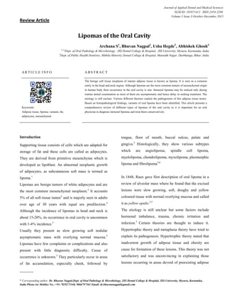
Figure 1: (A) Angiolipoma (B) Intramuscular lipoma (C)
Perineural fibrolipoma (D) Spindle Cell Lipoma
Figure 2: (A) Angiomyolipoma (B) Pleomorphic lipoma
(C) Myolipoma (D) Chondroid Lipoma
and is termed as cellular angiolipoma. These cellular
angiolipomas should be distinguished from
angiosarcoma and Kaposi sarcoma because they show
spindled endothelial appearance, with prominent
pericapillary pericytes. Long standing lesion show
degenerative changes like perivascular fibrosis,
hyalinization and stromal myxoid change. In the past,
deep seated intramuscular lipoma was termed as
infiltrating angiolipoma which is presently classified as
Figure 3: Fibrolipoma
intramuscular hemangioma with prominent adipocytic
component. This is important in view of very high local
recurrence rate in intramuscular hemangiomas.
Angiolipomas are always benign and show no tendency
to recur. Malignant transformation does not occur.4,10
(Figure 1A)
2) Intrmuscular/Infiltrating Lipoma: Adults are most
commonly affected and it may become very large &
measure upto 20 cm in diameter. Histologically, muscle
is replaced by adipose tissue which is mature, creating an
alternating checkboard pattern of fat and striated muscle
cells. Recurrence is more (about 15%) which is due to its
diffuse nature.11
(Figure 1B)
3) Perineural Fibrolipoma: It is a hamartoma of nerves
that causes disfiguring enlargement at a very young age
and sometimes multiple.12
There will be excess of mature
adipose tissue surrounding the nerves, which often
display concentric perineural fibrosis that is
characteristic for this subtype of lipoma.11
(Figure 1C)
4) Spindle Cell Lipoma: It is a circumscribed tumor,
commonly occuring in the subcutaneous tissue. It
4. LIPOMAS OF THE ORAL CAVITY 154
Journal Of Applied Dental and Medical Sciences 1(3);2015
consists of adipose tissue interspersed with short
fascicles of bland undifferentiated spindle cells in a
matrix containing bands of hyaline collagen and
occasional mast cells.4,11
(Figure 1D)
5) Angiomyolipoma: It is most probably a
hamartomatous lesion which is mostly asymptomatic.
Histologically, it consists of mature adipose tissue and
dilated blood vessels that are surrounded by sheets of
well differentiated smooth muscle.11
(Figure 2A)
6) Pleomorphic Lipoma: Pleomorphic lipoma and
spindle cell lipoma are considered as closely related
lesions because of significant proportion of cases
showing over lapping of clinicomorphologic features.
Now, it is considered as variations of single entity.4
But pleomorphic lipoma typically contains spindled,
rounded and multinucleated floret like giant cells
because of their multiple radially arranged nuclei which
resembles petals of flowers. Its superficial location and
sharp circumscription differentiates it from liposarcoma.
Transitional form between spindle cell and pleomorphic
is seen rarely.11
(Figure 2B)
7) Myolipoma: Rarest lesion with admixture of mature
adipose tissue and smooth muscle in varying proportions
in which the muscular component is predominant.13
Female predominance is seen and are often large.
Differential diagnosis include angiomyolipoma and well
differentiated liposarcoma with smooth muscle
component.4
(Figure 2C)
8) Chondroid Lipoma: Very uncommon and often
mistaken as sarcoma because of prominent population of
cells that closely resembles lipoblasts and chondroblasts.
Histologically, it is present with admixture of mature
adipose tissue, lipoblasts with bland nuclei, and
hibernoma like cells in a myxohyaline and
pseudochondroid matrix.4
(Figure 2D)
9) Fibrolipoma: Fibrolipoma is a microscopic variant of
lipoma characterized by a significant fibrous component
intermixed with lobules of fat cells. The consistency of
this lesion varies from soft to firm, depending on the
quantity and distribution of fibrous tissue and the depth
of the tumor.14
(Figure 3)
Generally, lipomas require no treatment unless they are
large, painful, in an inconvenient site, and/or unaesthetic.
Surgical excision is the usual mode of treatment. The
other modes include injection of steroid to reduce the
size of lipomas, which causes atrophy of adipose tissue.
Lidocaine and triamcinolone acetonide (1:1) is also used
for the regression of lipomas. Liposuction is also
recommended in order to avoid scarring. According to
some authors, IFN alpha can be used for infiltrating
angiolipoma. For large lipomas surgical excision is done
after regression therapy. Overall prognosis of lipomas is
good.3,6,15
Conclusion:
Intraoral lipomas are rare lesions which is asymptomatic
and seen during routine dental examination. As it is
painless in majority of the times, patients may visit for
aesthetic concern or due to any discomfort. It represents
about 1-4% of all neoplasms of the oral cavity.
Histological diagnosis and categorization is mandatory
because of its variants. Prognosis is good.
5. LIPOMAS OF THE ORAL CAVITY 155
Journal Of Applied Dental and Medical Sciences 1(3);2015
References:
1. Young Barbara, Lowe JS, Stevens A, Heath JW.
Wheater’s Functional Histology – A Text and Colour
Atlas. 5th
Ed. Noida:Elsevier; 2009. p.74-78.
2. Rajendran R and Sivapathasundharam B. Shafers’s
Textbook of Oral Pathology. 7th
Ed. Noida, India:
Elsevier; 2009.p.452-455.
3. Omisakin OO and Ajike SO. Oral lipomas: A report of
two cases. Int J Med Biomed Res 2014:3(1):58-62
4. Fletcher CDM. Diagnostic Histopathology of Tumors.
3rd
Ed. China:Elsevier; 2007.p.1528-1534.
5. Kumar LKS, Kurien NM, Raghavan VB, Menon PV,
and Khalam SA. Intraoral Lipoma: A Case Report. Case
Reports in Medicine. Volume 2014;1-4
6. Sharma M, Sharma GK, Bhullar RK. Noninfiltrating
Angiolipomas of Lip - A Case Report with Review of
Literature. Journal of Medical Research and Practice
2012;1(4):73-75.
7.http://www.pathologyoutlines.com/topic/softtissueadip
oselipoma.html
8. Neville BW, Damm DD, Allen CM, Bouquot JE. Oral
and Maxillofacial Pathology. 2nd
Ed. New Delhi:
Elsevier; 2005. p. 452.
9. Das S. A Concise Textbook of Surgery. 8th
Ed. CBS
Publishers & Distributors P Ltd; 2014.
10. Fletcher CDM, Unni KK, Mertens F. Pathology and
Genetics of Tumours of Soft Tissue and Bone.WHO
Classification of Tumors. Lyon:IARC Press;2002.
11. Damjanov I, Linder J. Anderson’s Pathology. 10th
Ed. Noida: Elsevier; 2009.
12. Dionne GP, Seemayer TA. Infiltrating lipomas and
angiolipomas revisited. Cancer 1974;33:732.
13. Meis JM, Enzinger FM. Myolpioma of soft tissue.
Am J Surg Patho 1991;l14:75-81.
14. Khubchandani M, Thosar NR, Bahadure RN, Baliga
MS, Gaikwad RN. Fibrolipoma of buccal mucosa.
Contemp Clin Dent 2012;3:112-114.
15. Sah K, Kadam A, Sunita JD, Chandra S.
Noninfiltrating angiolipoma of the upper lip: A rare
entity. J Oral Maxillofac Pathol 2012;16:103-106.
How to cite this article:
Archana S, Nagpal B, Hegde U, Ghosh A. Lipomas of the Oral Cavity
.JOADMS 2015;1(3):151-155.
Source of Support: Nil, Conflict of Interest: None declared