eeg basics in psychiatry
•Descargar como PPTX, PDF•
44 recomendaciones•7,554 vistas

EEG is a non-invasive method to measure electrical activity in the brain. It can help in psychiatry by ruling out physical causes for psychiatric symptoms, aiding in differential diagnosis and treatment selection, and predicting prognosis. EEG findings can provide clues to underlying conditions in disorders like schizophrenia, mood disorders, OCD, panic attacks, dementia, delirium, and substance abuse. However, EEG findings in psychiatry are often nonspecific and EEG has limitations due to only recording cortical activity from the scalp. It currently has no definitive role in diagnosing Axis I or II psychiatric disorders.

Recomendados
Más contenido relacionado
La actualidad más candente
La actualidad más candente (20)
Destacado
Destacado (20)
Similar a eeg basics in psychiatry
Similar a eeg basics in psychiatry (20)
Más de Deepika Singh
Último
Último (20)
eeg basics in psychiatry
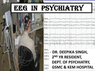
- 1. EEG IN PSYCHIATRY - DR. DEEPIKA SINGH, 2ND YR RESIDENT, DEPT. OF PSYCHIATRY, GSMC & KEM HOSPITAL
- 2. WHAT IS EEG Electroencephalography is non-invasive method for investigation of electrical activity of brain. Used to measure cortical neuronal activity through detection of potential differences across scalp
- 3. HOW IT CAN PSYCHIATRY: HELP IN To rule out physical or neurological causes before making psychiatric diagnosis May help in diagnosis and selection May help prognosis in differential treatment predicting
- 4. OVERVIEW How EEG is recorded Normal EEG findings Factors affecting EEG Application of EEG EEG in psychiatric disorders
- 5. How to record EEG Electrode Placement •10-20 percent system is used •This system measures the distance between readily identifiable landmarks on the head and then puts electrodes at 10 percent or 20 percent of that distance in an anteriorposterior or transverse direction •Even number for right hemisphere and odd number for left hemisphere.
- 6. How to record EEG Sensitivity: Frequency filter setting The amplification used in EEG recording can be adjusted to visualize low-voltage signals. Accepted standards across laboratories for most recordings are 7microvolts for each millimeter of pen deflection. For clinical purpose frequency falls within the range of 0.5 to 40.0 or 50.0 Hz. Setting frequency filter appropriately helps to rule out frequencies generated by non brain sources.
- 7. SPECIAL ACTIVATIONS: HYPERVENTILATION: Done with eyes closed and over breathing through mouth. It is especially effective in eliciting Petit Mal Seizure pattern. PHOTIC STIMULATION: Done by placing an intense strobe light 12 inches in front of subject’s closed eyes and flashing at frequency ranging from 1 to 50 Hz. More useful in detecting psychiatric disorders SLEEP: More useful in eliciting paroxysmal discharges SLEEP DEPRIVATION: can be useful in eliciting paroxysmal discharges
- 8. BRAIN WAVE It is the difference in electric potential measured between any two EEG electrodes which fluctuates rapidly, many times a second leading to a “squiggly line”.
- 10. NORMAL EEG TRACING ALPHA RHYTHM •Highly rhythmic with a frequency range from 8 to 13 Hz •Constitute the dominant brain wave frequency of the normal eyes-closed wake EEG. •Alpha activity is also most prominent over the posterior cortex, particularly the parietal, posterior temporal, and occipital cortex •Occipital region being best suited to show this activity.. •Alpha activity is abolished by eye opening, and alpha activity also disappears with drowsiness and sleep. •Alpha activity can be highly responsive to cognitive activity, such as focused attention or concentration. Example ,alpha can be blocked or attenuated by engaging in visual imagery, numeric calculation etc.
- 11. NORMAL EEG TRACING BETA RHYTHM •Frequencies that are faster than the upper 13 Hz limit •They are common in normal adult waking EEGs •Particularly seen over frontal-central regions. •The voltage of beta activity is also almost always lower than that of activity in the other frequency bands described previous
- 12. NORMAL EEG TRACING THETA RHYTHM •Waves with a frequency of 4.0 to 7.5 Hz •It is a prominent feature of the drowsy and sleep tracing •Although theta activity is limited in the waking EEG, •A small amount of sporadic, arrhythmic, and isolated theta activity can be seen in many normal waking EEGs, particularly in frontal-temporal regions •Excessive theta in wake, generalized or focal in nature, suggests a focal pathological process
- 13. NORMAL EEG TRACING NORMALRHYTHM DELTA EEG TRACING •Delta activity (equal to or less than 3.5 Hz) is not present in the normal waking EEG •Is a prominent feature of deeper stages of sleep. •The presence of significant generalized or focal delta in the wake EEG is strongly indicative of a Pathophysiological process GAMMA RHYTHM Evidence has been provided that high frequency oscillations within the gamma band (>30Hz) reflect mechanisms of cortical integration
- 14. FACTORS AFFECTING EEG Changes with Age •Preponderance of irregular medium- to highvoltage delta activity in the tracing of the infant •EEG activity gradually increases in frequency and becomes more rhythmic with increasing age •Rhythmic activity in the upper theta–lower alpha range (7~to 8 Hz) can be seen in posterior areas by early childhood •By mid-adolescence EEG has the appearance of an adult tracing.
- 15. FACTORS AFFECTING EEG Changes with sleep •The rhythmic posterior alpha activity of the waking state subsides during drowsiness and is replaced by irregular low-voltage theta activity. •As drowsiness deepens, slower frequencies emerge, and sporadic vertex sharp waves may appear at central electrode sites •The progression into sleep is marked by the appearance of 14-hz sleep spindles (also called sigma waves) •Which, in turn get replaced by high-voltage delta waves as deep sleep stages are reached.
- 17. FACTORS AFFECTING EEG Artifacts •Artifacts are electric potentials of nonbrain origin that are in the frequency and voltage range of EEG signals and that are detected by scalp electrodes. •Common artifacts include eye blinks, vertical or lateral eye movements, muscle potentials from jaw clenching, perspiration artifacts (galvanic skin response), and head movement. •Automatic artifact rejection programs exist for some computerized research applications, but they have not strongly entered the clinical arena.
- 18. FACTORS AFFECTING EEG Changes with Medications •Benzodiazepines always generate significant amount of diffuse beta activity. •The highest proportion of abnormal EEGs occurred with Clozapine followed by lithium •Lithium is capable of causing abnormal generalized slowing, paroxysmal activity, or both, including a 10 percent incidence of toxic delirium •The highest incidence of EEG abnormalities was associated with clozapine >olanzapine > risperidone, fluphenazine >haloperidol. There was no EEG abnormalities seen with quetiapine.
- 19. EEG IN PSYCHIATRIC DISORDERS Currently there is no accepted indication of EEG in diagnosing either axisI or II disorders
- 20. : EEG IN SCHIZOPHRENIA •EEG abnormalities have overall frequency of 20-60%. •May predict conversion of subjects at risk into psychosis •Their presence indicate worse outcome. •It helps to identify those with comorbid epileptic condition. •Epileptiform variants are found in affective disorder with psychotic feature and schizoaffective disorder but not in schizophrenia. EEG in Catatonia EEG abnormalities in schizophrenia : •Dysrhythmia •Spike and spike –and- wave •Generalized slowing EEG can help to find out specific etiology of catatonia as catatonia may be caused by several organic disorders
- 21. EEG IN MOOD DISORDERS: •Abnormal EEG found in 20-40% of patients •In bipolar patients increase in beta activity and decrease in alpha activity noted •Acute mania has increased posterior slow rhythms •An asymmetric alpha activity in left frontal region has been reported in depression •Unipolar and bipolar depression have sleeping EEG recording abnormality i.e., short REM latency, increased REM density and reduction in stage 3 and 4 of sleep. •Frequent increase sharp spikes,6/sec spike in patients with suicidal ideation
- 22. EEG in OCD: EEG in OCD EEG abnormalities present in varying frequency] Widespread increase in slow waves reported EEG in Panic disorders •25-30% of panic attack patients have EEG abnormalities •Helps in differentiating panic attack from epilepsy •focal paroxysms of sharp wave activity coinciding with spontaneous onset of panic attack is noted
- 23. EEG in DEMENTIA •Increased slow activity and decreased mean frequency are correlated with cognitive impairment and measures clinical severity of Alzheimer's dementia •The amount of theta activity shows the best correlation with cognitive deterioration •Increased delta appears to be correlate of severe advanced dementia, occurring subsequent to increased theta
- 24. EEG in DELIRIUM •Hallmark of delirium usually is the slowing of the background EEG rhythm •This is positively correlated with the degree of severity of the condition •Exception is in delirium tremens (DT), which usually shows a normal EEG record with fast rhythms. •Delirium accompanying the neuroleptic malignant syndrome shows only a mild diffuse slow wave. •Delirium can be differentiated from dementia, and the significant factors are an increased theta activity
- 25. EEG in Alcohol and Substance Abuse • Acute Alcoholic intoxication shows slowing in the EEG, seen as decreased alpha frequency and abundance & increased amount of theta, and even some generalized delta rhythm • These slow waves have a relationship with the degree of intoxication. The extent of the disturbance of consciousness is related to the amount of slow activity • Reports have appeared of an increased beta (relative power) in alcohol dependence • Increased alpha power, especially in anterior regions, has been reported in withdrawal, as well as after acute exposure to cannabis
- 26. PROBLEMS WITH EEG IN PSYCHIATRY •Nonspecificity of findings •Problem with placing electrodes in psychiatric patients •Limitations of scalp EEG i.e.,Only onethird of brain can be covered,EEG activity of sub-cortical area can’t be recorded •Currently there is no accepted indication of EEG in diagnosing either axis-I or II disorders
Notas del editor
- Nasopharyngeal and sphenoid electrodes can be used to improve readings from frontal, temporal regions.Sphenoidal electrode gives more positive results than regular electrodes but it is an invasive procedure