The document discusses various instruments used in microbiology labs, including their principles of operation and common uses. It describes analytical balances, autoclaves, Bunsen burners, centrifuges, colony counters, deep freezers, homogenizers, hot plates, hot air ovens, incubators, laminar flow hoods, magnetic stirrers, microscopes, pH meters, spectrophotometers, vortex mixers, water baths, water distillers, wire loops, Bijoh bottles, glassware, and Durham tubes. It also covers proper collection and transport of clinical specimens for microbiological analysis.
💸Cash Payment No Advance Call Girls Nagpur 🧿 9332606886 🧿 High Class Call Gir...
INSTRUMENTS USED IN MICROBIOLOGY LAB WITH PRINCIPLE AND.ppt
1. INSTRUMENTS USED IN MICROBIOLOGY
LAB WITH PRINCIPLE AND USES 441
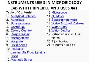
Table of Contents
1. Analytical Balance
2. Autoclave
3. Bunsen burner
4. Centrifuge
5. Colony Counter
6. Deep Freezer
7. Homogenizer
8. Hot plate
9. Hot air oven
10.Incubator
11.Laminar Air Flow/ Laminar
Hood
12.Magnetic Stirrer
13.Microscope
14.pH Meter
15.Spectrophotometer
16.Vortex Mixture/ Vortexer
17.Water Bath
18.Water Distiller
19.Petri-dish and culture
plates
20.Bijoh bottles
21.Duhams tubee.t.c.
2. • The instruments used in the microbiology labs include a
bunch of different kinds of instruments required for a lot of
different processes conducted within the laboratories.
1. Analytical Balance
• An analytical balance is a type of balance that is commonly
used for the measurement of mass in the sub-milligram range.
3. Working Principle
• These types of balances are made with a measuring pan
enclosed in a transparent covering that prevents small particles
or air currents from getting collected on the pan.
• An electric analytical balance uses the force necessary to
counteract the mass rather than measuring the mass itself.
• An electromagnet is used to create a force required to achieve a
balance with the mass of the substance, and the resulting force
is displayed.
Uses
• As they are highly precise and based on advanced technology,
analytical balances are explicitly used in laboratories for the
effective completion of tasks like weighing test materials and
sampling amounts, formulation, density determination, purity
analysis, quality control testing, and material and conformance
testing.
4. 2. Autoclave
• An autoclave is a pressurized chamber used for the process of
sterilization and disinfection by combining three factors: time,
pressure and steam.
5. Working Principle
• Autoclaves use steam as their sterilization agent. The
basic principle of an autoclave is that all the items
within the autoclave come in direct contact with the
steam for a particular period irrespective of the nature
of the material- whether it is liquid, plastic ware, or
glassware.
• The amount of time and the temperature depends on
the type of material being sterilized and the increase in
temperature of the cycle allows for shorter periods.
Uses
• Autoclaves are mostly used for the sterilization of
medical or laboratory equipment with the capacity of
sterilizing a large number of materials at once.
• They are commonly used for the preparation of culture
media during laboratory applications.
6. 3. Bunsen burner
Bunsen burner is a standard tool used in laboratories, named
after Robert Bunsen.
It is a gas-fueled single open flame.
7. Working Principle
• This burner is made with a metal tube on a flat base
with a gas inlet at the bottom of the tube, which
may have an adjustable valve. On the sides of the
tube are openings which can be adjusted with a
collar to control the amount of air that can enter.
• Once the burner is connected to a gas source, the
gas is forced by the gas pressure so that the gas
reaches the top where the flame is ignited with a
match or a lighter.
Uses
• It is commonly used for processes like sterilization,
combustion, and heating. In medical or
microbiology laboratories, it is commonly used for
micro-loop sterilization.
8. 4. Centrifuge
• A centrifuge is a device that allows the rotation of an object about a single
axis, where an outward force is applied perpendicularly to the axis.
• A laboratory centrifuge is motor-based and allows the rotation of a liquid
sample resulting in the separation of the components of the mixture.
9. Working Principle
• A centrifuge works on the principle of sedimentation,
where the high speed of the rotation causes the denser
particles to move away from the center while smaller,
less dense particles are forced towards the center.
• Thus, the denser particles settle at the bottom while the
lighter particles are collected at the top.
• In a laboratory tabletop centrifuge, the sample tubes are
aligned at an angle so that the particles have to travel a
shorter distance before they hit the bottom.
Uses
• The primary application of a centrifuge is the separation
of particles suspended in a suspension. It can be used for
the separation of cell organelles, nucleic acid, blood
components, and separation of isotopes.
10. 5. Colony Counter
• A colony counter is used to estimate the density of a liquid
culture by counting the number of CFU (colony forming units)
on an agar or culture plates.
11. Working Principle
• This instrument can accommodate different sizes of
plates which are scanned on top with UV, white light
and/or fluorescent illumination.
• One can accomplish the counting either manually
with the touch pressure or with a digital counter.
Uses
• A colony counter is primarily used for counting the
number of colonies present on a culture plate to
estimate the concentration of microorganisms in
liquid culture.
13. Working Principle
• Deep freezers are based on the principle that under
extremely low temperatures, there is minimum microbial
growth which allows for the protection and preservation of
different substances.
• Based on this principle, we can even preserve cultures over
a long period of time without any change in the
concentration of the microorganisms.
Uses
• A deep freeze can be used for the preservation of different
things used in the laboratories for a very long period of
time. Deep freezers are used in laboratories to store and
preserve medical equipment, food items, blood samples,
medicines, and injections, etc. for a more extended period
of time.
14. 7. Homogenizer
• Homogenizer is a device used in laboratories
for the mixing of various liquids and materials
like tissue, plant, food, soil, and many others.
15. Working Principle
• This instrument is based on the principle that when large
globules in coarse emulsion are passed under high
pressure through a narrow orifice, they break down into
smaller particles giving a more uniform and stable mixture.
• A homogenizer has a metal rod with narrow parallel
openings in the form of a comb at the end which acts as
the orifice for the homogenization process.
Uses
• A homogenizer is primarily used to disrupt cells to acquire
cell organelles for different microbiological processes.
• It is used in the preparation step before the extraction and
purification of different macromolecules like proteins,
nucleic acids, and lipids.
16. 8. Hot plate
• A hot plate is a stand-alone appliance used in
microbiology laboratories as a tabletop heating
system.
17. Working Principle
• Unlike the traditional ways of producing heat
through the fire, a hot plate produces heat by the
flow of electricity.
• On a hot plate, electricity runs through the coils
which have a high level of electrical resistance. The
resistance in the coils converts the electrical
energy into heat energy which causes the coils to
release heat.
Uses
• In a laboratory, hot plates are used to heat
glassware and their components.
• They are used over water baths as in water baths
might be hazardous in case of any spills or
overheat.
18. 9. Hot air oven
• A hot air oven is an electrical device that is
used for sterilization of medical equipment or
samples using dry heat.
19. Working Principle
• Hot air oven is a type of dry heat sterilization which is performed on dry
materials and on substances that do not melt or catch fire under high
temperature.
• There are two types of hot air oven based on the working principle
– Forced air hot air oven: In this type of hot air oven, the heated air inside
the oven is distributed throughout the oven with a fan. This prevents
the rising of hot air towards the top while keeping the cold air at the
bottom. This allows for the adequate heating of materials inside the
oven.
– Static air hot air oven: In this type of oven, the heat is produced by coils
present at the bottom of the oven with no fan. The hot air rises and
doesn’t allow the effective sterilization of the materials.
• The equipment inside the oven acquire heat and pass the heat towards the
center, one layer at a time which allows for effective dry heat sterilization.
Uses
• Hot air oven can be used to sterilize materials like glassware, metal
equipment, powders, etc.
• It allows for the destruction of microorganisms as well as bacterial spores.
20. 10. Incubator
• An incubator is a device that is used in the laboratories for the growth and
maintenance of microorganisms and cultures.
• Incubator provides an optimal temperature, pressure, moisture, among
other things required for the growth of microorganisms.
21. Working Principle
• The incubator is based on the principle of maintaining a
proper atmosphere for the growth of microorganisms.
• Incubators have a heating system that allows for the
temperature within the incubator to be adjusted according to
the type of organism cultivated inside.
• Similarly, they are provided with adjustments for maintaining
the concentration of CO2 to balance the pH and humidity
required for the growth of the organisms.
• Variation of the incubator like a shaking incubator is also
available, which allows for the continuous movement of the
culture required for cell aeration and solubility studies.
Uses
• Incubators have a wide range of applications including cell
culture, pharmaceutical studies, hematological studies, and
biochemical studies.
• Incubators can also be used in the steam cell research area.
22. 11. Laminar Air Flow/ Hood chamber
• Laminar Hood is a closed device primarily for processes or
instruments sensitive to microbial contamination.
23. Working Principle
• A Laminar Hood is made up of stainless steel, avoiding
joints and corners to prevent the accumulation of
bacterial spores.
• This device creates a sterile environment with the flow
of sterile air through a High-Efficiency Particulate Air
(HEPA) filter and shortwave ultraviolet germicidal lamp
that sterilizes the workstation.
• Laminar Air Flow has to be turn on 15 minutes before to
ensure complete sterilization and the workstation should
be cleaned with ethanol before and after use.
Uses
• Laminar Hood is commonly used to conduct processes
that are sensitive to contamination.
• It is used for experiments related to plant tissue culture
and for the experiments of genetic transformation.
24. • 12. Magnetic Stirrer
• Magnetic Stirrer is a device commonly used in
microbiology laboratories for the purpose of
mixing liquids.
25. Working Principle
• This device consists of a rotating magnetic or an
electromagnet creating a rotating magnetic field
that allows the stir bar (a piece of heavy metal) to
move around in the vessel.
• It is coupled with a heating system to heat the
liquid while it mixed.
Uses
• It is usually used for mixing various liquid
components in a mixture in a chemical or
microbiology laboratory.
• This device is used in place of other stirrers as it is
noise-free and because the size of the stir bar is so
tiny, there is less chance of contamination.
26. 13. Microscope
• Microscopes are devices that allow the observer to an
exceedingly close view of minute particles.
27. Working Principle
• There are many different types of microscopes, each of
which works on their respective principles. However, there
is some commonality in them.
• The basic principle in a microscope is magnification. Based
on the relative position of the object from the lens or
electromagnets, different positions, nature, and
magnification of the image can be achieved.
• Different types of microscopes are developed to cater to
the specific needs of the observation. However, the
common theme is magnification.
Uses
• Based on the type of microscopes, different microscopes
are used for different purposes.
• They are primarily used for the observation of minute
particles which cannot be observed with naked eyes.
28. 14. pH Meter
• pH meter is a device used in laboratories that measure the H-
ion concentration in water-based solutions to determine the
acidity or alkalinity of the solution.
• A pH meter is often termed as “potentiometric pH meter” as
it measures the difference in electric potential between the
reference and a pH electrode.
29. Working Principle
• In a potentiometric pH meter, single or multiple glass
electrodes, connected to a bulb selective to hydrogen
ions, are attached to a metal rod.
• When the bulb with the electrodes is dipped into a
solution, hydrogen ions in the solution exchange with
positive charges on the electrode generating an
electrochemical potential which is displayed in terms
of pH units on display.
Uses
• A pH meter is primarily used to measure the acidity
of pharmaceutical chemicals, cultures, soil, and water
treatment plant.
• It can be used to measure the acidity level in wine
and cheese during their production.
30. 15. Spectrophotometer
• The spectrophotometer is an optical instrument for
measuring the intensity of light in relation to the wavelength.
• Based on the amount of light absorbed by a colored solution,
a quantitative analysis of the solution can be done.
31. Working Principle
• Spectrophotometry is based on the Beer-Lambert Law,
which states the absorbance of light by a solution (of a
particular wavelength) is directly proportional to the
concentration of the substance.
• Different wavelengths of lights are passed through a
solution as different substances have better absorbance
at different wavelengths. Based on the absorbance of a
particular wavelength, the quantitative analysis of a
solution can be done.
Uses
• In a microbiology laboratory, a spectrophotometer is
applied for the measurement of substance concentration
of protein, nucleic acids, bacterial growth, and enzymatic
reactions.
32. 16. Vortex Mixture/ Vortexer
• A vortex mixture is one of the basic technologies used for the
mixing of samples in glass tubes or flasks in laboratories
33. Working Principle
• It is based on the simple principle of causing
reactions and homogenization by agitating the
mixture.
• Motorized draft shafts present on the mixer
oscillates and transfers the movement to the
sample tubes causing the sample fluids to
undergo turbulent flow.
Uses
• Vortex mixer is mostly used for the mixing of
various sample fluids in the sample tubes and
also allows for the homogenization of cells and
cell organelles
34. 17. Water Bath
• Water Bath is a conventional device that is used for chemical
reactions that required a controlled environment at a
constant temperature.
•
35. Working Principle
• A sensor in the device transfers water
temperature to a reference value which is
then amplified and a control system generates
a signal for the heating system which heats
the water to the desired temperature.
Uses
• Water baths are primarily used for heating
samples under a controlled temperature.
• These are suitable for heating chemicals that
might be flammable under direct ignition.
36. 18. Water Distiller
• A water distiller is a device that purifies water by the process
of distillation.
• This instrument is commonly used in medical laboratories,
microbiology laboratories, organic chemistry laboratories and
medical industries.
37. Working Principle
• A water distiller is based on the principle of
distillation.
• According to this process, water is first
brought to a boil and then condensed into
liquid form to obtain pure distilled water.
Uses
• It is used to obtain distilled water required for
many lab tests as well as for the preparation
of culture media.
38. 19. Wire loop on Petri dish for culture:
20. BIJOH BOTTLES:
Uses: for sugar fermentation tests
39. 21. GLASSWARES
22. DUHAM TUBES
Uses:-The are inserted upside down in bigger tubes and use to check for gas production in sugar
fermentation test in the lab.
40. COLLECTION AND TRANSPORT OF
CLINICAL SPECIMENS
• The proper collection and transport of clinical specimens is
critical for the isolation, identification, and characterization
of agents that cause bacterial infection.
• Optimally, clinical specimens should be obtained before
antimicrobial therapy commences in order to avoid loss of
viability of the etiological agents.
• Treatment of the patient, however, should not be delayed
while awaiting collection of specimens or results from the
laboratory and a specimen should be obtained in all
suspect cases as bacterial pathogens can still be detected
even after antimicrobial therapy has begun.
41. • Biosafety: It is important to adhere to proper
biosafety guidelines while handling potentially
infectious clinical specimens in order to maintain
a safe working environment for patients, health
care workers, and laboratorians.
• Infection may be transmitted from patient to staff
and from staff to patient during the procedures.
• Of particular importance are the viruses causing
hepatitis and acquired immunodeficiency
syndrome.
• To decrease the risk of transmission of these
agents, the recommendations below should be
followed:
42. –Wear latex or sterile gloves that are impermeable
to liquids and change gloves between every
patient.
–Dispose all syringes and needles in a puncture-
resistant, autoclavable discard container. Do not
attempt to re-cap or manipulate any needle. A
new sterile syringe and needle must be used for
each patient.
–For transport to a microbiology laboratory, place
the specimen in a container that can be securely
sealed. Wipe any bottles with CSF or blood on
the outside thoroughly with a disinfectant, such
as a 70% alcohol swab.
43. – Remove gloves and discard in an autoclavable
container.
– Wash hands with antibacterial soap and water
immediately after removing gloves.
– In the event of a needle-stick injury or other skin
puncture or wound, wash the wound liberally with
soap and water. Encourage bleeding.
– Report a needle-stick injury, any other skin puncture,
or any contamination of the hands or body with CSF to
the supervisor and appropriate health officials
immediately as prophylactic treatment of the
personnel performing the procedure may be indicated.
44. When working with patients sample, be careful,
articulate and reproducible because demonstration
of pathogenic organisms in the patient specimen is
the most definitive test in microbiology. However,
failure to demonstrate pathogens in a single
specimen is NOT definitive and may only indicate
that:
• The pathogen was absent or scanty in that
particular specimen;
• The sample was taken at a stage of the disease
when the pathogen was rare;
• Viability was lost between the times of collection
and arrival in the laboratory;
• The pathogen cannot be detected by this method
of testing.
45. SAMPLE COLLECTION, HANDLING AND TRANSPORT
• Collection kits are available for routine cultures upon request. Each kit
contains a transport system composed of a sterile swab and transport
medium.
• RESPIRATORY TRACT SPECIMENS:
A. Throat Culture
– Collect specimen under good lighting. Depress the tongue with a tongue
blade and pass the swab firmly over the back of the patient's throat,
tonsils or tonsillar fossae and any area of inflammation and or exudation.
– Return the swab to the transport tube and break the media ampule at the
base of the tube to moisten the swab.
– Label the swab transport tube with patient name.
– Complete the requisition form.
– Place specimen and requisition in pouch for pickup by lab courier or
arrange to have specimen taken to a courier pickup site.
– Culture into Blood agar, Chocolate agar, MacConkey Agar and any selective
media if any particular pathogen is being suspected to cause the infection.
46. B. Nasopharyngeal Culture:
For nasopharyngeal cultures, a special small tipped swab on
a flexible wire is required and may be obtained by contacting
the laboratory. Collect specimen under good lighting. Bend
the wire of the nasopharyngeal swab into a semi circle and
pass through the nostril to the pharynx, scrub back and forth
gently 2 to 3 times and remove.
– Return the swab to the transport tube and break
the media ampule at the base of the tube to
moisten the swab.
– Label the swab transport tube with a unique
patient identifier.
– Complete the requisition form. Place specimen and
requisition in pouch for pickup by lab courier.
47. C. Sputum Culture: An early morning specimen is
recommended. A volume of 5 to 10 ml is adequate
and there is no advantage in collecting a larger
volume. The sample should contain recently
discharged material from the bronchial tree with
minimal saliva content. It should be purulent.
• Upon rising in the morning, rinse mouth well
with water (not mouthwash)
• Inhale as deeply as possible. Expectorate into
sputum collection container, available from the
Laboratory, while coughing as deeply and
vigorously as possible into a tightly cap
container so no spillage occurs.
48. • Wash and dry outside of container and label the
SPECIMEN CONTAINER with name and date and time
of collection.
• Complete the requisition form.
• Place form and specimen container in pouch for
pickup by lab courier or Refrigerate specimen while
waiting for pickup. Specimens must be processed by
the laboratory within 24 hours.
• If more than one specimen is requested (for example,
3 AFB cultures are requested), collect only one
specimen per day, first thing in the morning. Have
specimens delivered to the Laboratory DAILY. Do not
wait until all three have been collected.
49. WOUND PUS AND ASPIRATES:
• Collect specimen under good lighting. Pass the
swab firmly over or into an area of suspected
infection and obtain a sample of exudate, drainage,
or purulent discharge if these are present. OR use
sterile syringe to aspirate the pus of fluid as the
case may be.
• Label the swab transport tube and syringe
• Complete the requisition form. Indicate the area of
the body from which the specimen was taken to
assist in distinguishing normal from abnormal flora
(ears, legs, etc).
• Place specimen and requisition in pouch and
promptly deliver to lab pickup site.
50. TISSUE:-
• Handle in same manner as Miscellaneous
specimens above; but place tissue specimen in
sterile container with a small amount of sterile
saline or sterile water to keep specimen from drying
out. Be sure container is labeled with name of
patient and source of specimen. Do not fix the
tissue, because fixing inactivates the bacteria.
SKIN :-To collect sample from skin cracked skin, the
skin can be scrapped with sterile surgical blade unto
a sterile white paper, that which is collected should
be taken to the laboratory for analysis.
51. STOOL SPECIMENS:-
• The recovery of bacterial pathogens from fecal
specimens will help confirm the diagnosis of
bacterial gastroenteritis as manifested by
diarrhea or dysentery. routinely screen stools
for Campylobacter, Salmonella, Shigella, and
Shigatoxin E.coli.
• If other pathogens are suspected please
indicate on request form. Obtain stool sample
into an open wide mouthed bottle containing
Cary Blair transport media from this laboratory.
52. • Collect feces from patients as soon after onset of illness as
possible, and before the start of treatment.
• Transfer a sample (no more than one ounce) of the
specimen using the spatula attached to the container lid
into the Cary Blair medium supplied in the kit and mix
thoroughly.
• For liquid stool specimens, no more than 10 ml (1/3 oz)
should be added to the Cary Blair medium and mixed.
• Complete the requisition form Place the specimen and
requisition form Specimens must be processed by the
laboratory within 72 hours of collection.
– NOTE: Do not ship stool cultures without using the Cary Blair
transport medium.
• If a rectal swab is used, be certain to insert swab into Cary
Blair and break off upper stem so that lid can be replaced.
53. • URINARY TRACT SPECIMENS:-
• 1. Wash hands with soap and water, rinse and
dry.
• 2. WASH area around urethra with soap.
• 3. RINSE area with warm water.
• 4. VOID- Pass the first portion of urine into the
toilet and then pass a portion (1 ounce) of the
remaining urine into a sterile container.
• Pass the rest of the urine into the toilet, close
and label the container with name and date.
Store samples in refrigerator or send to the lab
immediately.
54. GENITAL TRACT SPECIMENS
• Use swab to obtain a sample of endocervical,
vaginal, or urethral discharge. Return the swab to
the transport tube and break the media ampule
at the base of the tube to moisten the swab.
• Endocervical specimen can be collected using
sterile speculum, Label the swab transport tube
with the patient's name.
• Complete the requisition form and send
specimen to the lab. Send the specimen to the
lab. Special media and transport containers are
required when culturing for gonorrhea.
55. COLLECTION AND TRANSPORT OF BLOOD AND CSF
SPECIMENS
• To collect CSF:-The collection of CSF is an invasive
procedure and should only be performed by experienced
personnel under aseptic conditions. If bacterial meningitis
is suspected, CSF is the best clinical specimen to use for
isolation, identification, and characterization of the
etiological agents. Suspected agents should include N.
meningitidis, S. pneumoniae, and H. influenzae and other
pathogens in some cases.
• Cerebrospinal fluid (CSF) should be processed in a
microbiology laboratory within 1 hour after collection or
inoculated into Trans-Isolate (T-I) medium for transport to
the laboratory if processing within 1 hour is not feasible.
Blood specimens should be immediately inoculated into a
blood culture bottle and transported to a microbiology
laboratory as soon as possible for overnight incubation and
growth of bacteria.
56. • Inoculating and transporting in a T-I medium. T-I is a
biphasic medium that is useful for the primary culture of
meningococci and other etiological agents of bacterial
meningitis (S. pneumoniae and H. influenzae) from CSF. It
can be used as a growth medium as well as a holding and
transport medium. The preparation of T-I media should be
stored at 4°C and warmed to room temperature (25°C)
before use.
• Label the T-I bottle with appropriate information: patient
name, date and time of CSF inoculation, and Unique
Identification Number. Be sure this number matches the
number on both the request and report forms.
• Use a sterile syringe and needle to inoculate 0.5-1.0 ml of
CSF into the T-I medium. The remaining CSF should be kept
in the collection tube. It should not be refrigerated, but
should be maintained at room temperature (20-25°C)
before Gram staining and other tests and incubate for
24hrs at 35- 37°C.
57. • If turbidity is observed, subculture onto a blood agar plate (BAP) and a chocolate
agar plate (CAP) immediately give a Presumptive result. If no turbidity is observed,
culture onto a BAP and a CAP on day 4 and day 7. If T-I medium appears to be
contaminated, selective media such as Modified Thayer-Martin and chocolate agar
with backtracking may be used.
This is a picture of a bottle of Trans-Isolate (T-I) medium.
Transporting CSF specimens without T-I media.
CSF specimens should be transported to a microbiology laboratory as soon as
possible. Specimens for culture should not be refrigerated or exposed to extreme
cold, excessive heat, or sunlight. They should be transported at temperatures
between 20°C and 35°C. For proper culture results, CSF specimens must be plated
within 1 hour. If a delay of several hours in processing CSF specimens is anticipated
and T-I medium is not available, incubating the specimens (with screw-cap loosened)
at 35-37°C with ~5% CO2 (or in a candle-jar) may improve bacterial survival.
58. • Collection and transport of blood specimens:-
• Blood should be collected when bacteremia is suspected or when
CSF cannot be collected. Accuracy of the blood cultures depends on
the several variables affect the sensitivity of blood cultures: the
number of collections, the volume of each collection, the steps taken
to inhibit or neutralize bactericidal properties of blood, and the age
of the patient. It may be difficult to collect more than 3 ml of blood
from a child, but 1-3 ml is considered adequate. Collected blood
should be diluted in blood culture broth in order to obtain blood
cultures.
• Typically, 1-2 ml of blood from a child is added to 20 ml of blood
culture broth and 5-10 ml of blood from an adult is added to 50 ml of
blood culture broth. It is important to use appropriate ratios of blood
to culture broth for optimal bacterial growth.
• Blood should be cultured in tryptose soy broth (TSB) or brain heart
infusion (BHI) broth with a growth supplement (such as Vitox) to
support growth of other fastidious organisms such as H. influenzae.
59. • Venipuncture and inoculating blood culture
bottles,
• Label the blood culture bottle with appropriate
information: patient name, date and time of
blood culture bottle inoculation, and Unique
Identification Number. Be sure this number
matches the number on both the request and
report forms.
• Disinfect the rubber septum of the blood culture
bottle with a 70% alcohol swab and allow it to dry
and inoculate the blood into the blood culture
medium to prevent the blood from clotting in the
syringe.
60. • For blood from young children, add 1-2 ml of blood into 20 ml
of blood culture broth (approximately a 1:10 to 1:20 dilution).
• For blood from adults, add 5-10 ml of blood into 50 ml of
blood culture broth (approximately a 1:5 to 1:10 dilution).
• After inoculation, swirl the bottle several times to mix and
transport to a microbiology laboratory immediately
• If immediate transport to a microbiology laboratory is not
feasible, place the inoculated blood culture bottle in an
incubator at 35-37°C with ~5% CO2 (or in a candle-jar) until
transport to a microbiology laboratory is possible.
• Inoculated blood culture bottles should not be placed in the
refrigerator.
• Inoculated blood culture bottles should be transported to a
microbiology laboratory immediately for overnight incubation
at 35-37°C with ~5% CO2 (or in a candle-jar) and subsequent
culture onto a BAP and CAP.
61. • Collection of samples for serological tests:-
• Aseptically withdraw enough quantity of blood and pure
into a clean grease free glass bottle, it should be kept on
the in an up write position and allowed to clot, it will be
retracted and spurn at 3000rmp for 5mins , the clear
serum should be used for serological tests. can be stored
in the freeze when not in use.
• LABORATORY TESTINGS
• Routine cultures are plated on a variety of media
associated with the requirements for screening different
body sites. Cultures are incubated overnight, then
examined for normal flora and potential pathogens.
Potential pathogens are identified with a variety of
methods, and susceptibility testing is performed when
needed.
62. RESULTS
• Laboratory results are reported in lab forms when the test
results are available. Expected turnaround time is 24 -48
hours.
REJECTION OF SAMPLES:-
• Samples will be rejected if they are:
• Unlabeled - All specimens MUST have a unique patient
identifier.
• Insufficient in Quantity -Insufficient specimen to perform
testing.
• Improperly Preserved - Specimens must be preserved and
received in the transport media as defined by the
laboratory.
• Damaged - Specimen leaked or broken in transit.
• Too Old - Aged specimens are diagnostically unreliable.