SlideShare una empresa de Scribd logo
1 de 70
Development and growth of teeth and its
supporting structure
Dr. Dinraj Kulkarni
Some terminologies to be familiar
• Dental lamina
• Enamel organ
• Ectomesenchyme
• Dental papilla
• Dental sac
• OEE
• IEE
• Stellate reticulum
• Stratum intermedium
• Cell rests of serre’s
Embryological basis of dental tissue genesis
Lined by stratified
squamous epithelium
i.e. Oral ectoderm or
primitive oral epithelium
Connective tissue cells underlying oral
ectoderm are neural crest or
ectomesenchymal in origin , which
induce tooth development Ruptures at 27th day (4th week) of
gestation
Primary epithelial band /6th week (2-3
weeks after rupture of BF membrane)
Continuous band of
thickened epithelium
called “Primary
epithelial band”.
Roughly horse shoe shaped
Thickening first seen in anterior midline
Certain areas of basal cells of oral ectoderm
proliferate more rapidly than do cells of
adjacent areas
7th week
Buccaly
located
vestibular
lamina
Lingually dental lamina
Primary epithelial
band
Initiation of tooth development
• The initiation of tooth development begins at 37th day with
formation continuous horse shoe shaped band of thickened
epithelium in the location of upper and lower jaws– Primary
epithelial band
Each band of epithelium
gives rise to 2 subdivisions
Dental lamina
Vestibular lamina (Lip furrow
band)
Vestibular lamina – labial and
buccal to the dental lamina in
each dental arch, another
thickening develops apparently
and some what latter called
“Vestibular lamina”
8th week
Continued thickening of dental lamina in 10 areas of upper and
lower arch.
Thickenings corresponds to the position of future primary
dentition
• Development of 1st molar – 4th month in utero
• 2nd molar – 1st year after birth.
• 3rd molar – 4th or 5th years
• Succesional lamina – 5th month in utero
(permanent central inciosor) & (10th month –
2nd premolar)
Formation of vestibule in oral cavity
Cells of vestibular lamina
proliferate
Degeneration of central
epithelial cells
Sulcus of vestibule
Dental lamina
Is a band of epithelium
Serves as a primordium for
the ectodermal portion of the
deciduous teeth.
Dental lamina
• Within dental lamina localized and continuous proliferative
activity leads to formation of series of epithelial outgrowths
into the ectomesenchyme at the sites corresponding to the
future deciduous teeth which represents the beginning of
enamel organ.
• Successors of deciduous teeth arises from the lingual
extension of free end of dental lamina opposite to enamel
organ of each deciduous tooth, these lingual extensions are
referred to as “Successional lamina" and develops from 5th
month in utero to 10th month of age.
Functions of dental lamina
• Phase –I :- initiation of entire deciduous dentition (8th week).
• Phase – II :- Initiation successors of deciduous dentition (5th
to 10th month).
• Phase – III :- Initiation of permanent molars.
Fate of dental lamina
• The total activity of dental lamina extends over a period of at
least 5 years.
• Any particular portion of dental lamina functions for much
briefer period, since only a relatively short time elapses after
initiation of tooth development before dental lamina begins
to degenerate at particular location i.e. it might be still active
in 3rd molar area when it has disappeared elsewhere.
• Dental lamina looses its connection from enamel organ at the
start of bell stage.
• Remnants of dental lamina persists as epithelial pearls or
islands within jaw termed as “cell rests of Serre’s”
Rests of serre’s or epithelial cells
• Also called as “Epithelial Pearls or islands”.
• These are remnants of dental lamina.
• These are identified in jaws and gingival soft tissues.
• They results from early break up of dental lamina during bell
stage.
Clinical significance
- Gingival cyst of adult
- Gingival cyst of new
born
Rests of Malassez
• The break up of heartwig’s root sheath during root
formation i.e. these are remnants of Heartwig’s
epithelial root sheath.
• These are identified in periodontal ligament and are
responsible for the development of radicular cyst.
How development tooth begins??
1st arch and ectomesenchymal cells
derived from the neural crest cells
Up to 1st 12 days 1st arch epithelium retains the
ability to form tooth like structure when
combined with neural crest cells of regions
This potential is lost and transformed to neural crest
cells to produce tooth like structure
Interaction of epithelium derived
Afterwards
• Stages of tooth development
Stages of tooth development
Morphologic stage
Bud stage
Cap stage
Bell stage
 Early bell stage
 Advanced bell stage
Physiologic stages
Initiation
Proliferation
Histodiffrentiation
Marphodiffrentiation
Apposition
STAGES IN TOOTH GROWTH
Morphologic changes physiologic processes
• Dental lamina Initiation
• Bud stage Proliferation
• Cap stage
• Early bell stage Histodiffrentiation
• Advanced bell Marphodiffrentiation
• Enamel/dentin
formation apposition
Initiation
6th to 7th weeks
Ectoderm lining
stomodeum gives rise to
oral epithelium and dental
lamina
All the enamel organ
don’t start at same time,
first to start are those of
anterior mandibular
region
Bud stage
Bud stage or proliferative stage
8th week of prenatal development
Growth of dental lamina into bud that penetrates
growing ectomesenchyme
• Initial proliferation of oral epithelial cells and
adjacent mesenchymal cells is occurring and
the cells proliferate faster than the adjacent
cells giving rise to bud shaped enamel organ.
 Enamel organ – spherical to
ovoid epithelial condensation
 Cells of tooth bud have --
Increase RNA content,
Decrease Glycogen content
and increase oxidative
enzyme activity
 Cells undergo mitosis and
get condensed. Enamel organ surrounded
by mesenchyme
Bud stage at 8th week
Basement
membrane
Tooth bud
Histologically enamel organ at bud stage consists of,
• Peripherally located low columnar cells.
• Centrally located polygonal cells.
• Many cells of tooth bud and surrounding mesenchyme
undergoes mitosis, i.e. increased mitotic activity.
• Ectomesenchymal cells or neural crest cells migrates in the
area surrounding tooth bud.
• Ectomesenchymal condensation occurs surrounding the bud.
Cap stage
Cap stage
 9th to 10th week
 Unequal growth in
different parts of tooth bud
leads to concave surface
forming cap like structure.
 Enamel organ forms into
cap.
 There is a formation of
tooth germ
Early cap stage – 11 week
Peripherally located
cuboidal cells – Outer
enamel epithelium
Tall columnar cells in the
concavity – Inner enamel
epithelium
Polygonal cells in between
OEE and IEE called –
Stellate reticulum
(12th week)
Histologically
• Outer enamel epithelium
 Cuboidal cells covering convexity.
 Separated from dental sac by basal
lamina.
• Inner enamel epithelium
 Tall columnar cells lining concavity of
enamel organ.
 Separated from the dental papilla by
basal lamina.
Stellate reticulum
• Cells in the center of the enamel organ.
• These cells are star shaped which secrete GAG.
• Stellate reticulum cells synthesize and secrete GAG’s into
extracellular compartment between epithelial cells –>
hydrophilic -- > pull water into the enamel organ and central
cells are forced apart.
• They are so named because they retain connection with each
other through desmosomal contacts, they become star
shaped, the center of the enamel organ thus termed Stellate
reticulum.
Components of tooth germ during cap stage
Parts of cap
Dental papilla – dentin and pulp
Dental sac – cementum and periodontal ligament
What is tooth germ??
The enamel organ, the
dental papilla and the
dental sac together
called
“Tooth germ”.
 Enamel organ – enamel
 Dental papilla –Dentin
and Pulp
 Dental sac – PDL,
Cementum and bone
Transitory structures
Enamel knot
Localized mass of cells
produced by rapid
multiplication of cells in
the center of inner
enamel epithelium
Enamel cord
Strand of cells extending from stratum intermedium
into Stellate reticulum
These are vertical extensions of enamel knot.
These acts as reservoir of dividing cells for the
growing enamel organ
Enamel niche
- Double attachment of
enamel organ to overlying
epithelium.
- Funnel shaped area
between lateral and medial
strands of dental lamina
enclosing mesenchyme.
A. Lateral enamel strand
B. Medial enamel strand
C. Enamel niche
Enamel septum
• When the enamel cord extends to meet the outer enamel
epithelium is termed as enamel septum.
Enamel navel
• The outer enamel epithelium at the point of meeting shows a
small depression and is termed as enamel navel as it
resembles umbilicus.
What is enamel organ??
• The developing tooth as it progresses from
bud stage to advanced bell stage is called the
“enamel organ” or “dental organ”
13th week
Tooth
germ
Dental
lamina
Bell stage
• 11th to 12th weeks
• Proliferation,
differentiation,
morphogenesis
• Differentiation of
enamel organ into bell
with four cell types and
dental papilla into 2 cell
types.
Early bell stage (14th week)
Dental lamina break downs and degenerates
Shows 4 distinct layer
 Outer enamel epithelium
 Stellate reticulum
 Stratum intermedium
 Inner enamel epithelium
Outer enamel epithelium
• A single layer of very low cuboidal cells.
• At the end of this stage just before enamel formation, the
smooth surface of outer enamel epithelium get thrown into
folds.
• Between the folds the adjacent mesenchyme of the dental sac
forms papillae that contain capillary loops and thus provide a
rich nutritional supply for the intense metabolic activity of the
avascular enamel organ.
Outer enamel epithelium
Role of OEE
• Maintenance of the shape
of the enamel organ
• Exchange of substance
between enamel organ and
environment
• Holds the contents of
enamel organ
• As a constituent of
epithelial root sheath plays
a role in root formation.
Stellate reticulum
Mechanical
Acts as shock absorber
Maintains tooth shape
Nutritive
Supplies GAG’s to enamel forming cells
Stellate reticulum
• Expands mainly by an increase in the amount of intracellular
fluid.
• The star shaped cells with long processes anastomose with
those of adjacent cells.
• Before enamel formation begins, the Stellate reticulum
collapses, reducing the distance between centrally situated
ameloblasts and the nutrient capillaries near the outer
enamel epithelium.
• Changes begins at the height of the cusp or the incisal edge
and progresses cervically.
Stratum intermedium
Functions
Synthesis and storage of
proteins
Transport of material too
and from the inner enamel
epithelium (ameloblasts)
Induce inner enamel
epithelial cells to become
ameloblasts
Stratum intermedium
• These are 3-4 layers of squamous cells between inner enamel
epithelium and Stellate reticulum.
• These cells are attached by desmosomes and gap junctions.
• There are abundant cell organelles, acid mucopolysaccharides
and glycogen deposits in these cells, which indicate high
metabolic activity.
• This layer is essential for enamel formation.
• Are absent in the part of the tooth germ that outlines the root
portion of the tooth which doesn’t form enamel.
Inner enamel epithelium
Function
Its cells become ameloblasts
which form enamel.
It induces adjacent cells of
dental papilla to form
odontoblasts which in turn
form dentine.
Following amelogenesis, the
cells contribute to form the
REE together with OEE and
Stratum intermedium.
Important role in crown
formation due to intrinsic
growth pattern.
Inner enamel epithelium
Consists of a single layer of cells
differentiate prior to amelogenesis into
tall columnar cells
Ameloblasts
The cells of the inner enamel epithelium exert an
organizing influence on
mesenchymal cells in the dental papilla
later differentiate into odontoblasts.
• Are about 40 um high and 4-5 um in diameter.
• These cells attach to each other laterally by
junctional complexes and cells of stratum
intermedium by desmosomes.
Dental papilla
• Under the inductive influence of inner enamel epithelium, the
peripheral cells of the dental papilla differentiate into
odontoblasts.
• These cells initially become cuboidal and later assume
columnar shape.
• These cells now acquire the potential to produce dentine.
Membrana performativa
• The basement membrane that separates enamel organ and
the dental papilla just prior to dentin formation is called
“Membrana performativa.
Dental sac
• Just before the formation of dental tissues begins, the
collagen fibers of the dental sac are arranged in a circular
manner.
• The dental sac look like a capsule around the enamel organ
and dental papilla.
Changes before enamel formation
• The Stellate reticulum collapses and the
distance between inner enamel epithelium
and blood capillaries of the dental sac is
reduced, so that more nutrient reach the
ameloblasts during the period of high
metabolic activity.
• The formerly smooth surface of outer enamel
epithelium thrown into folds.
• Between the folds the mesenchyme of dental
sac form projections or dental papillae that
carry capillary loops closer to the ameloblast
prior toamelogenesis.
IEE become taller –
Preameloblasts
Peripheral cells of the
dental papilla –
Odontoblasts
Secrete ground
substance and collagen
fibers – Dentin matrix
There is change in cell
polarity.
With change in polarity
the cell called an
ameloblasts that begins
secretion of enamel matrix.
Boundary between inner
enamel epithelium and
odontoblasts outline the
future dentino-enamel
junction.
Enamel organ shows 4 different layers.
• Inner enamel epithelium (ameloblasts)
• Stratum intermedium
• Stellate reticulum
• Outer enamel epithelium
• The ameloblasts have fully differentiated from
the cells of inner enamel epithelium.
• The odontoblasts have fully differentiated
from dental papillae cells.
• The boundary between the ameloblasts and
odontoblasts define the future dentino-
enamel junction.
Histological difference from early bell stage
• Hard tissue (Enamel and Dentin) formation.
• Collapsed Stellate reticulum and folding of outer enamel
epithelium bringing capillaries of the dental follicle nearer to
ameloblasts.
• Dental papilla shows differentiated odontoblasts at periphery
• Dental follicle is distinct enclosing enamel organ and dental
papilla.
Reciprocal induction
• The inner enamel epithelium induces peripheral
cells of dental papilla to differentiate into
odontoblasts.
• The odontoblasts start secreting the organic
matrix of dentin.
• Only dentin matrix is formed, do the ameloblasts
in the area differentiate fully and start secreting
enamel matrix.
• This independence of tissue is an example of
“Reciprocal induction”.
INITIATION Lack of initiation may result in absence of single tooth or multiple
teeth (anodontia). The upper lateral incisor followed by 3rd molars
and lower 2nd premolar commonly involved.
Abnormal initiation result in development of supernumerary tooth.
E.g. – Mesiodens followed by 4th molar
Sometimes fused or geminated teeth are formed.
HISTODIFFERENTIATI
ON
This stage reaches its highest level in bell stage of enamel organ.
E.g. -- Dentinogenesis imperfecta or formation of atypical dentin
MORPHODIFFERENT
IATION
The advanced bell stage is important stage. This stage establishes the
morphologic pattern, or basic form and relative size of future tooth.
Supernumerary (talon’s) cusp, twinning, loss of cusps or roots, or
malformed or peg shaped tooth (Hutchinson’s incisor), dens in dente
and microdontia.
APPOSITION Both enamel hypoplasia and hypo calcification can occur as a result
of an insult to the cells responsible for the apposition stage.
Intrinsic staining and concrescence.

Más contenido relacionado

La actualidad más candente (20)

Dentinogenesis
DentinogenesisDentinogenesis
Dentinogenesis
 
Stages of tooth development
Stages of tooth developmentStages of tooth development
Stages of tooth development
 
Enamel
EnamelEnamel
Enamel
 
3. tooth development
3. tooth development3. tooth development
3. tooth development
 
Oral Histology - Dentine
Oral Histology -  Dentine Oral Histology -  Dentine
Oral Histology - Dentine
 
Enamel
EnamelEnamel
Enamel
 
AMELOGENESIS
AMELOGENESISAMELOGENESIS
AMELOGENESIS
 
development and growth of teeth
development and growth of teethdevelopment and growth of teeth
development and growth of teeth
 
Dentine
Dentine Dentine
Dentine
 
Enamel composition and structure
Enamel composition and structureEnamel composition and structure
Enamel composition and structure
 
Root formation
Root formationRoot formation
Root formation
 
Enamel
EnamelEnamel
Enamel
 
Development of tooth
Development of toothDevelopment of tooth
Development of tooth
 
Amelogenesis
Amelogenesis Amelogenesis
Amelogenesis
 
Histology of Pulp
Histology of PulpHistology of Pulp
Histology of Pulp
 
Dentinogenesis
DentinogenesisDentinogenesis
Dentinogenesis
 
Enamel
EnamelEnamel
Enamel
 
Enamel
EnamelEnamel
Enamel
 
Development of teeth
Development of teethDevelopment of teeth
Development of teeth
 
Dentin
DentinDentin
Dentin
 

Similar a 1.tooth development.pptx

Molecular Basis of Tooth Development
Molecular Basis of Tooth DevelopmentMolecular Basis of Tooth Development
Molecular Basis of Tooth DevelopmentSukesh Vangeti
 
Development of Tooth By Dr. Yashkumar Shah
Development of Tooth By Dr. Yashkumar ShahDevelopment of Tooth By Dr. Yashkumar Shah
Development of Tooth By Dr. Yashkumar ShahYashkumar Shah
 
Development of tooth
Development of toothDevelopment of tooth
Development of toothPiyush Verma
 
Early development of tooth.ppt
Early development of tooth.pptEarly development of tooth.ppt
Early development of tooth.pptridwana30
 
Development of tooth
Development of toothDevelopment of tooth
Development of toothkoilonychia
 
Development of teeth and its clinical significance
Development of teeth and its clinical significanceDevelopment of teeth and its clinical significance
Development of teeth and its clinical significanceaanmol
 
Development of Teeth by Dr.Smitha Rao
Development of Teeth by Dr.Smitha RaoDevelopment of Teeth by Dr.Smitha Rao
Development of Teeth by Dr.Smitha Raosmithanaik1980
 
DEVELOPMENT OF TOOTH.pptx
DEVELOPMENT OF TOOTH.pptxDEVELOPMENT OF TOOTH.pptx
DEVELOPMENT OF TOOTH.pptxDrPurvaPihulkar
 
Development of tooth - DCPS
Development of tooth - DCPSDevelopment of tooth - DCPS
Development of tooth - DCPSMuznah Sultan
 
2. Development of tooth.ppt ODONTOGENESIS
2. Development of tooth.ppt ODONTOGENESIS2. Development of tooth.ppt ODONTOGENESIS
2. Development of tooth.ppt ODONTOGENESISDrNonithaS
 
EMBRYOLOGY OF THE DENTITION.pptx
EMBRYOLOGY OF THE DENTITION.pptxEMBRYOLOGY OF THE DENTITION.pptx
EMBRYOLOGY OF THE DENTITION.pptxTolulaseYemitan1
 

Similar a 1.tooth development.pptx (20)

1 Development of teeth.pptx
1 Development of teeth.pptx1 Development of teeth.pptx
1 Development of teeth.pptx
 
Molecular Basis of Tooth Development
Molecular Basis of Tooth DevelopmentMolecular Basis of Tooth Development
Molecular Basis of Tooth Development
 
Development of tooth
Development of toothDevelopment of tooth
Development of tooth
 
Development of Tooth By Dr. Yashkumar Shah
Development of Tooth By Dr. Yashkumar ShahDevelopment of Tooth By Dr. Yashkumar Shah
Development of Tooth By Dr. Yashkumar Shah
 
Tooth
ToothTooth
Tooth
 
Development of tooth
Development of toothDevelopment of tooth
Development of tooth
 
Tooth development
Tooth development Tooth development
Tooth development
 
Tooth development
Tooth developmentTooth development
Tooth development
 
Development Of Teeth
Development  Of TeethDevelopment  Of Teeth
Development Of Teeth
 
Early development of tooth.ppt
Early development of tooth.pptEarly development of tooth.ppt
Early development of tooth.ppt
 
Development of tooth
Development of toothDevelopment of tooth
Development of tooth
 
Development of teeth and its clinical significance
Development of teeth and its clinical significanceDevelopment of teeth and its clinical significance
Development of teeth and its clinical significance
 
Development of Teeth by Dr.Smitha Rao
Development of Teeth by Dr.Smitha RaoDevelopment of Teeth by Dr.Smitha Rao
Development of Teeth by Dr.Smitha Rao
 
DEVELOPMENT OF TOOTH.pptx
DEVELOPMENT OF TOOTH.pptxDEVELOPMENT OF TOOTH.pptx
DEVELOPMENT OF TOOTH.pptx
 
Development of teeth
Development of teethDevelopment of teeth
Development of teeth
 
Development of Tooth
Development of ToothDevelopment of Tooth
Development of Tooth
 
Development of tooth - DCPS
Development of tooth - DCPSDevelopment of tooth - DCPS
Development of tooth - DCPS
 
2. Development of tooth.ppt ODONTOGENESIS
2. Development of tooth.ppt ODONTOGENESIS2. Development of tooth.ppt ODONTOGENESIS
2. Development of tooth.ppt ODONTOGENESIS
 
EMBRYOLOGY OF THE DENTITION.pptx
EMBRYOLOGY OF THE DENTITION.pptxEMBRYOLOGY OF THE DENTITION.pptx
EMBRYOLOGY OF THE DENTITION.pptx
 
Development of tooth
Development of toothDevelopment of tooth
Development of tooth
 

Último

Jaipur Call Girls 9257276172 Call Girl in Jaipur Rajasthan
Jaipur Call Girls 9257276172 Call Girl in Jaipur RajasthanJaipur Call Girls 9257276172 Call Girl in Jaipur Rajasthan
Jaipur Call Girls 9257276172 Call Girl in Jaipur Rajasthanindiancallgirl4rent
 
Mangalore Call Girls 👙 6297143586 👙 Genuine WhatsApp Number for Real Meet
Mangalore Call Girls 👙 6297143586 👙 Genuine WhatsApp Number for Real MeetMangalore Call Girls 👙 6297143586 👙 Genuine WhatsApp Number for Real Meet
Mangalore Call Girls 👙 6297143586 👙 Genuine WhatsApp Number for Real MeetCall Girls Service
 
Russian Call Girls in Noida Pallavi 9711199171 High Class Call Girl Near Me
Russian Call Girls in Noida Pallavi 9711199171 High Class Call Girl Near MeRussian Call Girls in Noida Pallavi 9711199171 High Class Call Girl Near Me
Russian Call Girls in Noida Pallavi 9711199171 High Class Call Girl Near Memriyagarg453
 
Call Girls Chandigarh 👙 7001035870 👙 Genuine WhatsApp Number for Real Meet
Call Girls Chandigarh 👙 7001035870 👙 Genuine WhatsApp Number for Real MeetCall Girls Chandigarh 👙 7001035870 👙 Genuine WhatsApp Number for Real Meet
Call Girls Chandigarh 👙 7001035870 👙 Genuine WhatsApp Number for Real Meetpriyashah722354
 
Call Girls Hyderabad Just Call 9907093804 Top Class Call Girl Service Available
Call Girls Hyderabad Just Call 9907093804 Top Class Call Girl Service AvailableCall Girls Hyderabad Just Call 9907093804 Top Class Call Girl Service Available
Call Girls Hyderabad Just Call 9907093804 Top Class Call Girl Service AvailableDipal Arora
 
VIP Call Girl DLF Phase 2 Gurgaon (Noida) Just Meet Me@ 9711199012
VIP Call Girl DLF Phase 2 Gurgaon (Noida) Just Meet Me@ 9711199012VIP Call Girl DLF Phase 2 Gurgaon (Noida) Just Meet Me@ 9711199012
VIP Call Girl DLF Phase 2 Gurgaon (Noida) Just Meet Me@ 9711199012adityaroy0215
 
Call Girl Raipur 📲 9999965857 whatsapp live cam sex service available
Call Girl Raipur 📲 9999965857 whatsapp live cam sex service availableCall Girl Raipur 📲 9999965857 whatsapp live cam sex service available
Call Girl Raipur 📲 9999965857 whatsapp live cam sex service availablegragmanisha42
 
❤️♀️@ Jaipur Call Girls ❤️♀️@ Meghna Jaipur Call Girls Number CRTHNR Call G...
❤️♀️@ Jaipur Call Girls ❤️♀️@ Meghna Jaipur Call Girls Number CRTHNR   Call G...❤️♀️@ Jaipur Call Girls ❤️♀️@ Meghna Jaipur Call Girls Number CRTHNR   Call G...
❤️♀️@ Jaipur Call Girls ❤️♀️@ Meghna Jaipur Call Girls Number CRTHNR Call G...Gfnyt.com
 
❤️♀️@ Jaipur Call Girl Agency ❤️♀️@ Manjeet Russian Call Girls Service in Jai...
❤️♀️@ Jaipur Call Girl Agency ❤️♀️@ Manjeet Russian Call Girls Service in Jai...❤️♀️@ Jaipur Call Girl Agency ❤️♀️@ Manjeet Russian Call Girls Service in Jai...
❤️♀️@ Jaipur Call Girl Agency ❤️♀️@ Manjeet Russian Call Girls Service in Jai...Gfnyt.com
 
Russian Call Girls Kota * 8250192130 Service starts from just ₹9999 ✅
Russian Call Girls Kota * 8250192130 Service starts from just ₹9999 ✅Russian Call Girls Kota * 8250192130 Service starts from just ₹9999 ✅
Russian Call Girls Kota * 8250192130 Service starts from just ₹9999 ✅gragmanisha42
 
Call Girl Amritsar ❤️♀️@ 8725944379 Amritsar Call Girls Near Me ❤️♀️@ Sexy Ca...
Call Girl Amritsar ❤️♀️@ 8725944379 Amritsar Call Girls Near Me ❤️♀️@ Sexy Ca...Call Girl Amritsar ❤️♀️@ 8725944379 Amritsar Call Girls Near Me ❤️♀️@ Sexy Ca...
Call Girl Amritsar ❤️♀️@ 8725944379 Amritsar Call Girls Near Me ❤️♀️@ Sexy Ca...Sheetaleventcompany
 
Ernakulam Call Girls 👙 6297143586 👙 Genuine WhatsApp Number for Real Meet
Ernakulam Call Girls 👙 6297143586 👙 Genuine WhatsApp Number for Real MeetErnakulam Call Girls 👙 6297143586 👙 Genuine WhatsApp Number for Real Meet
Ernakulam Call Girls 👙 6297143586 👙 Genuine WhatsApp Number for Real MeetCall Girls Chandigarh
 
Sambalpur Call Girls 👙 6297143586 👙 Genuine WhatsApp Number for Real Meet
Sambalpur Call Girls 👙 6297143586 👙 Genuine WhatsApp Number for Real MeetSambalpur Call Girls 👙 6297143586 👙 Genuine WhatsApp Number for Real Meet
Sambalpur Call Girls 👙 6297143586 👙 Genuine WhatsApp Number for Real MeetCall Girls Service
 
Call Girls Service In Goa 💋 9316020077💋 Goa Call Girls By Russian Call Girl...
Call Girls Service In Goa  💋 9316020077💋 Goa Call Girls  By Russian Call Girl...Call Girls Service In Goa  💋 9316020077💋 Goa Call Girls  By Russian Call Girl...
Call Girls Service In Goa 💋 9316020077💋 Goa Call Girls By Russian Call Girl...russian goa call girl and escorts service
 
Call Girl In Zirakpur ❤️♀️@ 9988299661 Zirakpur Call Girls Near Me ❤️♀️@ Sexy...
Call Girl In Zirakpur ❤️♀️@ 9988299661 Zirakpur Call Girls Near Me ❤️♀️@ Sexy...Call Girl In Zirakpur ❤️♀️@ 9988299661 Zirakpur Call Girls Near Me ❤️♀️@ Sexy...
Call Girl In Zirakpur ❤️♀️@ 9988299661 Zirakpur Call Girls Near Me ❤️♀️@ Sexy...Sheetaleventcompany
 
Chandigarh Escorts, 😋9988299661 😋50% off at Escort Service in Chandigarh
Chandigarh Escorts, 😋9988299661 😋50% off at Escort Service in ChandigarhChandigarh Escorts, 😋9988299661 😋50% off at Escort Service in Chandigarh
Chandigarh Escorts, 😋9988299661 😋50% off at Escort Service in ChandigarhSheetaleventcompany
 
Chandigarh Call Girls 👙 7001035870 👙 Genuine WhatsApp Number for Real Meet
Chandigarh Call Girls 👙 7001035870 👙 Genuine WhatsApp Number for Real MeetChandigarh Call Girls 👙 7001035870 👙 Genuine WhatsApp Number for Real Meet
Chandigarh Call Girls 👙 7001035870 👙 Genuine WhatsApp Number for Real Meetpriyashah722354
 
(Ajay) Call Girls in Dehradun- 8854095900 Escorts Service 50% Off with Cash O...
(Ajay) Call Girls in Dehradun- 8854095900 Escorts Service 50% Off with Cash O...(Ajay) Call Girls in Dehradun- 8854095900 Escorts Service 50% Off with Cash O...
(Ajay) Call Girls in Dehradun- 8854095900 Escorts Service 50% Off with Cash O...indiancallgirl4rent
 
VIP Call Girls Noida Jhanvi 9711199171 Best VIP Call Girls Near Me
VIP Call Girls Noida Jhanvi 9711199171 Best VIP Call Girls Near MeVIP Call Girls Noida Jhanvi 9711199171 Best VIP Call Girls Near Me
VIP Call Girls Noida Jhanvi 9711199171 Best VIP Call Girls Near Memriyagarg453
 
dhanbad Call Girls 👙 6297143586 👙 Genuine WhatsApp Number for Real Meet
dhanbad Call Girls 👙 6297143586 👙 Genuine WhatsApp Number for Real Meetdhanbad Call Girls 👙 6297143586 👙 Genuine WhatsApp Number for Real Meet
dhanbad Call Girls 👙 6297143586 👙 Genuine WhatsApp Number for Real MeetCall Girls Service
 

Último (20)

Jaipur Call Girls 9257276172 Call Girl in Jaipur Rajasthan
Jaipur Call Girls 9257276172 Call Girl in Jaipur RajasthanJaipur Call Girls 9257276172 Call Girl in Jaipur Rajasthan
Jaipur Call Girls 9257276172 Call Girl in Jaipur Rajasthan
 
Mangalore Call Girls 👙 6297143586 👙 Genuine WhatsApp Number for Real Meet
Mangalore Call Girls 👙 6297143586 👙 Genuine WhatsApp Number for Real MeetMangalore Call Girls 👙 6297143586 👙 Genuine WhatsApp Number for Real Meet
Mangalore Call Girls 👙 6297143586 👙 Genuine WhatsApp Number for Real Meet
 
Russian Call Girls in Noida Pallavi 9711199171 High Class Call Girl Near Me
Russian Call Girls in Noida Pallavi 9711199171 High Class Call Girl Near MeRussian Call Girls in Noida Pallavi 9711199171 High Class Call Girl Near Me
Russian Call Girls in Noida Pallavi 9711199171 High Class Call Girl Near Me
 
Call Girls Chandigarh 👙 7001035870 👙 Genuine WhatsApp Number for Real Meet
Call Girls Chandigarh 👙 7001035870 👙 Genuine WhatsApp Number for Real MeetCall Girls Chandigarh 👙 7001035870 👙 Genuine WhatsApp Number for Real Meet
Call Girls Chandigarh 👙 7001035870 👙 Genuine WhatsApp Number for Real Meet
 
Call Girls Hyderabad Just Call 9907093804 Top Class Call Girl Service Available
Call Girls Hyderabad Just Call 9907093804 Top Class Call Girl Service AvailableCall Girls Hyderabad Just Call 9907093804 Top Class Call Girl Service Available
Call Girls Hyderabad Just Call 9907093804 Top Class Call Girl Service Available
 
VIP Call Girl DLF Phase 2 Gurgaon (Noida) Just Meet Me@ 9711199012
VIP Call Girl DLF Phase 2 Gurgaon (Noida) Just Meet Me@ 9711199012VIP Call Girl DLF Phase 2 Gurgaon (Noida) Just Meet Me@ 9711199012
VIP Call Girl DLF Phase 2 Gurgaon (Noida) Just Meet Me@ 9711199012
 
Call Girl Raipur 📲 9999965857 whatsapp live cam sex service available
Call Girl Raipur 📲 9999965857 whatsapp live cam sex service availableCall Girl Raipur 📲 9999965857 whatsapp live cam sex service available
Call Girl Raipur 📲 9999965857 whatsapp live cam sex service available
 
❤️♀️@ Jaipur Call Girls ❤️♀️@ Meghna Jaipur Call Girls Number CRTHNR Call G...
❤️♀️@ Jaipur Call Girls ❤️♀️@ Meghna Jaipur Call Girls Number CRTHNR   Call G...❤️♀️@ Jaipur Call Girls ❤️♀️@ Meghna Jaipur Call Girls Number CRTHNR   Call G...
❤️♀️@ Jaipur Call Girls ❤️♀️@ Meghna Jaipur Call Girls Number CRTHNR Call G...
 
❤️♀️@ Jaipur Call Girl Agency ❤️♀️@ Manjeet Russian Call Girls Service in Jai...
❤️♀️@ Jaipur Call Girl Agency ❤️♀️@ Manjeet Russian Call Girls Service in Jai...❤️♀️@ Jaipur Call Girl Agency ❤️♀️@ Manjeet Russian Call Girls Service in Jai...
❤️♀️@ Jaipur Call Girl Agency ❤️♀️@ Manjeet Russian Call Girls Service in Jai...
 
Russian Call Girls Kota * 8250192130 Service starts from just ₹9999 ✅
Russian Call Girls Kota * 8250192130 Service starts from just ₹9999 ✅Russian Call Girls Kota * 8250192130 Service starts from just ₹9999 ✅
Russian Call Girls Kota * 8250192130 Service starts from just ₹9999 ✅
 
Call Girl Amritsar ❤️♀️@ 8725944379 Amritsar Call Girls Near Me ❤️♀️@ Sexy Ca...
Call Girl Amritsar ❤️♀️@ 8725944379 Amritsar Call Girls Near Me ❤️♀️@ Sexy Ca...Call Girl Amritsar ❤️♀️@ 8725944379 Amritsar Call Girls Near Me ❤️♀️@ Sexy Ca...
Call Girl Amritsar ❤️♀️@ 8725944379 Amritsar Call Girls Near Me ❤️♀️@ Sexy Ca...
 
Ernakulam Call Girls 👙 6297143586 👙 Genuine WhatsApp Number for Real Meet
Ernakulam Call Girls 👙 6297143586 👙 Genuine WhatsApp Number for Real MeetErnakulam Call Girls 👙 6297143586 👙 Genuine WhatsApp Number for Real Meet
Ernakulam Call Girls 👙 6297143586 👙 Genuine WhatsApp Number for Real Meet
 
Sambalpur Call Girls 👙 6297143586 👙 Genuine WhatsApp Number for Real Meet
Sambalpur Call Girls 👙 6297143586 👙 Genuine WhatsApp Number for Real MeetSambalpur Call Girls 👙 6297143586 👙 Genuine WhatsApp Number for Real Meet
Sambalpur Call Girls 👙 6297143586 👙 Genuine WhatsApp Number for Real Meet
 
Call Girls Service In Goa 💋 9316020077💋 Goa Call Girls By Russian Call Girl...
Call Girls Service In Goa  💋 9316020077💋 Goa Call Girls  By Russian Call Girl...Call Girls Service In Goa  💋 9316020077💋 Goa Call Girls  By Russian Call Girl...
Call Girls Service In Goa 💋 9316020077💋 Goa Call Girls By Russian Call Girl...
 
Call Girl In Zirakpur ❤️♀️@ 9988299661 Zirakpur Call Girls Near Me ❤️♀️@ Sexy...
Call Girl In Zirakpur ❤️♀️@ 9988299661 Zirakpur Call Girls Near Me ❤️♀️@ Sexy...Call Girl In Zirakpur ❤️♀️@ 9988299661 Zirakpur Call Girls Near Me ❤️♀️@ Sexy...
Call Girl In Zirakpur ❤️♀️@ 9988299661 Zirakpur Call Girls Near Me ❤️♀️@ Sexy...
 
Chandigarh Escorts, 😋9988299661 😋50% off at Escort Service in Chandigarh
Chandigarh Escorts, 😋9988299661 😋50% off at Escort Service in ChandigarhChandigarh Escorts, 😋9988299661 😋50% off at Escort Service in Chandigarh
Chandigarh Escorts, 😋9988299661 😋50% off at Escort Service in Chandigarh
 
Chandigarh Call Girls 👙 7001035870 👙 Genuine WhatsApp Number for Real Meet
Chandigarh Call Girls 👙 7001035870 👙 Genuine WhatsApp Number for Real MeetChandigarh Call Girls 👙 7001035870 👙 Genuine WhatsApp Number for Real Meet
Chandigarh Call Girls 👙 7001035870 👙 Genuine WhatsApp Number for Real Meet
 
(Ajay) Call Girls in Dehradun- 8854095900 Escorts Service 50% Off with Cash O...
(Ajay) Call Girls in Dehradun- 8854095900 Escorts Service 50% Off with Cash O...(Ajay) Call Girls in Dehradun- 8854095900 Escorts Service 50% Off with Cash O...
(Ajay) Call Girls in Dehradun- 8854095900 Escorts Service 50% Off with Cash O...
 
VIP Call Girls Noida Jhanvi 9711199171 Best VIP Call Girls Near Me
VIP Call Girls Noida Jhanvi 9711199171 Best VIP Call Girls Near MeVIP Call Girls Noida Jhanvi 9711199171 Best VIP Call Girls Near Me
VIP Call Girls Noida Jhanvi 9711199171 Best VIP Call Girls Near Me
 
dhanbad Call Girls 👙 6297143586 👙 Genuine WhatsApp Number for Real Meet
dhanbad Call Girls 👙 6297143586 👙 Genuine WhatsApp Number for Real Meetdhanbad Call Girls 👙 6297143586 👙 Genuine WhatsApp Number for Real Meet
dhanbad Call Girls 👙 6297143586 👙 Genuine WhatsApp Number for Real Meet
 

1.tooth development.pptx

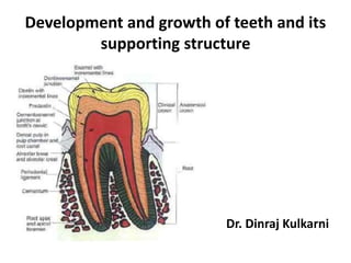
  • 1. Development and growth of teeth and its supporting structure Dr. Dinraj Kulkarni
  • 2. Some terminologies to be familiar • Dental lamina • Enamel organ • Ectomesenchyme • Dental papilla • Dental sac
  • 3. • OEE • IEE • Stellate reticulum • Stratum intermedium • Cell rests of serre’s
  • 4. Embryological basis of dental tissue genesis Lined by stratified squamous epithelium i.e. Oral ectoderm or primitive oral epithelium Connective tissue cells underlying oral ectoderm are neural crest or ectomesenchymal in origin , which induce tooth development Ruptures at 27th day (4th week) of gestation
  • 5. Primary epithelial band /6th week (2-3 weeks after rupture of BF membrane) Continuous band of thickened epithelium called “Primary epithelial band”. Roughly horse shoe shaped Thickening first seen in anterior midline Certain areas of basal cells of oral ectoderm proliferate more rapidly than do cells of adjacent areas
  • 7. Initiation of tooth development • The initiation of tooth development begins at 37th day with formation continuous horse shoe shaped band of thickened epithelium in the location of upper and lower jaws– Primary epithelial band Each band of epithelium gives rise to 2 subdivisions Dental lamina Vestibular lamina (Lip furrow band) Vestibular lamina – labial and buccal to the dental lamina in each dental arch, another thickening develops apparently and some what latter called “Vestibular lamina”
  • 8.
  • 9. 8th week Continued thickening of dental lamina in 10 areas of upper and lower arch. Thickenings corresponds to the position of future primary dentition
  • 10. • Development of 1st molar – 4th month in utero • 2nd molar – 1st year after birth. • 3rd molar – 4th or 5th years • Succesional lamina – 5th month in utero (permanent central inciosor) & (10th month – 2nd premolar)
  • 11. Formation of vestibule in oral cavity Cells of vestibular lamina proliferate Degeneration of central epithelial cells Sulcus of vestibule
  • 12. Dental lamina Is a band of epithelium Serves as a primordium for the ectodermal portion of the deciduous teeth.
  • 13. Dental lamina • Within dental lamina localized and continuous proliferative activity leads to formation of series of epithelial outgrowths into the ectomesenchyme at the sites corresponding to the future deciduous teeth which represents the beginning of enamel organ. • Successors of deciduous teeth arises from the lingual extension of free end of dental lamina opposite to enamel organ of each deciduous tooth, these lingual extensions are referred to as “Successional lamina" and develops from 5th month in utero to 10th month of age.
  • 14. Functions of dental lamina • Phase –I :- initiation of entire deciduous dentition (8th week). • Phase – II :- Initiation successors of deciduous dentition (5th to 10th month). • Phase – III :- Initiation of permanent molars.
  • 15. Fate of dental lamina • The total activity of dental lamina extends over a period of at least 5 years. • Any particular portion of dental lamina functions for much briefer period, since only a relatively short time elapses after initiation of tooth development before dental lamina begins to degenerate at particular location i.e. it might be still active in 3rd molar area when it has disappeared elsewhere. • Dental lamina looses its connection from enamel organ at the start of bell stage. • Remnants of dental lamina persists as epithelial pearls or islands within jaw termed as “cell rests of Serre’s”
  • 16. Rests of serre’s or epithelial cells • Also called as “Epithelial Pearls or islands”. • These are remnants of dental lamina. • These are identified in jaws and gingival soft tissues. • They results from early break up of dental lamina during bell stage. Clinical significance - Gingival cyst of adult - Gingival cyst of new born
  • 17. Rests of Malassez • The break up of heartwig’s root sheath during root formation i.e. these are remnants of Heartwig’s epithelial root sheath. • These are identified in periodontal ligament and are responsible for the development of radicular cyst.
  • 18. How development tooth begins?? 1st arch and ectomesenchymal cells derived from the neural crest cells Up to 1st 12 days 1st arch epithelium retains the ability to form tooth like structure when combined with neural crest cells of regions This potential is lost and transformed to neural crest cells to produce tooth like structure Interaction of epithelium derived Afterwards
  • 19. • Stages of tooth development
  • 20. Stages of tooth development Morphologic stage Bud stage Cap stage Bell stage  Early bell stage  Advanced bell stage Physiologic stages Initiation Proliferation Histodiffrentiation Marphodiffrentiation Apposition
  • 21. STAGES IN TOOTH GROWTH Morphologic changes physiologic processes • Dental lamina Initiation • Bud stage Proliferation • Cap stage • Early bell stage Histodiffrentiation • Advanced bell Marphodiffrentiation • Enamel/dentin formation apposition
  • 22. Initiation 6th to 7th weeks Ectoderm lining stomodeum gives rise to oral epithelium and dental lamina All the enamel organ don’t start at same time, first to start are those of anterior mandibular region
  • 24. Bud stage or proliferative stage 8th week of prenatal development Growth of dental lamina into bud that penetrates growing ectomesenchyme
  • 25. • Initial proliferation of oral epithelial cells and adjacent mesenchymal cells is occurring and the cells proliferate faster than the adjacent cells giving rise to bud shaped enamel organ.
  • 26.  Enamel organ – spherical to ovoid epithelial condensation  Cells of tooth bud have -- Increase RNA content, Decrease Glycogen content and increase oxidative enzyme activity  Cells undergo mitosis and get condensed. Enamel organ surrounded by mesenchyme
  • 27. Bud stage at 8th week Basement membrane Tooth bud
  • 28. Histologically enamel organ at bud stage consists of, • Peripherally located low columnar cells. • Centrally located polygonal cells. • Many cells of tooth bud and surrounding mesenchyme undergoes mitosis, i.e. increased mitotic activity. • Ectomesenchymal cells or neural crest cells migrates in the area surrounding tooth bud. • Ectomesenchymal condensation occurs surrounding the bud.
  • 30. Cap stage  9th to 10th week  Unequal growth in different parts of tooth bud leads to concave surface forming cap like structure.  Enamel organ forms into cap.  There is a formation of tooth germ
  • 31. Early cap stage – 11 week Peripherally located cuboidal cells – Outer enamel epithelium Tall columnar cells in the concavity – Inner enamel epithelium Polygonal cells in between OEE and IEE called – Stellate reticulum (12th week)
  • 32. Histologically • Outer enamel epithelium  Cuboidal cells covering convexity.  Separated from dental sac by basal lamina. • Inner enamel epithelium  Tall columnar cells lining concavity of enamel organ.  Separated from the dental papilla by basal lamina.
  • 33. Stellate reticulum • Cells in the center of the enamel organ. • These cells are star shaped which secrete GAG. • Stellate reticulum cells synthesize and secrete GAG’s into extracellular compartment between epithelial cells –> hydrophilic -- > pull water into the enamel organ and central cells are forced apart. • They are so named because they retain connection with each other through desmosomal contacts, they become star shaped, the center of the enamel organ thus termed Stellate reticulum.
  • 34. Components of tooth germ during cap stage
  • 36. Dental papilla – dentin and pulp Dental sac – cementum and periodontal ligament
  • 37. What is tooth germ?? The enamel organ, the dental papilla and the dental sac together called “Tooth germ”.  Enamel organ – enamel  Dental papilla –Dentin and Pulp  Dental sac – PDL, Cementum and bone
  • 38. Transitory structures Enamel knot Localized mass of cells produced by rapid multiplication of cells in the center of inner enamel epithelium
  • 39. Enamel cord Strand of cells extending from stratum intermedium into Stellate reticulum These are vertical extensions of enamel knot. These acts as reservoir of dividing cells for the growing enamel organ
  • 40. Enamel niche - Double attachment of enamel organ to overlying epithelium. - Funnel shaped area between lateral and medial strands of dental lamina enclosing mesenchyme. A. Lateral enamel strand B. Medial enamel strand C. Enamel niche
  • 41. Enamel septum • When the enamel cord extends to meet the outer enamel epithelium is termed as enamel septum. Enamel navel • The outer enamel epithelium at the point of meeting shows a small depression and is termed as enamel navel as it resembles umbilicus.
  • 42. What is enamel organ?? • The developing tooth as it progresses from bud stage to advanced bell stage is called the “enamel organ” or “dental organ”
  • 44.
  • 45.
  • 46. Bell stage • 11th to 12th weeks • Proliferation, differentiation, morphogenesis • Differentiation of enamel organ into bell with four cell types and dental papilla into 2 cell types.
  • 47. Early bell stage (14th week) Dental lamina break downs and degenerates Shows 4 distinct layer  Outer enamel epithelium  Stellate reticulum  Stratum intermedium  Inner enamel epithelium
  • 48. Outer enamel epithelium • A single layer of very low cuboidal cells. • At the end of this stage just before enamel formation, the smooth surface of outer enamel epithelium get thrown into folds. • Between the folds the adjacent mesenchyme of the dental sac forms papillae that contain capillary loops and thus provide a rich nutritional supply for the intense metabolic activity of the avascular enamel organ.
  • 49. Outer enamel epithelium Role of OEE • Maintenance of the shape of the enamel organ • Exchange of substance between enamel organ and environment • Holds the contents of enamel organ • As a constituent of epithelial root sheath plays a role in root formation.
  • 50. Stellate reticulum Mechanical Acts as shock absorber Maintains tooth shape Nutritive Supplies GAG’s to enamel forming cells
  • 51. Stellate reticulum • Expands mainly by an increase in the amount of intracellular fluid. • The star shaped cells with long processes anastomose with those of adjacent cells. • Before enamel formation begins, the Stellate reticulum collapses, reducing the distance between centrally situated ameloblasts and the nutrient capillaries near the outer enamel epithelium. • Changes begins at the height of the cusp or the incisal edge and progresses cervically.
  • 52. Stratum intermedium Functions Synthesis and storage of proteins Transport of material too and from the inner enamel epithelium (ameloblasts) Induce inner enamel epithelial cells to become ameloblasts
  • 53. Stratum intermedium • These are 3-4 layers of squamous cells between inner enamel epithelium and Stellate reticulum. • These cells are attached by desmosomes and gap junctions. • There are abundant cell organelles, acid mucopolysaccharides and glycogen deposits in these cells, which indicate high metabolic activity. • This layer is essential for enamel formation. • Are absent in the part of the tooth germ that outlines the root portion of the tooth which doesn’t form enamel.
  • 54. Inner enamel epithelium Function Its cells become ameloblasts which form enamel. It induces adjacent cells of dental papilla to form odontoblasts which in turn form dentine. Following amelogenesis, the cells contribute to form the REE together with OEE and Stratum intermedium. Important role in crown formation due to intrinsic growth pattern.
  • 55. Inner enamel epithelium Consists of a single layer of cells differentiate prior to amelogenesis into tall columnar cells Ameloblasts
  • 56. The cells of the inner enamel epithelium exert an organizing influence on mesenchymal cells in the dental papilla later differentiate into odontoblasts.
  • 57. • Are about 40 um high and 4-5 um in diameter. • These cells attach to each other laterally by junctional complexes and cells of stratum intermedium by desmosomes.
  • 58. Dental papilla • Under the inductive influence of inner enamel epithelium, the peripheral cells of the dental papilla differentiate into odontoblasts. • These cells initially become cuboidal and later assume columnar shape. • These cells now acquire the potential to produce dentine. Membrana performativa • The basement membrane that separates enamel organ and the dental papilla just prior to dentin formation is called “Membrana performativa.
  • 59. Dental sac • Just before the formation of dental tissues begins, the collagen fibers of the dental sac are arranged in a circular manner. • The dental sac look like a capsule around the enamel organ and dental papilla.
  • 60. Changes before enamel formation • The Stellate reticulum collapses and the distance between inner enamel epithelium and blood capillaries of the dental sac is reduced, so that more nutrient reach the ameloblasts during the period of high metabolic activity. • The formerly smooth surface of outer enamel epithelium thrown into folds.
  • 61. • Between the folds the mesenchyme of dental sac form projections or dental papillae that carry capillary loops closer to the ameloblast prior toamelogenesis.
  • 62.
  • 63. IEE become taller – Preameloblasts Peripheral cells of the dental papilla – Odontoblasts Secrete ground substance and collagen fibers – Dentin matrix There is change in cell polarity.
  • 64. With change in polarity the cell called an ameloblasts that begins secretion of enamel matrix. Boundary between inner enamel epithelium and odontoblasts outline the future dentino-enamel junction.
  • 65.
  • 66. Enamel organ shows 4 different layers. • Inner enamel epithelium (ameloblasts) • Stratum intermedium • Stellate reticulum • Outer enamel epithelium
  • 67. • The ameloblasts have fully differentiated from the cells of inner enamel epithelium. • The odontoblasts have fully differentiated from dental papillae cells. • The boundary between the ameloblasts and odontoblasts define the future dentino- enamel junction.
  • 68. Histological difference from early bell stage • Hard tissue (Enamel and Dentin) formation. • Collapsed Stellate reticulum and folding of outer enamel epithelium bringing capillaries of the dental follicle nearer to ameloblasts. • Dental papilla shows differentiated odontoblasts at periphery • Dental follicle is distinct enclosing enamel organ and dental papilla.
  • 69. Reciprocal induction • The inner enamel epithelium induces peripheral cells of dental papilla to differentiate into odontoblasts. • The odontoblasts start secreting the organic matrix of dentin. • Only dentin matrix is formed, do the ameloblasts in the area differentiate fully and start secreting enamel matrix. • This independence of tissue is an example of “Reciprocal induction”.
  • 70. INITIATION Lack of initiation may result in absence of single tooth or multiple teeth (anodontia). The upper lateral incisor followed by 3rd molars and lower 2nd premolar commonly involved. Abnormal initiation result in development of supernumerary tooth. E.g. – Mesiodens followed by 4th molar Sometimes fused or geminated teeth are formed. HISTODIFFERENTIATI ON This stage reaches its highest level in bell stage of enamel organ. E.g. -- Dentinogenesis imperfecta or formation of atypical dentin MORPHODIFFERENT IATION The advanced bell stage is important stage. This stage establishes the morphologic pattern, or basic form and relative size of future tooth. Supernumerary (talon’s) cusp, twinning, loss of cusps or roots, or malformed or peg shaped tooth (Hutchinson’s incisor), dens in dente and microdontia. APPOSITION Both enamel hypoplasia and hypo calcification can occur as a result of an insult to the cells responsible for the apposition stage. Intrinsic staining and concrescence.