Cellular organelles nucleus, mitochondria & plastids
•Descargar como PPTX, PDF•
33 recomendaciones•11,156 vistas

Nucleus, mitochondria and plastids.

Recomendados
Más contenido relacionado
La actualidad más candente
La actualidad más candente (20)
Similar a Cellular organelles nucleus, mitochondria & plastids
Similar a Cellular organelles nucleus, mitochondria & plastids (20)
Último
Último (20)
Cellular organelles nucleus, mitochondria & plastids
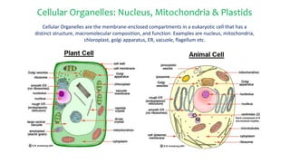
- 1. Cellular Organelles: Nucleus, Mitochondria & Plastids Cellular Organelles are the membrane-enclosed compartments in a eukaryotic cell that has a distinct structure, macromolecular composition, and function. Examples are nucleus, mitochondria, chloroplast, golgi apparatus, ER, vacuole, flagellum etc.
- 2. Nucleus: The control center
- 3. The nucleus was the first organelle to be discovered. We cannot know who first saw the nucleus but we do know that the father of optical microscopy, Antony van Leeuwenhoeck, who did so with amphibian and avian erythrocytes in 1710. He observed a "lumen", the nucleus, in the red blood cells of salmon. We do not know how and when the genome of an ancestral cell first became encased in a primitive nucleus. But we know that Franz Bauer, in 1802, sketched orchid cells and pointed out the nucleus. Nucleus was described in more detail in 1831 by Scottish botanist Robert Brown. We are uncertain about its evolutionary origin, but we know that the nucleus is bounded by a double membrane, the nuclear envelope, which in many cells is connecting with the endoplasmic reticulum. Now nucleus can be observed, isolated and is being utilized in the next generation molecular breeding.
- 4. Nucleus: Introduction The nucleus (pl. nuclei; from Latin nucleus or nuculeus, meaning kernel) is a membrane-enclosed organelle found in eukaryotic cells. Nucleus is most prominent organelle of eukaryotic cells which contains genetic material. Nucleus distinguishes the eukaryotic cell from prokaryotic cell. Eukaryotes usually have a single nucleus, but a few cell types, such as mammalian red blood cells, have no nuclei. The number of nuclei may vary, they may be uni-nucleate (single nucleus), bi-nucleate (two nuclei) or even multi-nucleate e.g.: osteoclasts (5 nucleus), coenocytes etc. The shape of the nucleus is mostly round, it may be oval, disc shaped depending on the type of cell.
- 5. Typical Structure of Nucleus Source: Bing Images
- 6. (a) Electron micrograph section of interphase nucleus (b) Cutway 3D view of nuclear envelope and pores Source: Microbiology A system approach
- 7. Internal Organization of the Nucleus • The nucleus is more than a container in which chromatin, RNAs, and nuclear proteins move freely in aqueous solution. Nucleus is the site for Central Dogma. • Chromosome, nuclear pore complex, nuclear matrix, nuclear lamina, nucleolus are the characteristics of nucleus. Source: Bing Images
- 9. 1. The Nuclear Envelope • The cell nucleus is bound by a double membrane called the nuclear envelope and is continuous with the endoplasmic reticulum. • This membrane separates the contents of the nucleus from the cytoplasm. • Like the cell membrane, the nuclear envelope consists of phospholipids that form a lipid bilayer. • Perinuclear space is the space between inner and outer nuclear membrane. • The envelope helps to maintain the shape of the nucleus and assists in regulating the flow of molecules into and out of the nucleus through nuclear pores. Source: https://www.ncbi.nlm.nih.gov
- 10. 2. Nuclear Pore Complex • The nuclear pore complexes are the only channels through which small polar molecules, ions, and macromolecules (proteins and RNAs) are able to travel between the nucleus and the cytoplasm. • The nuclear pore complex is an extremely large structure with a diameter of about 120 nm. 3. Nuclear Matrix and Nuclear Lamina • The nuclear matrix is the network of fibres found throughout the inside of a cell nucleus. • The nuclear lamina is a fibrous meshwork underlying the inner membrane. • The nuclear lamina is a structure near the inner nuclear membrane and the peripheral chromatin. • Nuclear lamina is involved in most nuclear activities including DNA replication, RNA transcription, nuclear and chromatin organization, cell cycle regulation, cell development and differentiation, nuclear migration, and apoptosis (programmed cell death).
- 11. 4. Chromosomes • Chromosomes are located within the nucleus. Chromosomes consists of DNA, which contains heredity information and instructions for cell growth, development, and reproduction. When a cell is “resting”, the chromosomes organized are called chromatin. • Chromatin becomes highly condensed during mitosis to form the compact metaphase chromosomes that are distributed to daughter nuclei. • During interphase, some of the chromatin (heterochromatin) remains highly condensed and is transcriptionally inactive; the remainder of the chromatin (euchromatin) is decondensed and distributed throughout the nucleus. • Much of the heterochromatin is localized to the periphery of the nucleus, possibly because one of the principal proteins associated with heterochromatin binds to a protein of the inner nuclear membrane.
- 12. 5. Nucleolus Contained within the nucleus is a dense structure composed of RNA and proteins called the nucleolus. The most obvious aspect of the internal organization of the nucleus is the nucleolus, which is the site at which the rRNA genes are transcribed and ribosomal subunits are assembled. The nucleolus is a ribosome production factory. The nucleolus contains nucleolar organizers, which are parts of chromosomes with the genes for ribosome synthesis on them. Source: Bing Images
- 13. Changes in the Nucleus during Mitosis • In the living interphase nucleus, no chromosomal structures are visible. Under certain conditions, this appearance of structure in the living interphase nucleus is reversible. It has been found that change in the interphase nucleus is the result of a change in the state of the chromosomes (Ris and Mirskey, 1949). In the living nucleus the chromosomes are in a greatly extended state, filling the entire nucleus. But, after injury to the interphase it showed condensed chromosomes. • At the beginning of mitosis (Prophase), the chromosomes condense, the nucleolus disappears, and the nuclear envelope breaks down, resulting in the release of most of the contents of the nucleus into the cytoplasm. At the end of mitosis (Telophase), the process is reversed: the chromosomes decondense, and nuclear envelopes re-form around the separated sets of daughter chromosomes.
- 14. Closed and open mitosis In closed mitosis (unicellular eukaryotes e.g.: yeasts), the nuclear envelope remains intact and chromosomes migrate to opposite poles of a spindle within the nucleus. In open mitosis (most of the multicellular eukaryotes), the nuclear envelope breaks down and then re-forms around the two sets of separated chromosomes. Source: https://www.ncbi.nlm.nih.gov
- 15. Breakdown of the nuclear membrane As the nuclear lamina dissociates, the nuclear membrane fragments into vesicles. The B-type lamins remain bound to these vesicles, while lamins A and C are released as free dimers. Source: https://www.ncbi.nlm.nih.gov
- 16. Re-formation of the nuclear envelop • The first step in reassembly of the nuclear envelope is the binding of membrane vesicles to chromosomes, which may be mediated by both integral membrane proteins and B-type lamins (intermediate filament proteins that form the nuclear lamina). • The vesicles then fuse, the nuclear lamina (provides structural support to the nucleus) reassembles, and the chromosomes decondense. Source: https://www.ncbi.nlm.nih.gov
- 17. Functions of Nucleus • It controls the heredity characteristics of an organism. • It is responsible for protein synthesis, cell division, growth and differentiation. • Stores heredity material in the form of deoxy-ribonucleic acid (DNA) strands. • Also stores proteins and ribonucleic acid (RNA) in the nucleolus. • It is a site for transcription process in which messenger RNA (m RNA) are produced for protein synthesis. • Aids in exchange of DNA and RNA (heredity materials) between the nucleus and the rest of the cell. • Nucleolus produces ribosomes and are known as protein factories. • It also regulates the integrity of genes and gene expression.
- 18. • Nucleus is the site for Central Dogma, the process by which the instructions in DNA are converted into a functional product . DNA replication, transcription, and RNA processing all take place within the nucleus, with only the final stage of gene expression (i.e. translation) occurs in the cytoplasm. Therefore, It is also called the control center of the cell. Source: https://www.ncbi.nlm.nih.gov
- 19. Mitochondria: Energy Generators of the Cell
- 20. • Most eukaryotic cells contain many mitochondria, which occupy up to 25 percent of the volume of the cytoplasm. • The term "mitochondria" was first coined by Carl Benda in 1898. In 1904, Friedrich Meves, made the first recorded observation of mitochondria in plants in cells of the white waterlily, Nymphaea alba. • Although the nucleus is the cell’s control center, none of the cellular activities it commands could proceed without a constant supply of energy. • The bulk of the energy is generated in most eukaryotes by mitochondria and called ‘The Power House of Cell’. • These organelles generate power by converting energy into forms i.e. ATP, that are usable by the cell through the cellular respiration. • When viewed with light microscopy, mitochondria appear as round or elongated particles scattered throughout the cytoplasm. • These complex organelles are among the largest organelles, generally exceeded in size only by the nucleus, vacuoles, and chloroplasts.
- 21. Mitochondrion Anatomy • The internal ultrastructure reveals that a single mitochondrion is bound by a double membrane. Each of these membranes is a phospholipid bilayer with embedded proteins. • The outermost membrane is smooth while the inner membrane has many folds. These folds are called cristae. The folds enhance the "productivity" of cellular respiration by increasing the available surface area. • The cristae membranes hold the enzymes and electron carriers of aerobic respiration and stores energy in the form of high-energy molecules, or ATP. • The spaces around the cristae are filled with a chemically complex fluid called the matrix, which holds ribosomes, DNA, and the pool of enzymes and other compounds involved in the metabolic cycle. • The mitochondrial matrix contains circular mitochondrial DNA (mtDNA), ribosomes (prokaryotic sized 70S type), and enzymes. Several of the steps in cellular respiration, including the Citric Acid Cycle and oxidative phosphorylation, occur in the matrix due to its high concentration of enzymes.
- 22. Fig: Mitochondrion Detailed 3D Structure Source: Microbiology A system approach
- 23. Mitochondria: Distinguishing Characteristics • Mitochondria have a distinctive oblong or oval shape and are bounded by a double membrane. • They are found in both animal and plant cells. • The number of mitochondria within a cell varies depending on the type and function of the cell. • Some cells, such as mature red blood cells, do not contain mitochondria at all. • The absence of mitochondria and other organelles leaves room for the millions of hemoglobin molecules needed in order to transport oxygen throughout the body. • Muscle cells, on the other hand, may contain thousands of mitochondria needed to provide the energy required for muscle activity. Mitochondria are also abundant in fat cells and liver cells. • Porins are found in mitochondria and chloroplasts which are beta barrel proteins that cross a cellular membrane and act as a pore through which molecules can diffuse.
- 24. Mitochondrial genome or DNA • Mitochondria contain their own genetic system, which is separate and distinct from the nuclear genome of the cell. • Mitochondria have their own circular DNA, ribosomes and can make their own proteins. • The genomes of human and most other animal mitochondria are only about 16 kb, but substantially larger mitochondrial genomes are found in yeasts (approximately 80 kb) and plants (more than 200 kb). • Mitochondrial DNA (mtDNA) encodes for proteins that are involved in electron transport and oxidative phosphorylation, which occur in cellular respiration. • Proteins synthesized from mtDNA also encode for the production of the RNA molecules: transfer RNA and ribosomal RNA. • Mitochondrial DNA differs from DNA found in the cell nucleus in that it does not possess the DNA repair mechanisms that help prevent mutations in nuclear DNA. • As a result, mtDNA has a much higher mutation rate than nuclear DNA. Exposure to reactive oxygen produced during oxidative phosphorylation also damages mtDNA. • Diseases caused by mutation in the mtDNA include Kearns-Sayre syndrome, MELAS syndrome and Leber's hereditary optic neuropathy etc.
- 25. Mitochondrial Replication and Inheritance • Mitochondria are semi-autonomous in that they are only partially dependent on the cell to replicate and grow. • They have their own DNA, ribosomes, make their own proteins, and have some control over their reproduction. Similar to bacteria, mitochondria have circular DNA and replicate by a reproductive process called binary fission. • Prior to replication, mitochondria merge together in a process called fusion. Fusion is needed in order to maintain stability, as without it, mitochondria will get smaller as they divide. These smaller mitochondria, if produced, are not able to produce sufficient amounts of energy needed for proper cell function. • An individual's mitochondrial genes are not inherited by the same mechanism as nuclear genes. Typically, the mitochondria are inherited from one parent only.
- 26. Mitochondrial Fission and Fusion (A) Generalized figure of fission and fusion (B) Electron Micrograph of Mitochondrial fission and fusion Source: https://www.ncbi.nlm.nih.gov
- 27. Functions of Mitochondria • The most important function of the mitochondria is to produce energy. The simpler molecules of nutrition are sent to the mitochondria to be processed and to produce charged molecules. These charged molecules combine with oxygen and produce ATP molecules. This process is known as oxidative phosphorylation. • Mitochondria help the cells to maintain proper concentration of calcium ions which increase mitochondrial uptake up to 10-1000folds and begin to shape dynamics within the compartments of the cell. • The mitochondria also help in building certain parts of blood and hormones like testosterone and estrogen. • The liver cells mitochondria have enzymes that detoxify ammonia. • The mitochondria also play important role in the process of apoptosis or programmed cell death. Abnormal death of cells due to the dysfunction of mitochondria can affect the function of organ.
- 28. Plastids: Sites for Photosynthesis and Storage and Pigmentation
- 29. • Plastids are thought to have originated from endosymbiotic cyanobacteria. • The term plastid was derived from the Greek word plastikas meaning formed or moulded. This term was coined by Schimper in 1885. • Plastids are membrane-bound organelles that convert energy from foodstuffs (non-photosynthetic plastids) or sunlight (chloroplasts) into cellular energy. • Chloroplasts and other non- photosynthetic plastids develop from cells called proplastids. • Proplastids are immature, undifferentiated cells that develop into different types of plastids. • A proplastid that develops into a chloroplast, only does so in the presence of light.
- 30. Types of Plastids The different types of plastids are frequently classified according to the kinds of pigments they contain. • Chloroplasts: Plastids which contain chlorophyll, takes part in photosynthesis and also called green plastids. • Chromoplasts: Plastids which lack chlorophyll but contain carotenoids; they are responsible for the yellow, orange, and red colors of some flowers and fruits, although their precise function in cell metabolism is not clear. • Leucoplasts: Plastids which are non-pigmented plastids, which store a variety of energy sources in non-photosynthetic tissues. Amyloplasts and elaioplasts are examples of leucoplasts that store starch and lipids, respectively. Proteinoplasts (sometimes called proteoplasts, aleuroplasts, and aleuronaplasts) are specialized organelles found only in plant cells. They contain crystalline bodies of protein and can be the sites of enzyme activity involving those proteins. Tannosomes are another kind of leucoplasts for synthesizing and producing tannins and polyphenols.
- 32. Chloroplast: Site for Photosynthesis
- 33. Chloroplast : Introduction • A chloroplast is a type of plant cell organelle in which photosynthesis occurs. It is 5 to 10 μm long (1μm = 10-6 m.) with double membrane envelope. • A chloroplast contains a green pigment called chlorophyll, which absorbs light energy for photosynthesis. Hence, the name chloroplast indicates that these structures are chlorophyll containing plastids. • Chloroplasts are also responsible for producing amino acids and lipid components needed for chloroplast membrane production. • Chloroplasts can also be found in other photosynthetic organisms such as algae.
- 34. Chloroplast: Structure • Plant chloroplasts develop mainly in cells located in plant leaves. Chloroplast consists following structural components with their specific functions: • Membrane Envelope - contains an inner and outer membranes that act as protective coverings and keep chloroplast structures enclosed. • Thylakoid Membrane - internal membrane system consisting of flattened sac-like membrane structures called thylakoids that serve as the sites of conversion of light energy to chemical energy. • Grana (singular granum) - dense layered stacks of thylakoid sacs that serve as the sites of conversion of light energy to chemical energy. • Stroma - dense fluid within the chloroplast that lies inside the envelope but outside the thylakoid membrane. This is the site of conversion of carbon dioxide to carbohydrates(sugar). • Chlorophyll - a green photosynthetic pigment within the chloroplast that absorbs light energy and plays a vital role in photosynthesis.
- 35. Chloroplast: Structure Source: Bing Images
- 36. Chloroplast: Photosynthesis • In photosynthesis, the sun's solar energy is converted to chemical energy. The chemical energy is stored in the form of glucose (sugar). • Carbon dioxide, water, and sunlight are used to produce glucose, oxygen, and water. Photosynthesis occurs in two stages. These stages are known as the light reaction stage and the dark reaction stage. • The light reaction stage takes place in the presence of light and occurs within the chloroplast grana. The primary pigment used to convert light energy into chemical energy is chlorophyll a. Other pigments involved in light absorption include chlorophyll b, xanthophyll, and carotene. In the light reaction stage, sunlight is converted to chemical energy in the form of ATP (free energy containing molecule) and NADPH (high energy electron carrying molecule). This process is done by electron transport chain reaction in thylakoid membranes of chloroplast. • Both ATP and NADPH are used in the dark reaction stage to produce sugar. The dark reaction stage is also known as the carbon fixation stage or the Calvin cycle. Dark reactions occur in the stroma. The stroma contains enzymes which facilitate a series of reactions that use ATP, NADPH, and carbon dioxide to produce sugar. The sugar can be stored in the form of starch, used during respiration, or used in the production of cellulose.
- 37. The reactions of photosynthesis in a chloroplast: • Water is oxidized and oxygen is released in the photosynthetic electron-transfer reactions (Light Reaction) while carbon dioxide is assimilated (fixed) to produce sugars and a variety of other organic molecules (Dark Reactions) in the carbon-fixation reactions. • The energy thus released during those reactions is stored in the form of proton gradient in the thylakoid membrane of chloroplast and is utilized later by cells. • A total of 3 molecules of ATP and 2 molecules of NADPH are consumed for each CO2 molecule converted into carbohydrate. • The net equation is: Source: https://www.ncbi.nlm.nih.gov
- 38. The Chloroplast Genome and Replication • Like mitochondria, chloroplasts contain their own genetic system, reflecting their evolutionary origins from photosynthetic bacteria. • The genomes of chloroplasts are similar to those of mitochondria in that they consist of circular DNA molecules present in multiple copies per organelle. • However, chloroplast genomes are larger and more complex than those of mitochondria, ranging from 120 to 160 kb and containing approximately 120 genes. • Like mitochondria, chloroplasts have their own DNA, are responsible for energy production, and reproduce independently from the rest of the cell through a division process similar to bacterial binary fission.
- 40. Protein Import and Sorting in Mitochondria and Plastids • The production of mitochondrial and chloroplast proteins occur by two separate genetic systems. Most of the proteins in these organelles are encoded by the nucleus and must be imported from the cytosol while other organelle proteins are encoded by organelle DNA. • But, no known proteins are exported from mitochondria or chloroplasts to the cytosol. • The multiple copies of mitochondrial and chloroplast DNA contained within the matrix or stroma of these organelles are usually distributed in several clusters, called nucleoids. • Nucleoids are thought to be attached to the inner mitochondrial membrane. Although it is not known how the DNA is packaged, the DNA structure in nucleoids is likely to resemble that in bacteria rather than that in eukaryotic chromatin. As in bacteria, for example, there are no histones. • Although chloroplasts encode more of their own proteins than mitochondria, about 90% of chloroplast proteins are still encoded by nuclear genes. Source: https://www.ncbi.nlm.nih.gov
- 41. Chemiosmotic generation of ATP in chloroplasts and mitochondria • In mitochondria, electron transport generates a proton gradient across the inner membrane, which is then used to drive ATP synthesis in the matrix. 34 ATP molecules are generated from single glucose molecule. • In chloroplasts, the proton gradient is generated across the thylakoid membrane and used to drive ATP synthesis in the stroma. • This proton gradient serves as an energy store that can be used to drive ATP synthesis by the ATP synthase enzyme. • Pump proteins maintains the proton gradient and harness the energy of electron transfer and ATP synthase synthesizes ATP from ADP in presence of inorganic phosphate (Pi). Source: https://www.ncbi.nlm.nih.gov
- 42. Characteristics: Mitochondria vs. Chloroplasts • Chloroplast contains thylakoid membranes and pigment molecules, whereas the mitochondria membranes contain respiratory enzymes not found in chloroplast membranes. • Chloroplasts are found in plants only while mitochondria are found in both plants and animals. • Besides production of ATP, chloroplasts help in photosynthesis and produce food molecules like sugars, amino acids, fatty acids, lipids whereas mitochondria generates energy in the form of ATP by degradation of food molecules.
- 43. References https://www.ncbi.nlm.nih.gov (date accessed: 19th to 24th September 2016) https://biology.about.com (date accessed: 19th to 24th September 2016) https://en.wikipedia.org (date accessed: 19th to 24th September 2016) Cowan, M. K., 2012. Microbiology - A Systems Approach (3rd Ed). McGraw-Hill Publications, New York. P. 108-138 Lodish, H., Berk, A., Zipursky, S.L. 2000. Molecular Cell Biology, 4th edition. New York: W. H. Freeman
- 44. Thank You !