
Recomendados
Más contenido relacionado
La actualidad más candente
La actualidad más candente (20)
Similar a Perforation of palate
Similar a Perforation of palate (20)
Más de DrGayatriMehrotra
Último
Último (20)
Perforation of palate
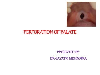
- 1. PERFORATIONOF PALATE PRESENTEDBY: DR GAYATRI MEHROTRA
- 2. INTRODUCTION • Palatal perforation can be defined as a communication between the nasal cavities and the oral cavity. • Palatal perforation is a rare condition encountered in the routine dental practice.
- 3. CLASSIFICATION • Congenital/Developmental- Cleft palate • Infection/Inflammatory Disease- Osteomyelitis Syphillitic gumma Tuberculosis Infection of traumatic and chronic wound
- 4. Leprosy Noma Mucormycosis Toxoplasmosis Blastomycosis • Autoimmune- Lupus erythematosus Sarcoidosis Crohns disease Wegener granulomatous
- 5. • Neoplastic- Tumor from maxillary sinus or nasal cavity Squamous cell carcinoma Mucoepidermoid carcinoma Adenocystic carcinoma Lymphoma Midline lethal granuloma Melanosarcoma
- 6. • Trauma- Fracture of maxilla Use of suction disc for denture • Drug related- Due to cocaine abuse Bisphosphonates
- 7. • Iatrogenic- Tooth extraction Maxillectomy • Rare causes- Rhinolithcan
- 8. CONGENITAL/DEVELOPMENTAL • CLEFTPALATE- It is a birth defect characterized by an opening in the roof of the mouth caused by a lack of tissue development. Cleft of hard palate and soft palate ; sometimes soft palate alone.
- 9. • Some syndromes, maternal alcohol consumption and cigarette smoking, folic acid deficiency, corticosteroid use and anticonvulsant therapy are some causative agents for this abnormality.
- 10. Eating and drinking are difficult due to regurgitation of food and liquid through the nose. Speech problem Defect in smelling Airway problems in some cases. Radiograph- missing, supernumerary, malpositioned teeth
- 11. • Management : Cheiloplasty Obturator Palatoplasty Bone grafting Orthodontic therapy Cleft rhinoplasty Speech therapy Pyschotherapy
- 12. INFECTION/INFLAMMATORY DISEASE • OSTEOMY ELITIS- It is defined as an inflammatory condition of the bone that begins as an infection of medullary cavity and the haversian system and extends to involve the periosteum of the affected area. Predisposing factors- diabetes mellitus, tuberculosis, severe anemia, leukemia, agranulocytosis,etc
- 13. Etiology- odontogenic infection, infection from pdl space , extraction wound. Compromised blood supply is a critical factor in the establishment of osteomyelitis. Virulent microorgansims Intense inflammatory reaction Localization of infection Disorganization of clot Mechanical trauma Accumulation of pus Elevation of periosteum Necrosis of bone Penetration of periosteum
- 14. • Management- Incision and drainage Irrigation and debridement of the necrotic area. Antibiotics Supportive therapy Sequestrectomy Saucerization Decortication Hyperbaric oxygen therapy
- 15. • SYPHILLITIC GUMMA- Classic disease with protean manifestations. 4 main clinical stages Gumma- tertiary syphillis Small raised area of mucosa ulcerates Large zone of necrosis Denudation of bone Perforate into nasal cavity
- 16. Common site- midline of hard palate Painless Appearance – solitary, deep,punched out mucosal ulcer Punched out ulcer with vertical walls and dull red granulomatous base Types- central gumma - cortical gumma Leutic glossitis Chronic superficial interstitial glossitis
- 17. Fig. 1 Perforation of the hard palate caused by tertiary syphilis Fig. 2 Abnormal scarring of throat and posterior oral cavity 70, yr old male pt, with c/o inability to eat & recent hoarseness of voice.pt was confirmed with edentulous arches , 2 perforations of palate of the hard palate and nasal speech…pt denied for any extramarital exposure…and was tested positive for acute infectious syphillis…. pts wife was invited for test…when she came to know that test was for veneral disease she left the hospital (british dental journal 2005)
- 18. • Management : Antibiotics- benzathine penicillin (2.4 million units IM). Allergic to penicillin- erythromycin (500 mg 4 times a day for 15 days). Obturator for managing the palatal perforation.
- 19. • Tuberculosis- Tuberculosis of hard palate is rare and usually secondary to pulmonary TB. Cough ,Low grade fever, loss of wt, chills, night sweats. Invasion of mycobacterium- break of natural barrier- trauma, inflammatory condition, Leukoplakia, tooth extraction, poor oral hygiene, dental abscess, periodontitis. Common site - tongue
- 21. Manifest as single, non-healing ulcers with an indurated, irregular, undermined margin, and a necrotic base. The palatal lesion of TB may be seen as granulomas, ulceration, or perforation. Usually seen in patients with strong immune response and due to hypersensitiviy to acid fast bacilli.
- 22. Oral cavity examination showing perforation of the hard palate, 3 × 3 cm in size with an irregular, undermined margin, and a necrotic base Plain axial computed tomography scan of midface with 3D reconstruction showing erosion and scalloping of the posterior part of the hard palate on the right side 7 yr old boy with difficulty in swallowing solid foods,low grade fever and loss of wt, wt was 13 kg and ht was 104 cm…perforation of hard palate was seen…chest xray showed homogenous opacity in upper rite lung …bilateral cervical lymphadenopathy was present.(Dental research journal 2012)
- 23. • Management: Timing Medications Months 1 and 2 INH, RIF, PZA,EMB Next 4 to 7 months depending on condition INH, RIF EMB= ethambutol; INH=isoniazid; PZA=pyrazinamide; RIF=rifampin
- 24. • INFECTIONOF TRAUMATIC WOUND- Traumatic lesions in the oral cavity occur frequently in clinical practice. Most of the acute or chronic injuries of soft tissues were aroused from incorrect hygienic procedures or iatrogenic injuries. Trauma is known to be one of the rarest causes leading to palatal perforation.
- 25. Figure 1: Perforation of the hard palate Figure 2: Closure of the palatal perforation. A 28-year-old male farmer reported to clinics with hole in the hard palate since 3 months. Patient had difficulty while eating and hoarseness of voice and change in speech. The patient revealed that, the perforation was resulted secondary to the injury led by a horn of a calf while working in his fields. ( journal trauma and treatment 2012)
- 26. • LEPROSY- Hansen’s disease usually presents as anaesthetic or hypoaesthetic skin lesions, tingling numbness with or without weakness of hand or feet. Major target organs- skin,peripheral nerves, nasal mucosa, upper respiratory tract,eyes. 2 types- tuberculoid and lepromatous
- 27. Various types of lesions observed are infiltration, ulceration, perforation, reddish or yellow reddish nodules, sessile or pedunculated, varying from 2 to 10 mm, some confluent and prone to ulceration .
- 28. As palate is a structure crossed by two air currents, the nasal and the oral , its temperature remains 1-2° c below the body temperature- explains location of palatal lesions in midline. Oral lesions are aymptomatic and secondary to nasal changes. Gums are usually affected behind the upper central incisors, often by contiguity of lesions of the hard palate
- 29. 35 yr old female pt with h/o multiple red raised skin lesions over upper limb, lower limb,trunk, back and face of 1 yr and hole in hard palate of 4 months duration (Eygyptian dematology journal 2014)
- 30. h/o lost upper central incisor with no h/o trauma..skin biopsy showed lepromatous leprosy
- 31. A patient with a history of leprosy. Look at depression of nasal bridge(A) and palatal perforation(B)
- 32. • Management –
- 33. • NOMA- Also known as cancrum oris It is a rapidly spreading mutilating gangrenous stomatitis. Begins as small ulcer of gingival mucosa which spreads and involves surrounding tissues of jaws, lips, and cheeks by gangrenous necrosis. Palate and occasionally tongue may become involved in this process, but this is not common. Last stage of ANUG.
- 35. • Management- Control of dehydration and electrolyte balance Antibiotics- drug of choice is penicillin Reconstructive surgery
- 36. • Mucormycosis- Also known as phycomycosis, zygomycosis Infection extending from sinus mouth lesions are ulcerative hard palate spreads Functional impairment of cranial nerves II, III, IV, VI are characteristic sequelae. Ptosis,proptosis, nasal obstruction, fever, swelling of cheek and paresthesia of face can occur. Appearance of reddish black nasal turbinate and septum.
- 37. Mucor is often recognized as a triad of symptoms, such as uncontrolled diabetes mellitus, periorbital infection and meningoencephalitis. Black nasal septum
- 39. 35 yr old female pt reported with c/o sudden exfoliation of maxillary teeth . Maxillary anterior teeth and premolars were missing with necrosed bony sockets…biopsy showed features of mucormycosis (JIAOMR 2010)
- 41. • Management- The gold standard antifungal agent is Amphotericin- B. 1.0-1.5 mg/kg/day dosage continued for 8-10 weeks. Control of underlying disease Prosthetic obturation of palatal defect Surgical debridement of infected and necrotic tissue
- 42. trauma • FRACTURE OF MAXILLA- Fracture of palate present a unique challenge to the surgeon in management of mid facial fracture. Most often palatal fracture are associated with other fractures of the midface. Rarely found isolated, usually found with lefort fracture ( usually in case of lefort II fracture)
- 43. 55 yr old male pt reported with c/o intraoral drainage from palate….the discomfort was of a few days duration and had its origin following a road traffic accident which led to Lefort II fracture. ( Journal of Medical and Dental sciences 2014)
- 45. • USE OF SUCTIONDISC FOR DENTURE- Construction of complete dentures with adequate retention is a complex procedure. For the past several years suction cups are used to get retention and stability of the compete dentures. But today they are not recommended one because of their destructive effects.
- 46. 65 yr old male pt with c/o hole in upper jaw, presence of nasal twang in voice ,loose fitting denture. H/o denture since 20 yrs & defect was present since 1 yr. (Journal of Clinical and Diagnostic Research 2013)
- 48. • Management- Educating the patient about the ill effects of continuous use of suction cup denture. The closure of palatal perforation is primarily by surgical means. These options are local advancement flaps viz. palatal flaps or tongue flap, distant pedicle flaps like buccal fat pad,temporoparietal fascial flap.
- 49. IATROGENIC • Maxillectomy- Maxillofacial orthopedic surgery is a safe, predictable and stable procedure. The number of life-threatening complications associated with this surgery appears to be very small. Usually, these complications are related to the degree of vascular compromise and occur in fewer than 1% of cases
- 50. Rupturing of the descending palatine artery(DPA) during surgery Postoperative vascular thrombosis Perforation of palatal mucosa when splitting the maxilla into segments Partial stripping of palatal soft tissues to increase maxillary expansion Impair blood supply to the maxillary segments
- 51. Pt reported after maxillectomy and radiotherapy (JSM Dentistry 2014)
- 52. Case of maxillary aseptic necrosis which was reported after lefort I osteotomy which was then treated with hyperbaric oxygen therapy and antibiotics. (Journal of Oral and Maxillofacial Surgery 2010)
- 53. Drug related • Due to cocaine abuse- Cocaine is an alkaloid extract derived from the Erythroxylon coca plant. Nowadays the most common route of administration is snorting. Smoking has increased in popularity with the advent of ‘crack’ cocaine. Also abused intravenously.
- 54. Local complications of intranasal cocaine abuse include chronic rhinitis, sinusitis, epistaxis, ossification or necrosis of the nasal septum, and in rare cases, palatal perforation. Use of cocaine, a potent vasoconstrictor, can lead to ischemia, necrosis, and ulceration.
- 55. A healthy 25-year-old man presented to the surgical clinic with a hole in the roof of his mouth that allowed nasal regurgitation of food when he ate. The hole had been present for a year, and he requested surgical correction. The patient reported a 5-year history of nasal cocaine use from which he had recently abstained. (New England Journal of medicine 2007)
- 56. The estimated incidence of complications associated with cocaine abuse is 4.8%. Oronasal palatine perforations are among the most extreme and rarely published side effects. The diagnosis of such lesions is based on the patient clinical history, including antecedents of sustained cocaine abuse. 50 yr old female with paranasal necrosis and perforation in hard palate as well as soft palate.( 1cm +3 cm ) ( J Clin Exp Dent 2010)
- 57. A 29 yr old male patient with a painless lesion measuring 2cm in diameter, located in hard palate and affecting the midline and presence of pionidal abscess (J Clin Exp Dent. 2010)
- 58. • Management- Sealing of the defect either surgically or with an obturator. Patient rehabilitation as regards cocaine abuse. The surgical technique is chosen according to the location and dimension of the lesion, the daily dose and duration of cocaine addiction.
- 59. • BISPHOSPHONATES- Bisphosphonates (BPs) are a class of drugs derived from pyrophosphates, endogenous inorganic regulators of mineralization, by substituting the oxygen atom in the basic pyrophosphate chain with a carbon; this leads to osteoclast inhibition, decreasing bone resorption
- 62. A 63 year old man presented with the complaint of an air leak through his mouth to his nose… had renal cell carcinoma…pt was on sunitinib and monthly pamidronate…exposed necrotic bone in the central-posterior part of the hard palate…diagnosed as osteonecrosis with a secondary oro-nasal fistula…..The patient was treated with antiseptic irrigation, conservative debridement, and antibiotics; the pamidronate and sunitinib treatments were discontinued (IMAJ 2012)
- 63. Rare causes • Rhinolithcan- Rhinoliths are calcareous concretions occuring in the nasal cavity. Rhinolithiasis is a disease caused by deposition of organic and inorganic compounds in the nasal cavity, leading to unilateral nasal obstruction, fetid rhinorrhea,epistaxis, and it may cause complications.
- 64. Rhinolithiasis is an uncommon disease that may present asymptomatically, characterized by presence of mineralized tumor in the nasal cavity, which may be large and deviate neighboring structures The presence of deviation and nasal septum perforation, destruction of nasal cavity lateral wall, involvement of maxillary sinus and production of oroantral or oronasal fistula are rare complications. Pinto et al reported a case of rhinolith in a 54 yr old woman causing palatal perforation.
- 65. Female 43-year-old patient, reported progressive nasal obstruction only on left nasal fossa…. 6 months before she had perforation of palate with drainage of nasal secretion into the oral cavity and regurgitation of fluids into the left nasal cavity. pt reported that at the age of 2 yrs she introduced a bean seed into the left nostril which was removed the next day and she had remained without complaints
- 67. • Management – Treatment consists of removal of rhinolith and the surgical approach chosen depends on location and size of the rhinolith and presence or not of complications, but most of them may be removed endonasally. External approaches may be necessary in cases of giant rhinoliths, and endoscopes are extremely helpful in both approaches.
- 68. Autoimmune disorders • Lupus erythematosus – 2 types- discoid lupus erythematosus systemic lupus erythematosus Patients develop antibodies to many of their own cells. Carpet track extension Butterfly distribution
- 69. In systemic, skin lesions are widespread, bilateral with signs of acute inflammation. Kidney and heart involvement in case of systemic lupus erythematosus. Oral manifestation- common site is buccal mucosa , tongue and palate 14 yr old girl with systemic lupus erythematosus has diffuse erythema of hard and soft palate associated with scattered vesicles, papules
- 70. Begins as erythematous area slighly elevated, typically with white spots with superficial painful ulceration. Lupus chelitis- lip lesion with central atrophic area with small dots surrounded by keratinized border. Lesions on palate progress to perforation in very aggressive cases and is very rarely found.
- 71. • Management – Coticosteroids- systemic and topical application also. Anti-malarial drugs like hydroxychloroquine combined with NSAID is also effective treatment modality. Avoid exposure to sunlight as it can aggravate the condition.
- 72. • Wegeners granulomatosis- It is a granulomatous involvement of blood vessels resulting in necrosis of tissue. 3 types- generalized or classic localized or limited superficial common symptom- nasal stuffiness with chronic discharge which is sometimes bloody Rhinitis, sinusitis, otitis
- 73. Oral lesion typically include ulceration of the palate by extension of nose lesion and destruction of nasal septum leading to perforation of palate Loosening of teeth Strawberry gingivitis Ulceration can occur on any surface which is perforating in nature. Cranial nerve palsy, oroantral fistulae, parotid swelling
- 75. A 26 yr old female pt….had an 18 month history of nasal discharge and nasal stiffness..examination revealed saddle nose deformity with destruction of nasal septum as well as palate ..pt was positive for classic antineutrophil cytoplasmic antibody (cANCA)
- 76. • Management- Cotrimoxazole- combination of trimethoprim and sulfamethoxazole Corticosteroids- regimen of cyclophosphamide 12 mg/kg body wt/day with prednisolone 1mg/kg body wt
- 77. • SARCOIDOSIS- Multisystem granulomatous disease. Impaired cell mediated immunity Most common in lungs,skin,lymph nodes,salivary glands, spleen and bones Bilateral hilar lyphadenopathy Oral features- rare in oral cavity Enlargement of salivary gland- causing xerostomia Small papular nodules on lip
- 78. On palate and buccal mucosa- bleb like containing a clear yellowish fluid or as solid nodules. Figure 1. Patient 4: clinical presentation of oral sarcoidosis showing a whitish lesion involving the left side of inferior part of the tongue. Figure 2. Patient 2: clinical presentation of oral sarcoidosis of the hard palate showing multiple nodules with a whitish central base.
- 79. • Management – Corticosteroids- only in symptomatic cases Asymptomatic- lesion resolves within 2 yrs of duration Doxycycline, hydrochloroquine,methotrexate are the other drugs used.
- 80. • Crohns disease- It is an inflammatory disease involving entire wall of a portion of small duct. Oral manifestation- Buccal mucosa- cobblestone appearance Vestibule – linear and hyperplastic folds which mimic denture induced hyperplasia Lips- swollen and indurated Palate- multiple ulcers Gingiva and alveolar mucosa- granular erythematous swelling
- 82. • Management – Patient can respond to sulfasalazine Corticosteroids like prednisolone can be used with azathioprine Oral lesions are treated with topical and intralesional corticosteroids.
- 84. • Squamous cell carcinoma- Tobacco particularly cigarettes and alcohol remains the primary factor in the etiology of squamous cell carcinoma.
- 85. Carcinoma of palate is common in pts with habit of reverse smoking. Palatal cancer usually manifests as a poorly defined ulcerated painful lesion on one side of the midline. Most of the lesions are exophytic and with broad base and nodular surface. Tumor of hard palate may invade the bone or occasionally the nasal cavity.
- 87. • CARCINOMA OF MAXILLARY SINUS- It is an uncommon malignancy of largely unknown cause. Among the paranasal sinus carcinoma, the maxillary sinus is the most common site. Affected pt generally complain of a chronic unilateral nasal stuffines or notice an ulceration or mass of the hard palate or alveolar bone. If extension is medial, then nasal obstruction or hemorrhage occurs.
- 88. • Involvement of floor of sinus leads to expansion of the alveolus, unexplained pain , numbness of teeth, loosening of teeth and swelling of palate leading to penetrating the oral cavity.
- 89. • NECROTIZING SIALOMETAPLASIA- It is an uncommon, locally destructive inflammatory condition of the salivary glands. It is the result of ischemia of the salivary tissue that leads to local infarction. Hard palate is affected more often than the soft palate. 2/3 cases are unilateral Common in adults- mean age = 46 yrs
- 90. • Initially as a non-ulcerated swelling often associated with pain or paresthesia • Within 2-3 weeks, necrotic tissue sloughs out leaving a crater like ulcer • Patient may report that “ a part of my palate fell out” • At this point, pain often subsides. • Rare instances, destruction of underlying palatal bone.
- 92. 40 yr old male pt who was a driver by occupation reported with c/o pain in palatal region since 14 days..h/o pus discharge..no h/o trauma, fever or paresthesia with h/o extraction of upper front teeth…I/O examination revealed swelling in hard palate extending posteriorly from palatal mucosa behind anterior teeth upto junction of hard and soft palate ..there was palatal perforation in mid palatal region…P/D of CGP with suppurative osteomyelitits was made ..biopsy was done which gave final diagnosis of necrotising sialometaplasia ( JIAOMR 2014)
- 94. • CARCINOMA EX PLEOMORPHIC ADENOMA- It represents a malignant transformation within what was previously a benign neoplasm. Mean age of pts with this tumor is about 15 yrs older than that for the benign pleomorphic adenoma. Primarily associated with parotid gland. Produce facial nerve palsy.
- 95. • MUCOEPIDERMOID CARCINOMA- 60 % occur in parotid and 30% in the minor salivary glands, especially those of palate. Slow enlarging painless mass. Pain and facial nerve palsy. Lacrimation, trismus, nasal discharge, and blood tinged saliva. Tumor of high grade malignancy tends to infiltrate the surrounding tissues.
- 96. • ADENOCARCINOMA- Adenocarcinoma of the salivary glands are rare but aggressive tumors. Polymorphous low grade adenocarcinoma is an indolent malignancy developed almost exclusively in minor salivary glands. Manifests as asymptomatic slow growing mass in the oral cavity. High density of glandular tissues in the palate and in particular the junction of hard and soft palates renders this location the most frequent site for minor salivary gland neoplasms
- 97. A 76 yr old lady presented for evaluation of an asymptomatic growth in the roof of her mouth gradually increasing over the past yr and interfering with the seating of her upper complete denture ( Archives of orofacial sciences 2012)
- 98. • ADENOID CYSTIC CARCINOMA- It is an epithelial malignant neoplasm of salivary glands. ACC is the second most common malignant neoplasm of salivary gland next only to mucoepidermoid carcinoma Approximately 31% of lesions affect minor salivary glands, particularly the palate. Typical clinical findings include slow growth, local recurrence, perineural invasion and distant metastasis
- 99. 38 yr old female..with c/o swelling in upper back jaw region for past 6 mths..had difficulty in mastication and deglutition..swelling obliterated the nasolabial fold…swelling extended from 24 to 28 which was crossing the midline and filled the palatal vault…..incisional biopsy confirmed the diagnosis of adenoid cystic carcinoma (JIADS 2010)
- 100. • MIDLINE LETHAL CARCINOMA- Described as idiopathic progressive destruction of nose, palate, face and pharynx. It is due to a dysfunction of the immune mechanism normally responsible for granuloma formation. Begins as superficial ulceration of the palate or nasal septum. Eventually ulceration spreads from palate to inside of nose.
- 101. • Palatal, nasal and malar bones may become involved. • Loss of entire palate is common. • Destructive lesion in the midline of palate will diagnose this condition.
- 103. 21 yr old male pt..with 2 month h/o pain and foul smelling discharge from his hard palate..asociated with low grade fever, chills and sweating along with regurgitation of food and fluid through nostrils…with possibility of bacterial osteomyelitis , pt was started on iv clindamycin but did not work…then voriconazole was given due to possibility of fungal infection..immunophenotyping of the biopsy revealed focal zones of neoplastic lynphocytic infiltrate involving minor salivary glands ..thus giving diagnosis of T cell lymphoma (BMJ CASE REPORT 2011)
- 104. • MELANOCARCINOMA- Melanoma is the malignant neoplasm of melanocytic origin. Common oral site of occurrence- palate and maxillary gingiva Oral lesion typically begins as a brown to black macule with irregular borders. Ulceration may develop early Pain is not a common feature
- 107. CONCLUSION
- 108. refernces • Palatal Perforation: Causes And Features • Unusual Traumatic Injury Causing Hard Palate Perforation J Trauma Treat Volume 1 Issue 4 • Palatal Perforations: Past And Present. Two Case Reports And A Literature Review British Dental Journal Volume 199 No. 5 Sept 10 2005 267 • Suction Cup Induced Oronasal Communication JIADS VOL -1 Issue 3 July - September,2010 • Surgical And Prosthetic Management of Suction Cup Induced Palatal Perforation: Case Report Journal of Clinical and Diagnostic Research. 2013 Sept, Vol-7(9)
- 109. • Oral Involvement In Sarcoidosis: Report Of 12 Cases • Rhinolithiasis As Cause Of Oronasal Fistula • Palatal Perforation: A Rare Presenting Sign Of Leprosy • Palatal polymorphous low grade adenocarcinoma: case report and review of diagnostic challenges. • Adenoid Cystic Carcinoma of Palate – A Case report with Review of Literature. • Bisphosphonate and nonbisphosphonate associated osteonecrosis of the jaw- A review
- 110. QUESTIONS • Causes of perforation of palate? • Management of perforation of palate? • Difference between perforation due to syphillis and osteomyelitis?
- 111. • Most common tumor which causes perforation of palate? • Complications of palatal perforation seen?
Notas del editor
- Pierre-robin syndrome ,goldenhar syndrome, perts syndrome, nagar syndrome, downs syndrome, marfan syndrome
- Cheiloplasty- surgical closure of lip, palatoplasty- performed to close the opening of the palate, cleft rhinoplasty to improve the nasal function.
- Osteomyelitis is an inflammatory reaction of bone to infection which originates from either a tooth, fracture site, soft tissue wound or surgery site. The dental infection may be from a root canal, a periodontal ligament or an extraction site.
- Mandible is affected more than maxilla. If maxilla is affected there may be chances of perforation of palate in severe cases.
- The incubation period lasts from 10 to 90 days after which the first two stages are highly contagious. Tertiary syphilis develops 3-10 years later in 30-40% of patients. It results from reactivation in an untreated or inadequately treated person or from reinfection in somebody. Chronic necrosis destroys the palatal bone to leave a clean perforation
- Palatal lesions are usually midline. Gumma are painless and no infectivity. Interstitial glossitis- atrophy of fliform and fungiform papilla.
- He confirmed that he had been married to his wife since the age of 29 years and they had one son.
- Tuberculosis (TB) is a chronic granulomatous disease caused by Mycobacterium tuberculosis. . Other sites include the soft palate, hard palate, lip, cheek, tonsils, gingiva, floor of mouth, uvula, and alveolar mucosa
- . His mother was on antituberculous therapy for pulmonary TB. There was no history of cough, abdominal pain, vomiting, diarrhea, or urinary complaints. On admission, his heart rate was 104/min, the respiratory rate was 28/min and blood pressure was 98/60 mmHg.
- a case of submucous cleft palate in a 27-year-old woman because of ingestion of hot food (Thermal injury). Perforation and midline notching at the posterior edge of the hard palate was seen noted .
- It may also involve mouth, the reticuloendothelial system, eyes, bones, testis, liver and kidney
- There was hypoesthesia in the hands and feet i.e. glove and stocking area for temperature, touch and pain sensations after testing with hot and cold water, cotton wool and pin prick respectively.
- biopsy from hard palate could not be done as patient refused for a second biopsy and shewas treated for multibaciliary leprosy
- It is caused by fusiform bacilli nd spirochate. Pts have high temp during course of disease, suffer secondary infection and may die from toxemia or pneumonia.first involvement of tip of interdental papilla, den entire papilla, den extending to gingival margin, den also to attached gingiva, followed by buccal or labial mucosa, den exposing alveolar bone and finally noma where der is necrosis perforating skin of cheek
- Parental fluid is given.
- This drug alters membrane permeability of fungi by binding to ergosterol and acts as fungistatic agent.
- For retention suction cups produces negative pressure on the mucosa which it contacts. This negative pressure induced by them produces destructive effect on the palatal tissues.
- Pt was wearing the denture day and nite and removed only during cleaning
- Sequellae of compromised vasculature include loss of tooth vitality, development of periodontal defects, tooth loss, or loss of major segments of alveolar bone or the entire maxilla.
- In normal bone homeostasis, osteoclastic resorption is tightly linked to osteoblastic bone deposition and both functions are essential for repair of physiologic microdamage. Prolonged use of bisphosphonates may suppress bone turnover to the point that such microdamage persists and accumulates (15). The result is hypodynamic bone with decreased biomechanical competence. Although osteoblastic function is also reduced during bisphosphonate therapy,continued mineralization yields a hard, brittle bone with an osteopetrotic appearance and an increased risk for fracture. Thus, some experts caution that the benefits of prolonged use of bisphosphonates must be carefully weighed against the potential negative effects of oversuppression of bone metabolism
- Bisphosphonates are used to treat osteoporosis, Paget disease of bone and other metabolic bone diseases, multiple myeloma, and skeletal events associated with metastatic neoplasms. Their primary mechanism of action is inhibition of osteoclastic resorption of bone.
- Etiology is not always detected, and it may be exogenous (such as grains, small stone fragments, plastic parts, seeds, insects, glass, wood and others), or endogenous, resulting from dry secretion, clots, cell lysis products, mucosa necrosis and tooth fragments, which operate as foreign body
- Generalised- involves upper respiratory tract,pulmonary and renal lesions….localised affects oral an nasal cavity and the lungs…superficial exhibits lesion of skin and mucosa
- Involvement of gingiva is the most common manifesttaio. Inflammed, hyperplastic appearing and hemorrhagic gingiva may be found
- Heerfordts syndrome- consist of parotid enlargement, anterior uveitis, facial paralysis and fever
- Frequently crosses the midline and may extend laerally to include tonsillar pillars or even the uvula
- The diagnosis may not be made untill the lesion has perforated through the surrounding bone
- Ulceration in plate with radiopacity in maxillary antrum may suggest maxillary antrum malignancy.
- Etiology- traumatic injuries,dental injection, ill fitting denture, upper respiratory infection, previoys surgery
- Iopa showed horizontal bone loss
- PLGA typically presents as a firm, indolent mass in the oral cavity. Intraorally, the palate is the most common site of PLGA in upto 80% of caes.Although locally invasive, pain and ulceration are not frequent features of PLGA but may be the consequence of trauma or pressure from oral prosthesis . Diagnosis is by overlap of histopathological features with pleomorphic adenoma (PA) and adenoid cystic carcinoma (ACC)
- Also associated with T cell lympho proliferative disorders. It is considered a form of Extranodal NK T cell lymphoma
- Patient ultimately die of exhaustion or of haemorrhage if a large blood vessel is eroded.
- 1)Congenital, inflammatory, drug induced, autoimmune disordr, tumor induced, trauma
- Nasal stiffness…speech is affected..hoarseness of voice..nasal regurgitation…