
Recomendados
Más contenido relacionado
Similar a Burns_2_.pptx
Similar a Burns_2_.pptx (20)
Más de DrSachinPandey2
Más de DrSachinPandey2 (20)
Último
Último (20)
Burns_2_.pptx
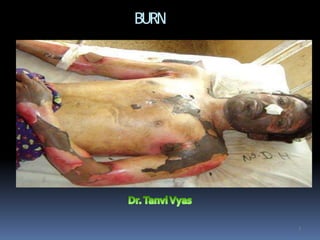
- 1. BURN 1
- 2. 2 INTRODUCTION Majorty of burns in childrenare SCALDScaused by accidents with kettles,pans,hot drinks and bath water In young males burn causedbyexperimenting With mathes and inflamable liquides Electrical and chemical injuries occur in adultswith Associated conditions suchasmental disease, EpilepsyAlcohal and drug abuse
- 3. Risk Factors Fire/Combustion Firefighter IndustrialWorker Occupant of burning structures Chemical Exposure IndustrialWorker Electrical Exposure Electrician Electrical Power DistributionWorker
- 4. Types of Burn Injuries Thermal burn Skin injury Inhalation injury Chemical burn Skin injury Inhalation injury Mucous membrane injury Electrical burn Lightning Radiation burn
- 6. Effects Burn injury causes destruction of tissue, usually the skin, from exposure to thermal extremes (either hot or cold), electricity, chemicals, and/or radiation The mucosa of the upper GI system (mouth, esophagus, stomach) can be burned with ingestion of chemicals The respiratory system can be damaged if hot gases, smoke, or toxic chemical fumes are inhaled Fat, muscle, bone, and peripheral nerves can be affected in electrical injuries or prolonged thermal or chemical exposure Skin damage can result in altered ability to sense pain, touch, and temperature
- 7. Skin Largest body organ. Much more than a passive organ. Protects underlying tissues from injury Temperature regulation Acts as water tight seal, keeping body fluids in Sensory organ
- 8. Skin Injuries to skin which result in loss, have problems with: Infection Inability to maintain normal water balance Inability to maintain body temperature
- 9. Skin Two layers Epidermis Dermis Epidermis Outer cells are dead Act as protection and form water tight seal
- 10. Skin Epidermis Deeper layers divide to produce the stratum corneum and also contain pigment to protect against UV radiation Dermis Consists of tough, elastic connective tissue which contains specialized structures
- 11. Skin Dermis - Specialized Structures Nerve endings Blood vessels Sweat glands Oil glands - keep skin waterproof,usually discharges around hair shafts Hair follicles - produce hair from hair root or papilla Each follicle has a small muscle (arrectus pillorum) which can pull the hair upright and cause goose flesh
- 13. Burn Classification - Depth New terminology Superficial: only the epidermis Superficial partial thickness: epidermis and dermis, excluding all the dermal appendages Deep partial thickness: epidermis and most ofthe dermis Full thickness: epidermis and all of the dermis Old terminology 1st degree: only the epidermis 2nd degree: epidermis and dermis, excluding all the dermal appendages 3rd degree: epidermis and all of the dermis 4th degree: epidermis, dermis, and subcutaneous tissues (fat, muscle, bone, and peripheral nerves)
- 14. Very painful, dry, red burns which blanch with pressure. They usually take 3 to 7 days to heal without scarring. Also known as first-degree burns. The most common type of first-degree burn is sunburn.
- 15. Very painful burns sensitive to temperature change and air exposure. More commonly referred to as second-degree burns. Typically, they blister and are moist, red, weeping burns which blanch with pressure. They heal in 7 to 21 days.
- 16. Blistering or easily unroofed burns which are wet or waxy dry, and are painful to pressure. Their color may range from patchy, cheesy white to red, and they do not blanch with pressure.
- 17. Burns which cause the skin to be waxy white to a charred black and tend to be painless. Healing is very slow, if at all, and may require skin grafting.
- 19. Burn Classifications 1st degree (Superficial burn) Involves the epidermis Characterized by reddening Tenderness and Pain Increased warmth Edema may occur, but no blistering Burn blanches under pressure Example - sunburn Usually heal in ~ 7 days
- 20. Burn Classifications First Degree Burn (Superficial Burn)
- 21. Burn Classifications 2nd degree Damage extends through the epidermis and involves the dermis. Not enough to interfere with regeneration of the epithelium Moist, shiny appearance Salmon pink to red color Painful Does not have to blister everytime Usually heal in ~7-21 days
- 22. Burn Classifications 2nd Degree Burn (Partial Thickness Burn)
- 23. Classification(cont …..) b.2nd degreeburns affected area isred,mottled,painfull Blister formation Healsin 14-21 daysby epithelialization With scaring 2 3
- 24. CLASSIFICATION(CONT……) al 3rddegree burns Affected area is painless and insensetive with Thrombosis of superfici vessels It requiresgrafting 10
- 25. Burn Classifications 3rd degree Both epidermis and dermis are destroyed with burning into SQ fat Thick, dry appearance Pearly gray or charred black color Painless - nerve endings are destroyed Pain is due to intermixing of 2nd degree May be minor bleeding Cannot heal and require grafting
- 26. Burn Classifications 3rd Degree Burn (FullThicknessburn)
- 29. CLASSIFICATION(CONT……) 4th degreeburns It involvesunderlying Tissues Muscles bones 10
- 30. Degrees of burns. Partial thickness and full thickness discussin asessmentofburn 30
- 31. 3 1 CLASSIFICATION OF BURNS Depending on the percentage of burns MILD o Partial thickness burns <15%in adults and <10% In children or o Full thickness <2% o Canbe treated on outpatient department
- 32. 3 2 Classification (cont……. MODERA TE oSeconddegree burn of 15-25%burns oThird degree burn between 2-10%burns oBurns which are not involvingeyes,ears Face,hand,feet and perineum
- 33. 3 3 CLASSIFICATION (CONT……) SEVERE oSeconddegree burns more then 25%in adults and More then 20%in children oAll third degree burns more then10% oAll electrical burns and inhalation burns oBurns with fracture oBurns involving eyes,ears,feet,handsand perineum
- 34. Pathophysiology of burn injury 34 Most common organ affected is the skin Burn canalso damage airways and lungs with life Threatening consequenses Respiratory system injuries occure if person trapped In a burning vehicle,house,car and is forced to inhale Thehot and poisonous gases Hot gasesburn the lining of airway above thelarynx And lining start to swell later on block the airway Steam causesdamage to the lowerairways,respiratory Epithelium swells and detach from bronchialtree
- 35. Pathophysiology(cont……) Metabolic poisoning Carbon monoxide is a product of incomplete combustion That is often produced by fires in a closed space is one of Many poisonous gases Cobinds to hb with an affinity of 240> O2soblock Transport Of O2 Level of carboxyhaemoglobin in blood canbemeasure Conc>10%dangerous and need treatment with pure Oxygenfor more then 24hours Hydrogen cynide causesmetabolic acidosis by interefering with 1 mitochondrial respiration 13
- 36. Pathophysiology(cont……) Inhalational injury causedby minute particles within thick smokebecause Of their small sizeand are not filtered by the upper Airwayand are carried down to lung parenchyma Stick to moist liningcauses intense reaction in alveoli Causeschemical pneumonitis followed by oedema within Alveolar sacand dec gaseous exchange 1 0/2 B 2/2 a 01 c 6 terial pneumonia occures 14
- 37. Pathophysiology(cont……) 10/22/2016 37 Inflamtion and circulatory changes Burn skin release of neuropeptides activation of Complement are intiated by stimulation of pain fibersand Alteration of proteins byheat Activation of hageman factor alter archidonicacid Thrombin and kallikrein pathways
- 38. Pathophysiology(cont……) 10/22/2016 38 At cellular level Complement causesdegranulation of mast cells Attracts neutrophils which also degranulate and releases Largeamount of free radicals andproteases Mast cells also releases TNF@which act aschemotactic Agent to inflamatory cells Theseinflamatory factors alter permeability of bldvessels Largeprotein molecules canalso escapewith ease Damaged collagen and extravasated proteins oncotic Pressurefurther increase flow of water fromintravascular T oextravascularspace
- 39. Pathophysiology Heat causes coagulation necrosis of skin and subcutaneous tissue. 10/22/2016 39 ↓ Releaseof vasoactivepeptides ↓ Altered capillary permeability ↓ Lossof fluid →Severe hypovolaemia ↓ Decreasedcardiac →Decreasedmyocardial output function ↓ Decreasedrenal blood →Oliguria flow (Renalfailure) ↓ Altered pulmonary resistance causing pulmonary Oedema infection SystemicInflammatory ResponseSyndrome (SIRS) ↓ Multi OrganDysfunction Syndrome(MODS).
- 40. BURN ASSESSMENT
- 41. ASSESSMENT OF BURNS ASSESSING SIZE Burn sizeshould be assessedin acontrolled environment T o avoid hypothermia In smaller burns just cut apiece aclean paper the sizeof patient ,swhole hand (digit and palm)which present 1%TBSA And match this to thearea Another accurate way of measuring the size of burns is to draw Theburn on a Ruleof9or LUND AND BROWDER CHART 10/22/2 18
- 42. ASSESSMENT(CONT…..) 10/22/2016 42 RULEOF9 (wallace,s rule of9 Eachupper limb is 9%TBSA Eachlower limb is 18%TBSA Torso18%each side Head and neck 9% Perineum 1% In children head and neck is 18%and Lower limb is 13.5%each=13.5*2=27%
- 45. ASSESSMENT(CONT…..) 10/22/2016 45 2.Assessing depth from the history Burning of human skin is temperature and timedependent It takes 6 hours for skin maintained at 44c* for irreversible changes Asurface teperature of 70c* for 1 second produceepidermal destruction example of exposure of hot water at 65 degree C temperature 45 second exposure produce full thicknessburn 15 second exposure produce deep partial thicknessburn 7 second exposure produce superficial partial thiknessburn
- 46. BURN ASSESSMENT
- 47. ASSESSMENT(CONT…..) 10/22/2016 47 a) Superficial partial thickness burns No deeper then papillary dermis Blister formation Lossof epidermis Capillary return visible When blanched Dermis is pink and moist Pin prick sensation normal Heal without scarring In 2 weeks Treatment is non surgical
- 48. Superficial partial thickness burns 10/22/2016 48 After 24hours afterburn After 2weeks
- 49. Superficial partial thickness burn after 3months Pigment returning 10/22/2016 49
- 50. ASSESSMENT(CONT…..) 10/22/2016 50 b)Deep partial thickness burn Damageto deeper parts ofdermis Epidermis is usually lost Fixedcapillary staining Colour does not blanch with pressure Sensation is reduced Pt is unable todistinguish sharp from blunt pressure T akes3 or more weeks to heal without surgery Leadsto hypertrophic scarring
- 51. ASSESSMENT(CONT…..) 10/22/2016 51 c)Full thicknessburns Whole of the dermisdestroyed Hard and leathery feel No capillary return Often thrombosed vesselscanbe seenunder The skin Theseare completely anesthetised No pain and no bleeding
- 52. Burn Patient Severity Factors to Consider Depth or Classification Body Surface area burned Age: Adult vs Pediatric Preexisting medical conditions AssociatedTrauma blast injury fall injury airway compromise child abuse
- 53. Burn Criteria
- 54. Critical Burn Criteria 30 > 10% BSA 20 > 30% BSA >20% pediatric Burns with respiratory injury Hands, face, feet, or genitalia Burns complicated by other trauma Underlying health problems Electrical and deep chemical burns
- 55. Moderate Burn Criteria 30 2-10% BSA 20 15-30% BSA 10-20% pediatric Excluding hands, face, feet, or genitalia Without complicating factors
- 56. Minor Burn Criteria 30 < 2% BSA 20 < 15% BSA <10% pediatric 10 < 20% BSA
- 57. Causes of death inburns 10/22/2016 57 a. Hypovolaemia and shock b. Renalfailure c. Pulmonary oedema andARDS d. Septicaemia e. Multiorgan failure
- 58. Full thicknessburn Wraped in clingfilm 10/22/2016 58
- 59. Deep partial thicknessburn tangential shavingto removedead dermis Dermis remove layer by layer untill bleeding 10/22/2016 59
- 60. Hypertrophicscarring following a deep dermal burn. After 3 or moreweeks 10/22/2016 60
- 62. Management of burns 10/22/2016 62 Management of burns is consist of prehospital care & hospital care
- 63. Management of burns Prehospital care Stop the burning process Stop,drop and roll is agood method of extinguishing Fire Cool the burn wound Thisprovide analgesia and slow the delayed micro- -vascular damage which occure after aburn injury Cooling should be for minimum 10 mintues and up to Onehour to avoidhypothermia Give oxygen Giveoxygen especialy if there is altered conciousness_ level 32
- 64. Management of burns 64 Elevate Sitting apatient up with aburned airway may provelife Saving Elevation of burned limbs reduce swelling anddiscomfort Check for other injuries AstandardABCcheck followed by asecondarysurvey Patients burned in explosions may have head and spineinjuries And other life threateningproblems
- 65. Management of burns 65 Indications for admission in burns Susectedairway or inhalational injury Any burn require fluide resusciation Any burn in extreme of ages All electrical and chemicalburns Any burn which require surgery Burn of any significance to hands,face,feet orperineum Suspiciousof non accidentalinjury
- 66. HYPOVOLAEMIC SHOCK Hypotension Oliguria Tachy cardia Sweating Pallor Hyper ventilation Clouding of consciousness
- 67. SEPTIC SHOCK IncreasedT* Hypotension < 90 mmhg Oliguria < 30 ml Dry &pink exremities Altered pulmonary functions
- 68. SKIN ASSESSMENT Inspection Appearance Temperature Moisture Dryness Texture Color Size Palpate lymph nodes Cyanosis Pulses Area Duration Itching/burning
- 69. Management of burns 69 Hospital care Admit the patient Airway control Breathing and ventilation Circulation Disability Exposure with environmentcontrol Fluid resuscitation Assessthe %age,degreeand type of burn Keepthe patient in cleanenvironment Sedation and proper analgesia
- 70. Management of burns 10/22/2016 70 A.AIRWAY CONTROL Burned airway creates problems by swelling and cancompletely Occlude the airway Secureairway with an endotracheal tubeuntil swelling subsided which is Usually 48hours Delayed diagnosis of airway burn make difficult to intubate the Patient in presence of lyrangeal oedema socricothyroidectomy Should be done Early intubation of suspected airway burn is the treatment of Choice in suchpatients
- 71. Management of burns 10/22/2016 71 B.BREATHING Aprogressive increase in respiratory rate and effort,anxiety Risingpulse and confusion with decreasing o2saturation Thesesymptoms take 24 hours to 5 daysto appear Treatment starts assoon aspossible including Physiotherapy Nebulisers Warm humidified oxygen
- 72. Management of burns 10/22/2016 72 Fluide resuscitation Iv volume must be maintained following aburn in order to provide sufficient circulation to perfuse not only the organs but also the peripheral tissues,especially damagedskin Iv resuscitation is appropriate for any child withaburn greater Then 10%and 15%for TBSAfor adults If oral resuscitation is to be commenced then water is given Should not be saltfree It is appropriate to give oral rehydration with asolution such as DIORALYTE* Most common fluid used is ringer lactate
- 73. Management of burns Fluid volume is relatively constant in proportion to the area Of body burned therefore there are formulate that calculate The approximate volume of fluid needed for the pt of agiven Body weight with agiven %ageof the bodyburned Formulas to calculate the fluid replacement 1.parkland regime (commonly used) 4ML/%burn/kg body weight/24 hours 4*50*60=12000ml in 24hours Half this volume is given in the frist 8 hours Secondhalf is given in the subsequent 16hours Others 1. Evan,s formula 2. Muir and barclay 10/223/2.016Modified brook formula 39
- 75. Management (cont…) 10/22/2016 75 Fluids used Crystalloid resuscitation Ringer lactate is the most commonly usedcrystalloid Theseare aseffective ascolloids for maintaining intra- -vascular volume Lessexpensive In children Dextrose saline given for maintanaince 100ml/kg for 24 hours for frist10kg 50ml/kg for 24 hours fornext 10kg 20ml/kg for 24 hours for each kgabove 20kg bodyweight
- 76. Management (cont…) 10/22/2016 76 hypertonic saline it produces hyperosmolarity andhypernatremia Reducesshift of intracellular water to extracellular space Advantages Include less tissue oedema and aresultant decrease in Escharotomies and intubations
- 77. Management (cont…) 10/22/2016 77 Colloid resuscitation Plasmaproteins are responsible for the inward oncotic Pressure that counteracts the outward capillaryhydrostatic Pressure. Without proteins there willbe oedema Proteins should be given after frist 12 hours of burn before This time proteins will leak out of cells Given through muir and barclay formula 0.5*%agebsa burn*weight=one portion Periods of 4/4/4, 6/6,12 hours respectively Oneportion to b given ineach period
- 78. Management (cont…) 10/22/2016 78 Monitoring of resuscitation Thekey to monitoring of resuscitation is urinaryoutput Output should be between 0.5ml and1.0ml/kg/hour If urine output is below this infusion rate should increase By50% If still output is inadequate then abolus of 10ml/kggiven 2ml/kg/hr urinary output signals decrease in the rate of Perfusion Haematocrit measurement is ausefull tool in confirming Suspected under or overhydration
- 79. Management (cont…) 10/22/2016 44 Asan absorbent Treating the burn wound Dressings Paraffine guaze Hydrocolloids (duoderm) Biological dressings synthetic (biobrane) natural (amniotic membrane) Full-thickness and deep dermal burns needantibacterial dressings to delay colonisation prior tosurgery Open method Silver sulfadiazine application without dressingscommonly Usedin burns of face,head andneck. Closed method Dressing done to soothen and to protect the wound T oreduce thepain
- 80. Management (cont…) 10/22/2016 Treating the burn wound (cont…… Tangential excision Canbe done within 48 hours with skin grafting in patients with less Then 25% burn Usually done in deep dermal burns Deaddermis is removed layer by layer Untill fresh bleeding occurs Later skin grafting done 45
- 81. Treating the burn wound (cont…… Treating the burn wound (cont…… escharotomy Circumferential full-thickness burns tothe limbs require emergency Surgery Thetourniquet effect of thisinjury is easily treated by incising the whole length of full-thickness burns. Thisshould be done in the mid-axial line,avoiding major Nerves Theburn needs to be cleaned and the sizeand depth need tobe Full thickness burns and deep partial-thickness burns thatwill requireoperative treatment will need to be dressed with an antibacterialdressing to delay the onset of colonisation of the wo1 0 u/ 2 2 n/ 2 d0 1 6 46
- 83. Afull-thickness burn to the upper limb with a mid-axial escharotomy. Thesoot and debris have been washedoff. 10/22/2016 83
- 84. 10/22/2016 84 Topicalagent advantages problems Silver sulfadiazine 1 -Antiseptic (G+ve -Neutropenia, % and G–ve pseudoeschar -Soothening, good -- Causeswound penetration maceration - Hydration and softening of eschar occurs Sulfamylon – 5%- Antipseudomonal, Very irritant,painful (Mafenide acetate) anticlostridial Causesacidosis - Penetrates very well in to tissues Silver nitrate –0.5% -Antiseptic Stainsburn area Povidone iodine Irritant (5%) - Usedon Painfull granulation tissue - Not used in partial after eschar burns separation Silver sulphadiazine - Boostscell and cerium nitrate mediated immunity and forms sterile eschar
- 85. Additional aspects of treating burn patient 10/22/2016 85 Analgesia Oral form of paracetamol and nsaids insuperficial burns Iv opiates for largeburns Im should not be given in over 10%of TBSAasabsorption is Unpredictable Short acting analgesia given beforedressing Energy balance Feeding should start within 6 hrs of injury to reduce gut mucosal damag Burns patients need extra feeding Anasogastric tube should be used in all patients withburns over 15% of TBSAand 10%in caseofchildren Burn injuries are catabolic in theacute episode. Removing the burn and achieving healing stops thecatabolic drive
- 86. Additional aspects of treating burn patient control of infection Patients with major burns areimmunocompromised, pathogenic and opportunistic bacteria and fungi enter via theburn wound,cathetars and iv lines Theyhave compromised local defences in the lungs and gut dueto oedema Sterile precautions must berigorous Swabsshould be taken regularly Arise in white blood cell count, thrombocytosisand increased catabolism are warnings of infection Nursing care Physiotherapy Psy 10c /2h 2/2o 016 logical support 50
- 87. SURGERY FORTHEACUTEBURN 10/22/2016 87 Any deep partial-thickness and full-thickness burnsexcept those that are less than about 4 cm2, needsurgery Atopical solution of 1:500 000 adrenaline also helps toreduce bleeding, deep dermal burns, the top layer of dead dermisis shaved off untilpunctate bleeding is observed and the dermis can be seento be free of any small thrombosedvessels Full-thickness burns require full-thickness excision ofthe Skin Postoperative management of these patientsobviously requires careful evaluation of fluid balance and levels ofhaemoglobin.
- 88. Delayed reconstruction and scar management 10/22/2016 88 is common for large Full thicknessburns Eyelids must be treated before exposure keratitisarises Transposition flaps and Z-plasties with or withouttissue expansion are useful Full-thickness grafts and free flaps may be needed forlarge or difficult areas Hypertrophy is treated with pressure garments tobe worn for 6-18months Smaller areas of hypertrophy, silicone patches will speed scar maturation,as will intralesional injection of steroid. Pharmacological treatment of itchis important
- 89. Pediatric Burns Thin skin increases severity of burning relative to adults Large surface/volume ratio rapid fluid loss increased heat loss hypothermia Delicate balance between dehydration and over hydration Immature immunological response sepsis Always consider possibility of child abuse
- 90. Geriatric Burns Decreased myocardial reserve fluid resuscitation difficulty Peripheral vascular disease, diabetes slow healing COPD increases complications of airway injury Poor immunological response - Sepsis % mortality ~= age + % BSA burned
- 91. Complication of burn 10/22/2016 91 Shockdue to hypovolaemia Renalfailure(toxins from burn&myoglobin) Pulmonary oedema,resp infections,ARDS,resp failure Infection by staph aureus,pseudomonas,klebsella leadsto Septicemia Fungaland viral infections of dangerous type canoccure GIT :Hypovolaemia, ischaemia of mucosa,erosive gastritis—Curling’s ulcer (seen in burns >35%). Fluid and electrolyte imbalance. immunosuppression predisposes to severe opportunistic infection. Escharformation and its problems likedefective circulation, ischaemia when it is circumferential. Electrical injuries often causefractures,major internal organ injury, convulsions.
- 92. Complications (cont…..) 10/22/2016 . 92 Inhalation burn causespulmonary oedema, respiratory arrest,ARDS. Chemical injury causessevere GITdisturbances like erosions, perforation, stricture oesophagus (alkali), pyloric stenosis (acid), mediastinalinjury. Other problems DVT,pulmonaryembolism bed-sores, severe malnutrition with catabolicstatus, Toxic shock syndrome: It is a life-threatening exotoxin mediated disease causedby Staphylococcusaureus. It is common in children, presentswith rashes, myalgia, diarrhoea, vomiting, andmultiorgan failure with highmortality
- 93. Complications (cont…..) 10/22/2016 93 Development of contracture is alate problem.It leads to ectropion, microstomia, disability of different joints, defective hand functions, growth retardation causingshortening COMPLICATIONS OFBURNSCONTRACTURE Ectropion of eyelid causing keratitis andcorneal ulcer. Disfigurement in face. Narrowing of mouthmicrostomia. Contracture in the neck causing restricted neckmovements. Disability and nonfunctioning of joints due to contracture Hypertrophic scarand keloid formation.
- 94. COMPLICATIONS OFBURNS CONTRACTURE Marjolin’s ulcer It is avery well-differentiated squamous cell carcinoma occurring in ascarulcer due to repeated breakdown(unstable scar of longduration). •It is locallymalignant. •Asthere are no lymphatics in the scar,sothere is no spread to lymphnodes. • Asthere are no nerves in the scar itis painless. •It hasraised and everted edge with induration. •Biopsy confirms the diagnosis. Treatment Radiotherapy is not given forMarjolin’sulcer . Treatment is either wide excision or amputation. It is curable. Once it spreads out of the scar tissue it behaves like any other squamous cell carcinoma and socanspreadto 10/22/20r1e6gional lymphnodes 56
- 95. Marjolin’s ulcer developed over burnscontracture 10/22/2016 95
- 96. Contracture at different parts of body chest and neck 10/22/2016 96
- 97. Contracture at different parts of body 10/22/2016 97 Severe contracture at knee joint causing deformity Contractureatface
- 98. Complication of contracture 10/22/2016 98 Hypertrophicscar Keloid formation
- 99. Treatment of burn contracture 10/22/2016 99 • Releaseof contracture surgically and useof skingraft or “Z” plasty or differentflaps. • Proper physiotherapy and rehabilitation is essential. • Pressuregarments to prevent hypertrophicscars. • Management of itching in the scarusing aloevera, antihistamines and moisturizing creams.
- 100. Prevention of developmentof contracture 10/22/2016 100 •Joint exercise in full range during recovery periodof burns • Pressuregarments for along period •Topical silicon sheeting •Saline expanders for scars
- 101. NON THERMALINJURIES 10/22/2016 101 Electrical burns Chemical burn Cold injuries Ionising radiation
- 102. Non thermal(cont……) 10/22/2016 102 Electrical burns 1000v Low tension injuries Low tension injuries do not have enough energy to cause Significant destruction Entry and exit points normally in the fingers suffers small Deepburns may damage underlying nerves and vessels Accreates atetany within musclessopatient unable to Releasethe device untill the power wasturnedoff May interfere with normal cardiac pacing and cancause Cardiac arrest
- 103. Electrical burns(cont…..) high tension injuries 3 sources of damage 1) The flash 2) The flame 3) The current When ahigh tension line is earthed itcanarc over the pt And causesaflash burn Extremely rapid heating of the aircausesan explosion That propel the victimbackward It is always amajor burn There is awound of entry and wound ofexit Major internal organ injuriesoccures Convulsions candevelope 10/22/2016 65
- 104. Electrical burns(cont…..) Management Depending on injury it is managedaccordingly Patient should always be admitted and should be assessedby i. ECG ii. u/s abdomen iii. Chestx-ray iv. Ctscanhead sometimes v. Cardiac enzyme analysis Acidosis is common sobicarbonate infusion needed Fractures and dislocations common somanaged accordingly Releaseof myoglobin cancauserenal tubular damage andrenal Failure somanitol is used to prevent myoglobin inducedrenal failure (compartment syndrome) 10/22/2016 66
- 105. Wound of entry in anelectricburn. Electric burn 10/22/2016 105
- 106. Electrical burns(cont…..) 10/22/2016 106 An exit wound of ahigh-tension injury, Amputation and coverwith the lateral portion of the secondtoe.
- 107. Chemical burns 10/22/2016 107 There are 70000 different chemicals in regular usewithinindustry Occasionally these causeburns There are two aspects to achemicalinjury 1. Physicaldestruction to theskin 2. Systemicabsorption Theinitial management of any chemical injury is copious lavage With water but some need tobe remove physically eg.phosphorus Acomponent of millitary devices Themore common injuries are causedbyeither 1. Alkalis 2. acids
- 108. Chemical burns(cont…) 10/22/2016 108 Alkalis alkalis are more destructive and especially when come incontact With eyes Commonly used alkalis are sodium hydroxide,lime,bleach Theycausefat sponification,fluide loss,release of alkali proteinase Alkali burns occur in oral cavity andoesophagus which leads to multiple oesophagealstrictures. After copious lavage, the next step in themanagement of any chemical injury is toidentify the chemical and its concentration and to elucidate whether there is any underlying threat to the patient’s life if absorbed systemically
- 109. Acidaemia should be corrected by IVsodiumbicarbonate. 10/22/2016 Chemical burns(cont…) 109 Acids Acid burn occurs in skin, soft tissues and GIT .In GIT , Burns affecting the fingers and causedby dilute acid arerelatively common. Theinitial management is with calcium gluconate geltopically severe burns or burns to large areas of the hand canbe subsequently treated with Bier’s blocks containing calcium gluconate 10 per centgel it is common in stomach either due to nitric acid or sulphuric acid which may lead tosevere gastritis or pyloric stenosis. Other acids are formic acid, hydrofluoricacid. Theycausemetabolic acidosis, renal failure, ARDS,haemolysis.
- 110. Cold injuries inflammatory reactionisnot asmarked. Thetissueismoreresistanttocoldinjurythantoheatinjury Theassessmentof depth of injuryismoredifficult, Frostbite injuries affectthe peripheriesincoldclimates coldinjury producesdelayedmicrovasculardamagesimilarto that of cardiacreperfusioninjury. Theinitial treatmentiswith rapidrewarminginabathat42°C. Thelevelof damageisdifficult toassess surgery usuallydoesnot playarole in itsmanagement Cold injuries are principally divided into twotypes 1. Acute cold injuries fromindustries 2. Frost bite 72
- 111. Ionising radiation 111 Theseinjuries canbe divided into 1. Localised 2. Whole body exposure Themanagement of localised radiation damage is usually Conservative until the true extent of the tissue injury is apparent. If damage have causedan ulcer, then excision and coveragewith vascularised tissue is required. Apatient who hassuffered whole-body irradiation and issuffering From acute desquamation of the skin hasreceived alethal dose of Radiation which cancauseaparticularly slow and unpleasantdeath Dosemay be lethal and may not belethal Giving iodine tablets, the management of these injuries is supportive