SlideShare una empresa de Scribd logo
1 de 78
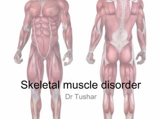
Skeletal muscle disorder
Dr Tushar
Organization of Skeletal Muscle Including
Connective Tissue (CT) Compartments
EPIMYSIU
M•Loose CT
•Blood vessels
PERIMYSIUM
•Septa
•Nerve branches
•Muscle spindles
•Fat
•Blood vessels
ENDOMYSIUM
•Muscle fibers
•Capillaries
•Small nerve fibers
Perimyseal
connective tissue
Endomyseal
connective tissue
Normal H&E-Stained Frozen Cross-Section of Skeletal Muscle
 Note uniform sizes, polygonal shapes, and eccentric nuclei.
Normal H&E-Stained
Longitudinal Paraffin Section
• Note the banding pattern.
• Nuclei are eccentrically placed.
 Can be identified by the esterase
reaction due to the presence of
acetylcholinesterase.
Neuromuscular
Junctions
Normal Structures: Muscle Spindle
and Associated Nerve Fibers
(Gomori trichrome)
 Type I fibers are light
 Type II fibers are dark (pattern reverses at ATPase pH 4.3)
Normal (ATPase pH
9.4)
Skeletal Muscle atrophy
• common features of many
disorders
• causes:- loss of innervation ,
disuse, cachexia, old age, and
primary myopathies
• patterns:-
– clusters or groups of atrophic
fibers are seen in neurogenic
disease
– perifascicular atrophy is seen
in dermatomyositis
– type ii fiber atrophy with
sparing of type i fibers is seen
with prolonged corticosteroid
therapy or disuse.
Classification of
Myopathies
ACQUIRED INHERITED
Inflammatory Myopathies Dystrophies
Polymyositis (PM) Dystrophinopathies
Dermatomyositis (DM) Limb-Girdle
Inclusion body myositis (IBM) Myotonic
Granulomatous myositis Facioscapulohumeral (FSHD)
Infectious myositis Oculopharyngeal (OPD)
Toxic Distal
Endocrine Congenital
Metabolic
Mitochondrial
Glycogen & lipid storage
Muscle Biopsy
• Often necessary for final diagnosis of myopathy
• Choose site based on clinical, electrodiagnostic, or
imaging features
• avoid “end-stage” fatty muscle
• Frozen sections most useful
• routine stains
• histochemistry
• immunohistochemistry
ACQUIRED
Inflammatory Myopathies
Polymyositis
• Adult-onset inflammatory myopathy that shares myalgia and weakness
with dermatomyositis but lacks its distinctive cutaneous features.
• Pathogenesis:
– Believed to have an immunologic basis.
– CD8-positve cytotoxic T cells are a prominent part of the
inflammatory infiltrate in affected muscle (mediators of tissue
damage)
– Vascular injury does not play major role (unlike dermatomyositis)
• Morphology:
– Endomysial mononuclear inflammatory cell infiltrates
– Degenerating necrotic, regenerating and atrophic myofibers are
typically found in a random or patchy distribution
– Absent perifascicular pattern of atrophy (characteristic of
Polymyositis
(Longitudinal Paraffin-Embedded Section)
• in all myopathies, degenerating fibers stain pale initially
and then become digested by macrophages.
• mononuclear inflammatory cell infiltrates and many
basophilic regenerating fibers (arrow)
Polymyositis
(Longitudinal Paraffin-Embedded Section-Higher Power)
• regenerating fiber (non-specific)
• fiber is basophilic due to presence of increased
RNA and RNA.
• activated plump nuclei and prominent nucleoli
Invasion of a Non-necrotic Fiber by
Inflammatory Cells
• Seen in polymyositis, inclusion body myositis, and a
few dystrophies.
Myophagocytosis
(Esterase Stain)
• macrophages are ingesting the remnants of a degenerating
fiber. this is a non-specific myopathic finding.
Dermatomyositis
• Immunologic disease in which damage to small blood vessels contributes to muscle
injury.
• Vasculopathic changes – Telangiectasias
• Pathogenesis :
– Inflammatory signature enriched for genes that are unregulated by type I
interferons is seen in muscle and in leukocytes (prominence – disease activity)
– Autoantibodies:
• Anti-Mi2 antibodies – Directed against a helicase implicated in nucleosome
remodeling. Strong association with prominent Gottron papules and heliotrope
rash.
• Anti-Jo1 antibodies – Directed against the enzyme histidyl t-RNA synthetase,
associated with interstitial lung disease, nonerosive arthritis and a skin rash
(Mechanic’s hand)
• Anti-P155/P140 antibodies – Directed against several transcriptional
• Morphology:
– Perimysial mononuclear inflammatory infiltrates in connective
tissue and around blood vessels.
– Myofiber atrophy is accentuated at the edges of the fascicles –
Perifascicular atrophy
– Segmental fiber necrosis and regeneration.
– Deposition of CD4+ T-helper cells and C5b-9 (MAC) in capillary
vessels.
– EM: tubuloreticular endothelial cell inclusion
Dermatomyositis
• perifascicular atrophy & degeneration
• perimysial nflammatory cells surround a blood vessel.
• inflammatory cells tend to be b-cells.
• vasculitis with bowel infarction and subcutaneous
calcifications sometimes occur in the childhood form.
Perifascicular Atrophy
(NADH-Reacted Section)
Membrane Attack Complex
(MAC)
(Immunohistochemical Stain)
• MAC is the terminal component of the complement
pathway.
• It is often deposited in capillaries in dermatomyositis.
INCLUSION BODY MYOSITIS
• Disease of late adulthood that typically affects patients older than 50
years and is the most common inflammatory myopathy in patients older
than age 65 years.
• Slowly progressive muscle weakness – m/c feature
– Most severe in quadriceps and distal upper extremity muscles.
– Dysphasia from esophageal and pharyngeal muscle involvement
• Lab investigation:
– S. creatine kinase level increased
– Myositis associated autoantibodies are absent.
• Morphology:
– Patchy often endomysial mononuclear inflammatory cell infiltrate rich in
CD8+ T- cells
– Increased sarcolemmal expression of MHC class I antigens
– Focal invasion of normal appearing myofibers by inflammatory cells
– Admixed degenerating and regenrating myofibers
– Abnormal cytoplasmic inclusions described as “rimmed vacuoles”
– Tubolofilamentous inclusions in myofibers – EM
– Cytoplasmic inclusions containing proteins typically associated with
neurodegenerative disease, like beta-amyloid, TDP-43, and ubiquintin
– Endomysial fibrosis and fatty replacement, reflective of a chronic disease
course.
Inclusion Body Myositis (IBM)
• Features of chronic myopathy with endomysial inflammation and
rimmed vacuoles are characteristic.
Vacuole
Invaded fiber
Lymphocytic inflammation
“Rimmed vacuoles”
• IBM: Vacuoles contain amyloid.
(Congo Red)
IBM Intracytoplasmic (within Vacuoles) or
Intranuclear Filamentous Inclusions
Giant cell
 Granulomas tend not to cause significant damage to adjacent
myofibers.
Granulomatous Myositis in a Patient with
Sarcoidosis
Endocrine Disturbance Type II Fiber Atrophy
(ATPase pH9.4)
• Characteristic of most endocrine myopathies and steroid
myopathy
Toxic myopathies
• Statin induced
• Chloroquine & hydroxychloroquine (Drug induced lysosomal storage
myopathy)
• ICU myopathy (corticosteroid therapy)
– Degradation of sarcomeric myosin thick filaments leading
to profound weakness
• Thyrotoxic myopathy
– Proximal muscle weakness, exophthalamic
ophthalmoplegia
• Alcohol
INHERITED
Congenital Myopathies
Central Core Myopathy
(NADH)
• Central areas of absent staining in the dark type I fibers
• Mitochondria absent
Congenital Myopathies: Central Core Myopathy
(NADH)
 The core consists of disorganized myofibrils and the area is devoid
of mitochondria.
 Eosinophilic inclusions present.
Nemaline Myopathy
Nemaline Myopathy
(Gomori Trichrome)
• Eosinophilic inclusions stain
darkly.
Nemaline Myopathy
(Electron Microscopy)
• Named for thread-like appearance
• Inclusions extend from Z-band to Z-band
Centronuclear myopathy
 Internalized nuclei predominant.
 Consistent with centronuclear myopathy.
 Can be seen in other disorders such as myotonic dystrophy with
congenital onset.
Muscle Biopsy from an Infant:
Centronuclear Myopathy
• Central position of the nucleus
resembling an embryonic
Congenital Fiber Type
Disproportion
(H&E)
• Bimodal size
population
Muscular
Dystrophies
X linked muscular dystrophy
with dystrophin mutation
Duchenne and Becker musclar
dystrophy
• Most common muscular dystrophies x-linked and stem from mutations that disrupt the
function of a large structural protein called dystrophin.
• Early onset form – Duchenne muscular dystrophy
– Severe progressive phenotype
• Late onset form - Becker muscular dystrophy
– Isolated cardiomyopathy, asymptomatic elevation of creatine kinase, exercise
intolerance
• Pathogenesis:
– Loss of function mutations in the dystrophin gene on X- chormosome
– Dystrophin provide mechanical stability to the myofiber and its cell complex
• Morphology:
– Chnages in Duchenne and Becker muscular dystrophy are similar, but differ in
degree.
– Chronic muscle damage that outpaces the capacity for repair.
– Segmental myofiber degeneration and regeneration with an admixture of atrophic
myofirbers.
– Fatty replacement as disease progress
– IHC studies show absence of the normal sarcolemmal staining pattern in
Duchenne muscular dystrophin and reduced stationing in Becker muscular
• Clinical Feature
– Duchenne muscular dystrphy
• Normal at birth
• Early motor milestones
• Walking is delayed
• Clumsiness & inability to keep up with peers
• Pseudohyperthrophy of muscle
• Mean age of wheel chair dependence around 9.5 years
• Cardiomyopathy & arrhythmias
• Frank mental retardation
• Mean age of death 25 to 30 years
– Becker muscular dystrophy
• Later onset and slowly progressive
Frozen Section from a Patient with
Duchenne Muscular Dystrophy
• Opaque or hyaline fibers (arrows)
• Increase in endomysial connective tissue
Group of basophilic regenerating fibers
Normal Immunohistochemical Stain for
Dystrophin
(Subsarcolemmal Staining)
Duchenne Muscular Dystrophy
(Absent Staining for Dystrophin)
split fiber
(non-specific chronic change)
Becker Muscular Dystrophy
(Reduced but Present Staining)
Female Carrier of Duchenne Muscular
Dystrophy
(A Mosaic Staining Pattern)
Myotonic Dystrophy
• Autosomal dominant multisystem disorder associated with skeletal
muscle weakness, cataracts, endocrinopathy, and cardiomyopathy.
• Myotonia key feature
• Pathogenesis
– Expansions of CTG triplet repeats in 3’-noncoding region of
myotonic dystrophy protein kinase (DMPK)
– Toxic gain of function
– CUG-repeats in the DMPK mRNA transcript appear to bind and
sequester a protein called muscleblind-like1 – Important role in
RNA splicing.
– This inhibits muscleblind-like-1function leading to missplicing of
other RNA transcripts including transcript for a chloride channel
called CLC1and is responsible for characteristic myotonia.
Myotonic Dystrophy
• Chronic changes
• Marked excess in internalized nuclei
• Variation in fiber sizes
• Nuclear clumps (not shown)
(H & E, Paraffin)
 The excess of internalized nuclei can lead to nuclear
chains.
Myotonic Dystrophy
(NADH-Reacted Section)
• Ring fibers in which myofilaments are organized
in different directions
Emery-Dreifuss Muscular
Dystrophy
• Caused by mutation in genes that encode nuclear lamina proteins.
• Triad
– Slowly progressive humeroperoneal weakness
– Cardiomyopathy
– Early contractures of the Achilles tendon, spine & elbow
• X-linked form [EMD1]– mutation in genes encoding emerin
• Autosomal form [EMD2] – mutation in genes encoding lamin
• These protein helps in maintaining the shape and mechanical stability of the
nucleus during muscle contraction.
Emery-Dreifuss Muscular Dystrophy
(Gomori Trichrome-Stained Frozen Section)
Necrotic fiber
 Variation in fiber size with many hypertrophic fibers
 Increase in endomysial connective tissue
 Nonspecific so-called dystrophic changes seen in many of the muscular
dystrophies.
 Can also be seen in any chronic myopathic disorder.
 This disorder is due to loss of the protein emerin.
Fascioscapulohumeral
Dystrophy
• Characteristic pattern of muscle involvement that includes prominent
weakness of facial muscles and muscles of the shoulder girdle.
• Autosomal Dominant
• Pathogenesis:
– Overexpression of a gene called DUX4, located in a region of
subtelomeric repeats on the long arm of chromosome 4.
– Deletion in flanking repeats causes changes in chromatin that derepress
the remaining copies of DUX4 thus leading to overexpression.
Fascioscapulohumeral Dystrophy (FSHD)
• The majority of dystrophies do not have a specific histopathologic appearance.
• Clinical features are also very important.
• For example, winging of the scapula is characteristic of FSHD.
FSH Dystrophy
• Variable non-specific changes
• Range from scattered atrophy to “dystrophic” features.
• Inflammation can be present (arrow).
Limb-Girdle Muscular Dystrophy
• Heterogeneous group
– 6 AD
– 15 AT
• Characterized by muscle weakness that preferentially involves proximal
muscle groups.
• Pathogenesis
– Genes encoding structural components (sarcoglycans) of the
dystrophin glycoprotein complex
– Genes encoding enzymes that are responsible for glycosylation of a-
dystroglycan, a component of the dystrophin glycoprotein complex
– Genes encoding proteins that associate with the Z-disks of sarcomeres
– Genes encoding proteins involved in vesicle trafficking and cell
signaling
– Genes that seemingly stand alone, such as those encoding the
protease calpain 3 and laminA/C (which is also mutated in some
patients with Emery-Dreifuss muscular dystrophy)
INHERITANCE
GENETIC
ABNORMALITY
DISORDER
X-linked
Dystrophin
Emerin
Duchenne, Becker MD
Emery-Dreifuss MD
AD
Myotilin
Lamin A/C
Caveolin – 3
PABP2
αβ-crystallin/Desmin
Limb-Girdle MD (LGMD 1A)
LGMD 1B
LGMD 1C
Oculopharyngeal
Myofibrillar Myopathy
AR
Calpain – 3
Dysferlin
g Sarcoglycan
a Sarcoglycan
β Sarcoglycan
Δ Sarcoglycan
Telethonin
LGMD 2A
LGMD 2B
LGMD 2C
LGMD 2D
LGMD 2E
LGMD 2F
LGMD 2G
Mutations in “Limb-Girdle” and Other
Dystrophies
Mitochondrial Myopathies
• Complex systemic conditions that can involve many organ systems,
including skeletal muscle.
• Manifest as weakness, elevations in serum creatine kinase levels, or
rhabdomyolysis.
• Morphology:
– Most consistent pathologic change in skeletal muscle is abnormal
aggregates of mitochondria that are seen preferentially in the
subsarcolemmal area.
– Ragged red fibers appearance.
– Morphologically abnormal mitochondria – EM
• Clinical feature
– Chronic progressive external ophthalmoplegia
– Mitochondrial encephalomyopathy with lactic acidosis and strokelike
episodes
– Kearns-Sayre syndrome
– Myoclonic epilepsy with ragged red fibers
– Leber hereditary optic neuropathy
Metabolic: Inherited – Mitochondrial
Myopathy
 Ragged red fiber present (Gomori trichrome)
 Due to proliferation of abnormal mitochondria
 SDH-rich fibers are seen with mitochondrial proliferation.
 SDH is a respiratory chain enzyme encoded by nuclear DNA.
Mitochondrial Myopathy
(Succinic Dehydrogenase
Reaction)
Cytochrome Oxidase (COX) Respiratory Chain
Enzyme
Normal Fibers
Many COX-Negative
Fibers
 COX-negative fibers are usually seen with mtDNA mutations.
Mitochondrial Disorders
(Electron Microscopy)
Higher power view of paracrystalline inclusion
Disease of Lipid or Glycogen
Metabolism
• Inborn errors of lipid or glycogen metabolism
• Severe muscle cramping and pain
• Extensive muscle necrosis (rhabdomyolysis)
• Example
– Carnitine palmitoyltransferace II deficiency (m/c)
– Myophosphorylase deficiency (McArdle ds)
– Acid maltase deficiency
• Increased lipid storage
• Seen in carnitine deficiency states (primary or secondary)
• Sometimes as a consequence of certain toxins
• Focal increases can be non-specific.
(Oil-Red-O
Stain)
Lipid Storage Myopathy
(Electron Microscopy)
Glycogen Storage Myopathies
• Some glycogen storage myopathies, such as myophosphorylase deficiency
(McArdle’s Disease), cause subsarcolemmal blebs.
• PAS-positive due to the presence of glycogen.
• Only with acid maltase deficiency is glycogen deposited in lysomsomes.
Acid Maltase Deficiency
(Acid Phosphatase)
• Due to the intralysosomal activity of this enzyme
• Prominent staining with acid phosphatase in vacuoles
Vacuolarmyopathy noted.
Normal Glycogen
(PAS Stain) Control
Increased Glycogen
 Acid maltase deficiency
 Increased glycogen (diffusely and in vacuoles)
Ion Channel Myopathies
(Channelopathies)
• KCNJ2 :
– mutations affecting this potassium channel cause Andersen-Twail
syndrome
• AD, Periodic paralysis, Heart arrhythmias, skeletal abnormalities
• SCN4A :
– Mutations affecting this sodium channel cause several AD with
presentations ranging from myotonia to periodic paralysis.
• CACNA1S :
– Missense mutations in this protein, a subunit of a muscle calcium
channel, are the most common cause of hypokalemic paralysis.
• CLC1:
– Mutations affecting this chloride channel cuases myotonia congenita
– Expression decreased in myotonic dystrophy
• RYR1:
– Mutation in the RYR1 gene disrupt the funciton of the ryanodine
receptor , which regulated calcium release from the sarcoplasmic
reticulum
– Central core myopathy
Thank you for listening.
Have a Good Day.

Más contenido relacionado

La actualidad más candente

La actualidad más candente (20)

Approach to peripheral neuropathy
Approach to peripheral neuropathyApproach to peripheral neuropathy
Approach to peripheral neuropathy
 
Muscle biopsy
Muscle biopsyMuscle biopsy
Muscle biopsy
 
Congenital myopathy
Congenital myopathyCongenital myopathy
Congenital myopathy
 
Myopathies
MyopathiesMyopathies
Myopathies
 
Non compressive myelopathy
Non compressive myelopathyNon compressive myelopathy
Non compressive myelopathy
 
Myopathies
MyopathiesMyopathies
Myopathies
 
Hereditary Ataxia
Hereditary AtaxiaHereditary Ataxia
Hereditary Ataxia
 
Myopathies and myotonic dystrophies
Myopathies and myotonic dystrophiesMyopathies and myotonic dystrophies
Myopathies and myotonic dystrophies
 
Myopathies
MyopathiesMyopathies
Myopathies
 
Paraparesis
ParaparesisParaparesis
Paraparesis
 
Neuropathy ..paras
Neuropathy ..parasNeuropathy ..paras
Neuropathy ..paras
 
Polyarteritis nodosa
Polyarteritis nodosaPolyarteritis nodosa
Polyarteritis nodosa
 
SPINAL MUSCULAR ATROPHY
SPINAL MUSCULAR ATROPHYSPINAL MUSCULAR ATROPHY
SPINAL MUSCULAR ATROPHY
 
Approach to Peripheral neuropathy
Approach to Peripheral neuropathyApproach to Peripheral neuropathy
Approach to Peripheral neuropathy
 
Neuropathies & myopathies - an overview
Neuropathies &  myopathies - an overviewNeuropathies &  myopathies - an overview
Neuropathies & myopathies - an overview
 
Inflammatory myositis
Inflammatory myositis Inflammatory myositis
Inflammatory myositis
 
Ataxia
AtaxiaAtaxia
Ataxia
 
Peripheral nerve biopsy
Peripheral nerve biopsyPeripheral nerve biopsy
Peripheral nerve biopsy
 
Peripheral Neuritis by Dr. Sookun Rajeev Kumar
Peripheral Neuritis by Dr. Sookun Rajeev KumarPeripheral Neuritis by Dr. Sookun Rajeev Kumar
Peripheral Neuritis by Dr. Sookun Rajeev Kumar
 
Neuromuscular Disorders
Neuromuscular DisordersNeuromuscular Disorders
Neuromuscular Disorders
 

Similar a Myopathies - In detail (Classification and images)

muscle biopsy site indications staining processing of muscle biopsy.pptx
muscle biopsy site indications staining processing of muscle biopsy.pptxmuscle biopsy site indications staining processing of muscle biopsy.pptx
muscle biopsy site indications staining processing of muscle biopsy.pptxDrDivitasaxena1
 
Muscle biopsy –INDICATIONS, processing and morphology.pptx
Muscle biopsy –INDICATIONS, processing and morphology.pptxMuscle biopsy –INDICATIONS, processing and morphology.pptx
Muscle biopsy –INDICATIONS, processing and morphology.pptxDrAbhisekkumar
 
muscle biopsy (3).ppt, diagnosis, types, biopsy
muscle biopsy (3).ppt, diagnosis, types, biopsymuscle biopsy (3).ppt, diagnosis, types, biopsy
muscle biopsy (3).ppt, diagnosis, types, biopsytejaswi71117
 
approachtoneuromusculardisorders-180318071230.pptx
approachtoneuromusculardisorders-180318071230.pptxapproachtoneuromusculardisorders-180318071230.pptx
approachtoneuromusculardisorders-180318071230.pptxjomns
 
Muscle biopsy adrija
Muscle biopsy  adrijaMuscle biopsy  adrija
Muscle biopsy adrijapathakadrija
 
Musculoskeletal system revision notes
Musculoskeletal system revision notes Musculoskeletal system revision notes
Musculoskeletal system revision notes TONY SCARIA
 
Ch 2 adaptations, cell injury, cell death
Ch 2 adaptations, cell injury, cell deathCh 2 adaptations, cell injury, cell death
Ch 2 adaptations, cell injury, cell deathAshish Jawarkar
 
NECROSIS FOR MBBS FIRST YEAR STUDENTS MADE EASY.pptx
NECROSIS FOR MBBS FIRST YEAR STUDENTS MADE EASY.pptxNECROSIS FOR MBBS FIRST YEAR STUDENTS MADE EASY.pptx
NECROSIS FOR MBBS FIRST YEAR STUDENTS MADE EASY.pptxSizan Thapa
 
Muscle disorders
Muscle disordersMuscle disorders
Muscle disordersAnu Priya
 

Similar a Myopathies - In detail (Classification and images) (20)

muscle biopsy site indications staining processing of muscle biopsy.pptx
muscle biopsy site indications staining processing of muscle biopsy.pptxmuscle biopsy site indications staining processing of muscle biopsy.pptx
muscle biopsy site indications staining processing of muscle biopsy.pptx
 
MUSCLE BIOPSY.pptx
MUSCLE BIOPSY.pptxMUSCLE BIOPSY.pptx
MUSCLE BIOPSY.pptx
 
Myopathies
MyopathiesMyopathies
Myopathies
 
Musle biopsy
Musle biopsyMusle biopsy
Musle biopsy
 
Muscle biopsy –INDICATIONS, processing and morphology.pptx
Muscle biopsy –INDICATIONS, processing and morphology.pptxMuscle biopsy –INDICATIONS, processing and morphology.pptx
Muscle biopsy –INDICATIONS, processing and morphology.pptx
 
Muscle bx
Muscle bxMuscle bx
Muscle bx
 
muscle biopsy (3).ppt, diagnosis, types, biopsy
muscle biopsy (3).ppt, diagnosis, types, biopsymuscle biopsy (3).ppt, diagnosis, types, biopsy
muscle biopsy (3).ppt, diagnosis, types, biopsy
 
approachtoneuromusculardisorders-180318071230.pptx
approachtoneuromusculardisorders-180318071230.pptxapproachtoneuromusculardisorders-180318071230.pptx
approachtoneuromusculardisorders-180318071230.pptx
 
Approach to neuromuscular disorders
Approach to neuromuscular disordersApproach to neuromuscular disorders
Approach to neuromuscular disorders
 
Muscle biopsy adrija
Muscle biopsy  adrijaMuscle biopsy  adrija
Muscle biopsy adrija
 
68. myopathies
68. myopathies68. myopathies
68. myopathies
 
Musculoskeletal system revision notes
Musculoskeletal system revision notes Musculoskeletal system revision notes
Musculoskeletal system revision notes
 
Cell injury(1)
Cell injury(1)Cell injury(1)
Cell injury(1)
 
Ch 2 adaptations, cell injury, cell death
Ch 2 adaptations, cell injury, cell deathCh 2 adaptations, cell injury, cell death
Ch 2 adaptations, cell injury, cell death
 
NECROSIS FOR MBBS FIRST YEAR STUDENTS MADE EASY.pptx
NECROSIS FOR MBBS FIRST YEAR STUDENTS MADE EASY.pptxNECROSIS FOR MBBS FIRST YEAR STUDENTS MADE EASY.pptx
NECROSIS FOR MBBS FIRST YEAR STUDENTS MADE EASY.pptx
 
Muscle disorders
Muscle disordersMuscle disorders
Muscle disorders
 
Myopathies
MyopathiesMyopathies
Myopathies
 
Muscle biopsy interpretation
Muscle biopsy interpretationMuscle biopsy interpretation
Muscle biopsy interpretation
 
Myopathy
MyopathyMyopathy
Myopathy
 
NECROSIS by Shabistan.pptx
NECROSIS by Shabistan.pptxNECROSIS by Shabistan.pptx
NECROSIS by Shabistan.pptx
 

Último

Case presentation on Antibody screening- how to solve 3 cell and 11 cell panel?
Case presentation on Antibody screening- how to solve 3 cell and 11 cell panel?Case presentation on Antibody screening- how to solve 3 cell and 11 cell panel?
Case presentation on Antibody screening- how to solve 3 cell and 11 cell panel?DrShinyKajal
 
Renal Replacement Therapy in Acute Kidney Injury -time modality -Dr Ayman Se...
Renal Replacement Therapy in Acute Kidney Injury -time  modality -Dr Ayman Se...Renal Replacement Therapy in Acute Kidney Injury -time  modality -Dr Ayman Se...
Renal Replacement Therapy in Acute Kidney Injury -time modality -Dr Ayman Se...Ayman Seddik
 
Is Rheumatoid Arthritis a Metabolic Disorder.pptx
Is Rheumatoid Arthritis a Metabolic Disorder.pptxIs Rheumatoid Arthritis a Metabolic Disorder.pptx
Is Rheumatoid Arthritis a Metabolic Disorder.pptxSamar Tharwat
 
ROSE CASE SPINAL SBRT BY DR KANHU CHARAN PATRO
ROSE  CASE SPINAL SBRT BY DR KANHU CHARAN PATROROSE  CASE SPINAL SBRT BY DR KANHU CHARAN PATRO
ROSE CASE SPINAL SBRT BY DR KANHU CHARAN PATROKanhu Charan
 
Video capsule endoscopy (VCE ) in children
Video capsule endoscopy (VCE ) in childrenVideo capsule endoscopy (VCE ) in children
Video capsule endoscopy (VCE ) in childrenRaju678948
 
TEST BANK For Lewis's Medical Surgical Nursing in Canada, 4th Edition by Jane...
TEST BANK For Lewis's Medical Surgical Nursing in Canada, 4th Edition by Jane...TEST BANK For Lewis's Medical Surgical Nursing in Canada, 4th Edition by Jane...
TEST BANK For Lewis's Medical Surgical Nursing in Canada, 4th Edition by Jane...marcuskenyatta275
 
Premium ℂall Girls In Mira Road👉 Dail ℂALL ME: 📞9004268417 📲 ℂall Richa VIP ℂ...
Premium ℂall Girls In Mira Road👉 Dail ℂALL ME: 📞9004268417 📲 ℂall Richa VIP ℂ...Premium ℂall Girls In Mira Road👉 Dail ℂALL ME: 📞9004268417 📲 ℂall Richa VIP ℂ...
Premium ℂall Girls In Mira Road👉 Dail ℂALL ME: 📞9004268417 📲 ℂall Richa VIP ℂ...Rabia Malik
 
Tips to Choose the Best Psychiatrists in Indore
Tips to Choose the Best Psychiatrists in IndoreTips to Choose the Best Psychiatrists in Indore
Tips to Choose the Best Psychiatrists in IndoreGokuldas Hospital
 
Denture base resins materials and its mechanism of action
Denture base resins materials and its mechanism of actionDenture base resins materials and its mechanism of action
Denture base resins materials and its mechanism of actionDr.shiva sai vemula
 
Cardiovascular Physiology - Regulation of Cardiac Pumping
Cardiovascular Physiology - Regulation of Cardiac PumpingCardiovascular Physiology - Regulation of Cardiac Pumping
Cardiovascular Physiology - Regulation of Cardiac PumpingMedicoseAcademics
 
Cervical screening – taking care of your health flipchart (Vietnamese)
Cervical screening – taking care of your health flipchart (Vietnamese)Cervical screening – taking care of your health flipchart (Vietnamese)
Cervical screening – taking care of your health flipchart (Vietnamese)Cancer Institute NSW
 
5cladba raw material 5CL-ADB-A precursor raw
5cladba raw material 5CL-ADB-A precursor raw5cladba raw material 5CL-ADB-A precursor raw
5cladba raw material 5CL-ADB-A precursor rawSherrylee83
 
Sonia Journal club presentation (2).pptx
Sonia Journal club presentation (2).pptxSonia Journal club presentation (2).pptx
Sonia Journal club presentation (2).pptxpalsonia139
 
CONGENITAL HYPERTROPHIC PYLORIC STENOSIS by Dr M.KARTHIK EMMANUEL
CONGENITAL HYPERTROPHIC PYLORIC STENOSIS  by Dr M.KARTHIK EMMANUELCONGENITAL HYPERTROPHIC PYLORIC STENOSIS  by Dr M.KARTHIK EMMANUEL
CONGENITAL HYPERTROPHIC PYLORIC STENOSIS by Dr M.KARTHIK EMMANUELMKARTHIKEMMANUEL
 
ANAPHYLAXIS BY DR.SOHAN BISWAS,MBBS,DNB(INTERNAL MEDICINE) RESIDENT.pptx
ANAPHYLAXIS BY DR.SOHAN BISWAS,MBBS,DNB(INTERNAL MEDICINE) RESIDENT.pptxANAPHYLAXIS BY DR.SOHAN BISWAS,MBBS,DNB(INTERNAL MEDICINE) RESIDENT.pptx
ANAPHYLAXIS BY DR.SOHAN BISWAS,MBBS,DNB(INTERNAL MEDICINE) RESIDENT.pptxDr. Sohan Biswas
 
Unlocking Holistic Wellness: Addressing Depression, Mental Well-Being, and St...
Unlocking Holistic Wellness: Addressing Depression, Mental Well-Being, and St...Unlocking Holistic Wellness: Addressing Depression, Mental Well-Being, and St...
Unlocking Holistic Wellness: Addressing Depression, Mental Well-Being, and St...Health Kinesiology Natural Bioenergetics
 
Unveiling Alcohol Withdrawal Syndrome: exploring it's hidden depths
Unveiling Alcohol Withdrawal Syndrome: exploring it's hidden depthsUnveiling Alcohol Withdrawal Syndrome: exploring it's hidden depths
Unveiling Alcohol Withdrawal Syndrome: exploring it's hidden depthsYash Garg
 
VVIP Whitefield ℂall Girls 6350482085 Heat-flaring { Bangalore } Worthy Girl ...
VVIP Whitefield ℂall Girls 6350482085 Heat-flaring { Bangalore } Worthy Girl ...VVIP Whitefield ℂall Girls 6350482085 Heat-flaring { Bangalore } Worthy Girl ...
VVIP Whitefield ℂall Girls 6350482085 Heat-flaring { Bangalore } Worthy Girl ...janusa9823#S0007
 

Último (20)

Case presentation on Antibody screening- how to solve 3 cell and 11 cell panel?
Case presentation on Antibody screening- how to solve 3 cell and 11 cell panel?Case presentation on Antibody screening- how to solve 3 cell and 11 cell panel?
Case presentation on Antibody screening- how to solve 3 cell and 11 cell panel?
 
Renal Replacement Therapy in Acute Kidney Injury -time modality -Dr Ayman Se...
Renal Replacement Therapy in Acute Kidney Injury -time  modality -Dr Ayman Se...Renal Replacement Therapy in Acute Kidney Injury -time  modality -Dr Ayman Se...
Renal Replacement Therapy in Acute Kidney Injury -time modality -Dr Ayman Se...
 
Is Rheumatoid Arthritis a Metabolic Disorder.pptx
Is Rheumatoid Arthritis a Metabolic Disorder.pptxIs Rheumatoid Arthritis a Metabolic Disorder.pptx
Is Rheumatoid Arthritis a Metabolic Disorder.pptx
 
ROSE CASE SPINAL SBRT BY DR KANHU CHARAN PATRO
ROSE  CASE SPINAL SBRT BY DR KANHU CHARAN PATROROSE  CASE SPINAL SBRT BY DR KANHU CHARAN PATRO
ROSE CASE SPINAL SBRT BY DR KANHU CHARAN PATRO
 
Video capsule endoscopy (VCE ) in children
Video capsule endoscopy (VCE ) in childrenVideo capsule endoscopy (VCE ) in children
Video capsule endoscopy (VCE ) in children
 
TEST BANK For Lewis's Medical Surgical Nursing in Canada, 4th Edition by Jane...
TEST BANK For Lewis's Medical Surgical Nursing in Canada, 4th Edition by Jane...TEST BANK For Lewis's Medical Surgical Nursing in Canada, 4th Edition by Jane...
TEST BANK For Lewis's Medical Surgical Nursing in Canada, 4th Edition by Jane...
 
Premium ℂall Girls In Mira Road👉 Dail ℂALL ME: 📞9004268417 📲 ℂall Richa VIP ℂ...
Premium ℂall Girls In Mira Road👉 Dail ℂALL ME: 📞9004268417 📲 ℂall Richa VIP ℂ...Premium ℂall Girls In Mira Road👉 Dail ℂALL ME: 📞9004268417 📲 ℂall Richa VIP ℂ...
Premium ℂall Girls In Mira Road👉 Dail ℂALL ME: 📞9004268417 📲 ℂall Richa VIP ℂ...
 
Tips to Choose the Best Psychiatrists in Indore
Tips to Choose the Best Psychiatrists in IndoreTips to Choose the Best Psychiatrists in Indore
Tips to Choose the Best Psychiatrists in Indore
 
Denture base resins materials and its mechanism of action
Denture base resins materials and its mechanism of actionDenture base resins materials and its mechanism of action
Denture base resins materials and its mechanism of action
 
Cardiovascular Physiology - Regulation of Cardiac Pumping
Cardiovascular Physiology - Regulation of Cardiac PumpingCardiovascular Physiology - Regulation of Cardiac Pumping
Cardiovascular Physiology - Regulation of Cardiac Pumping
 
In Kuwait Abortion pills (+918133066128)@Safe abortion pills in Kuwait City
In Kuwait Abortion pills (+918133066128)@Safe abortion pills in Kuwait CityIn Kuwait Abortion pills (+918133066128)@Safe abortion pills in Kuwait City
In Kuwait Abortion pills (+918133066128)@Safe abortion pills in Kuwait City
 
Cervical screening – taking care of your health flipchart (Vietnamese)
Cervical screening – taking care of your health flipchart (Vietnamese)Cervical screening – taking care of your health flipchart (Vietnamese)
Cervical screening – taking care of your health flipchart (Vietnamese)
 
5cladba raw material 5CL-ADB-A precursor raw
5cladba raw material 5CL-ADB-A precursor raw5cladba raw material 5CL-ADB-A precursor raw
5cladba raw material 5CL-ADB-A precursor raw
 
Sonia Journal club presentation (2).pptx
Sonia Journal club presentation (2).pptxSonia Journal club presentation (2).pptx
Sonia Journal club presentation (2).pptx
 
CONGENITAL HYPERTROPHIC PYLORIC STENOSIS by Dr M.KARTHIK EMMANUEL
CONGENITAL HYPERTROPHIC PYLORIC STENOSIS  by Dr M.KARTHIK EMMANUELCONGENITAL HYPERTROPHIC PYLORIC STENOSIS  by Dr M.KARTHIK EMMANUEL
CONGENITAL HYPERTROPHIC PYLORIC STENOSIS by Dr M.KARTHIK EMMANUEL
 
ANAPHYLAXIS BY DR.SOHAN BISWAS,MBBS,DNB(INTERNAL MEDICINE) RESIDENT.pptx
ANAPHYLAXIS BY DR.SOHAN BISWAS,MBBS,DNB(INTERNAL MEDICINE) RESIDENT.pptxANAPHYLAXIS BY DR.SOHAN BISWAS,MBBS,DNB(INTERNAL MEDICINE) RESIDENT.pptx
ANAPHYLAXIS BY DR.SOHAN BISWAS,MBBS,DNB(INTERNAL MEDICINE) RESIDENT.pptx
 
Unlocking Holistic Wellness: Addressing Depression, Mental Well-Being, and St...
Unlocking Holistic Wellness: Addressing Depression, Mental Well-Being, and St...Unlocking Holistic Wellness: Addressing Depression, Mental Well-Being, and St...
Unlocking Holistic Wellness: Addressing Depression, Mental Well-Being, and St...
 
Making Patient-Centric Immunotherapy a Reality in Lung Cancer: Best Practices...
Making Patient-Centric Immunotherapy a Reality in Lung Cancer: Best Practices...Making Patient-Centric Immunotherapy a Reality in Lung Cancer: Best Practices...
Making Patient-Centric Immunotherapy a Reality in Lung Cancer: Best Practices...
 
Unveiling Alcohol Withdrawal Syndrome: exploring it's hidden depths
Unveiling Alcohol Withdrawal Syndrome: exploring it's hidden depthsUnveiling Alcohol Withdrawal Syndrome: exploring it's hidden depths
Unveiling Alcohol Withdrawal Syndrome: exploring it's hidden depths
 
VVIP Whitefield ℂall Girls 6350482085 Heat-flaring { Bangalore } Worthy Girl ...
VVIP Whitefield ℂall Girls 6350482085 Heat-flaring { Bangalore } Worthy Girl ...VVIP Whitefield ℂall Girls 6350482085 Heat-flaring { Bangalore } Worthy Girl ...
VVIP Whitefield ℂall Girls 6350482085 Heat-flaring { Bangalore } Worthy Girl ...
 

Myopathies - In detail (Classification and images)

  • 2. Organization of Skeletal Muscle Including Connective Tissue (CT) Compartments EPIMYSIU M•Loose CT •Blood vessels PERIMYSIUM •Septa •Nerve branches •Muscle spindles •Fat •Blood vessels ENDOMYSIUM •Muscle fibers •Capillaries •Small nerve fibers
  • 3. Perimyseal connective tissue Endomyseal connective tissue Normal H&E-Stained Frozen Cross-Section of Skeletal Muscle  Note uniform sizes, polygonal shapes, and eccentric nuclei.
  • 4. Normal H&E-Stained Longitudinal Paraffin Section • Note the banding pattern. • Nuclei are eccentrically placed.
  • 5.  Can be identified by the esterase reaction due to the presence of acetylcholinesterase. Neuromuscular Junctions Normal Structures: Muscle Spindle and Associated Nerve Fibers (Gomori trichrome)
  • 6.  Type I fibers are light  Type II fibers are dark (pattern reverses at ATPase pH 4.3) Normal (ATPase pH 9.4)
  • 7. Skeletal Muscle atrophy • common features of many disorders • causes:- loss of innervation , disuse, cachexia, old age, and primary myopathies • patterns:- – clusters or groups of atrophic fibers are seen in neurogenic disease – perifascicular atrophy is seen in dermatomyositis – type ii fiber atrophy with sparing of type i fibers is seen with prolonged corticosteroid therapy or disuse.
  • 8. Classification of Myopathies ACQUIRED INHERITED Inflammatory Myopathies Dystrophies Polymyositis (PM) Dystrophinopathies Dermatomyositis (DM) Limb-Girdle Inclusion body myositis (IBM) Myotonic Granulomatous myositis Facioscapulohumeral (FSHD) Infectious myositis Oculopharyngeal (OPD) Toxic Distal Endocrine Congenital Metabolic Mitochondrial Glycogen & lipid storage
  • 9. Muscle Biopsy • Often necessary for final diagnosis of myopathy • Choose site based on clinical, electrodiagnostic, or imaging features • avoid “end-stage” fatty muscle • Frozen sections most useful • routine stains • histochemistry • immunohistochemistry
  • 12. Polymyositis • Adult-onset inflammatory myopathy that shares myalgia and weakness with dermatomyositis but lacks its distinctive cutaneous features. • Pathogenesis: – Believed to have an immunologic basis. – CD8-positve cytotoxic T cells are a prominent part of the inflammatory infiltrate in affected muscle (mediators of tissue damage) – Vascular injury does not play major role (unlike dermatomyositis) • Morphology: – Endomysial mononuclear inflammatory cell infiltrates – Degenerating necrotic, regenerating and atrophic myofibers are typically found in a random or patchy distribution – Absent perifascicular pattern of atrophy (characteristic of
  • 13. Polymyositis (Longitudinal Paraffin-Embedded Section) • in all myopathies, degenerating fibers stain pale initially and then become digested by macrophages. • mononuclear inflammatory cell infiltrates and many basophilic regenerating fibers (arrow)
  • 14. Polymyositis (Longitudinal Paraffin-Embedded Section-Higher Power) • regenerating fiber (non-specific) • fiber is basophilic due to presence of increased RNA and RNA. • activated plump nuclei and prominent nucleoli
  • 15. Invasion of a Non-necrotic Fiber by Inflammatory Cells • Seen in polymyositis, inclusion body myositis, and a few dystrophies.
  • 16. Myophagocytosis (Esterase Stain) • macrophages are ingesting the remnants of a degenerating fiber. this is a non-specific myopathic finding.
  • 17. Dermatomyositis • Immunologic disease in which damage to small blood vessels contributes to muscle injury. • Vasculopathic changes – Telangiectasias • Pathogenesis : – Inflammatory signature enriched for genes that are unregulated by type I interferons is seen in muscle and in leukocytes (prominence – disease activity) – Autoantibodies: • Anti-Mi2 antibodies – Directed against a helicase implicated in nucleosome remodeling. Strong association with prominent Gottron papules and heliotrope rash. • Anti-Jo1 antibodies – Directed against the enzyme histidyl t-RNA synthetase, associated with interstitial lung disease, nonerosive arthritis and a skin rash (Mechanic’s hand) • Anti-P155/P140 antibodies – Directed against several transcriptional
  • 18. • Morphology: – Perimysial mononuclear inflammatory infiltrates in connective tissue and around blood vessels. – Myofiber atrophy is accentuated at the edges of the fascicles – Perifascicular atrophy – Segmental fiber necrosis and regeneration. – Deposition of CD4+ T-helper cells and C5b-9 (MAC) in capillary vessels. – EM: tubuloreticular endothelial cell inclusion
  • 19. Dermatomyositis • perifascicular atrophy & degeneration • perimysial nflammatory cells surround a blood vessel. • inflammatory cells tend to be b-cells. • vasculitis with bowel infarction and subcutaneous calcifications sometimes occur in the childhood form.
  • 21.
  • 22. Membrane Attack Complex (MAC) (Immunohistochemical Stain) • MAC is the terminal component of the complement pathway. • It is often deposited in capillaries in dermatomyositis.
  • 23. INCLUSION BODY MYOSITIS • Disease of late adulthood that typically affects patients older than 50 years and is the most common inflammatory myopathy in patients older than age 65 years. • Slowly progressive muscle weakness – m/c feature – Most severe in quadriceps and distal upper extremity muscles. – Dysphasia from esophageal and pharyngeal muscle involvement • Lab investigation: – S. creatine kinase level increased – Myositis associated autoantibodies are absent.
  • 24. • Morphology: – Patchy often endomysial mononuclear inflammatory cell infiltrate rich in CD8+ T- cells – Increased sarcolemmal expression of MHC class I antigens – Focal invasion of normal appearing myofibers by inflammatory cells – Admixed degenerating and regenrating myofibers – Abnormal cytoplasmic inclusions described as “rimmed vacuoles” – Tubolofilamentous inclusions in myofibers – EM – Cytoplasmic inclusions containing proteins typically associated with neurodegenerative disease, like beta-amyloid, TDP-43, and ubiquintin – Endomysial fibrosis and fatty replacement, reflective of a chronic disease course.
  • 25. Inclusion Body Myositis (IBM) • Features of chronic myopathy with endomysial inflammation and rimmed vacuoles are characteristic. Vacuole Invaded fiber
  • 27. • IBM: Vacuoles contain amyloid. (Congo Red)
  • 28. IBM Intracytoplasmic (within Vacuoles) or Intranuclear Filamentous Inclusions
  • 29.
  • 30. Giant cell  Granulomas tend not to cause significant damage to adjacent myofibers. Granulomatous Myositis in a Patient with Sarcoidosis
  • 31. Endocrine Disturbance Type II Fiber Atrophy (ATPase pH9.4) • Characteristic of most endocrine myopathies and steroid myopathy
  • 32. Toxic myopathies • Statin induced • Chloroquine & hydroxychloroquine (Drug induced lysosomal storage myopathy) • ICU myopathy (corticosteroid therapy) – Degradation of sarcomeric myosin thick filaments leading to profound weakness • Thyrotoxic myopathy – Proximal muscle weakness, exophthalamic ophthalmoplegia • Alcohol
  • 35.
  • 36. Central Core Myopathy (NADH) • Central areas of absent staining in the dark type I fibers • Mitochondria absent
  • 37. Congenital Myopathies: Central Core Myopathy (NADH)  The core consists of disorganized myofibrils and the area is devoid of mitochondria.
  • 38.  Eosinophilic inclusions present. Nemaline Myopathy
  • 39. Nemaline Myopathy (Gomori Trichrome) • Eosinophilic inclusions stain darkly.
  • 40. Nemaline Myopathy (Electron Microscopy) • Named for thread-like appearance • Inclusions extend from Z-band to Z-band
  • 41. Centronuclear myopathy  Internalized nuclei predominant.  Consistent with centronuclear myopathy.  Can be seen in other disorders such as myotonic dystrophy with congenital onset.
  • 42. Muscle Biopsy from an Infant: Centronuclear Myopathy • Central position of the nucleus resembling an embryonic
  • 45. X linked muscular dystrophy with dystrophin mutation
  • 46. Duchenne and Becker musclar dystrophy • Most common muscular dystrophies x-linked and stem from mutations that disrupt the function of a large structural protein called dystrophin. • Early onset form – Duchenne muscular dystrophy – Severe progressive phenotype • Late onset form - Becker muscular dystrophy – Isolated cardiomyopathy, asymptomatic elevation of creatine kinase, exercise intolerance • Pathogenesis: – Loss of function mutations in the dystrophin gene on X- chormosome – Dystrophin provide mechanical stability to the myofiber and its cell complex • Morphology: – Chnages in Duchenne and Becker muscular dystrophy are similar, but differ in degree. – Chronic muscle damage that outpaces the capacity for repair. – Segmental myofiber degeneration and regeneration with an admixture of atrophic myofirbers. – Fatty replacement as disease progress – IHC studies show absence of the normal sarcolemmal staining pattern in Duchenne muscular dystrophin and reduced stationing in Becker muscular
  • 47. • Clinical Feature – Duchenne muscular dystrphy • Normal at birth • Early motor milestones • Walking is delayed • Clumsiness & inability to keep up with peers • Pseudohyperthrophy of muscle • Mean age of wheel chair dependence around 9.5 years • Cardiomyopathy & arrhythmias • Frank mental retardation • Mean age of death 25 to 30 years – Becker muscular dystrophy • Later onset and slowly progressive
  • 48. Frozen Section from a Patient with Duchenne Muscular Dystrophy • Opaque or hyaline fibers (arrows) • Increase in endomysial connective tissue Group of basophilic regenerating fibers
  • 49. Normal Immunohistochemical Stain for Dystrophin (Subsarcolemmal Staining)
  • 50. Duchenne Muscular Dystrophy (Absent Staining for Dystrophin)
  • 51. split fiber (non-specific chronic change) Becker Muscular Dystrophy (Reduced but Present Staining)
  • 52. Female Carrier of Duchenne Muscular Dystrophy (A Mosaic Staining Pattern)
  • 53. Myotonic Dystrophy • Autosomal dominant multisystem disorder associated with skeletal muscle weakness, cataracts, endocrinopathy, and cardiomyopathy. • Myotonia key feature • Pathogenesis – Expansions of CTG triplet repeats in 3’-noncoding region of myotonic dystrophy protein kinase (DMPK) – Toxic gain of function – CUG-repeats in the DMPK mRNA transcript appear to bind and sequester a protein called muscleblind-like1 – Important role in RNA splicing. – This inhibits muscleblind-like-1function leading to missplicing of other RNA transcripts including transcript for a chloride channel called CLC1and is responsible for characteristic myotonia.
  • 54. Myotonic Dystrophy • Chronic changes • Marked excess in internalized nuclei • Variation in fiber sizes • Nuclear clumps (not shown)
  • 55. (H & E, Paraffin)  The excess of internalized nuclei can lead to nuclear chains.
  • 56. Myotonic Dystrophy (NADH-Reacted Section) • Ring fibers in which myofilaments are organized in different directions
  • 57. Emery-Dreifuss Muscular Dystrophy • Caused by mutation in genes that encode nuclear lamina proteins. • Triad – Slowly progressive humeroperoneal weakness – Cardiomyopathy – Early contractures of the Achilles tendon, spine & elbow • X-linked form [EMD1]– mutation in genes encoding emerin • Autosomal form [EMD2] – mutation in genes encoding lamin • These protein helps in maintaining the shape and mechanical stability of the nucleus during muscle contraction.
  • 58. Emery-Dreifuss Muscular Dystrophy (Gomori Trichrome-Stained Frozen Section) Necrotic fiber  Variation in fiber size with many hypertrophic fibers  Increase in endomysial connective tissue  Nonspecific so-called dystrophic changes seen in many of the muscular dystrophies.  Can also be seen in any chronic myopathic disorder.  This disorder is due to loss of the protein emerin.
  • 59. Fascioscapulohumeral Dystrophy • Characteristic pattern of muscle involvement that includes prominent weakness of facial muscles and muscles of the shoulder girdle. • Autosomal Dominant • Pathogenesis: – Overexpression of a gene called DUX4, located in a region of subtelomeric repeats on the long arm of chromosome 4. – Deletion in flanking repeats causes changes in chromatin that derepress the remaining copies of DUX4 thus leading to overexpression.
  • 60. Fascioscapulohumeral Dystrophy (FSHD) • The majority of dystrophies do not have a specific histopathologic appearance. • Clinical features are also very important. • For example, winging of the scapula is characteristic of FSHD.
  • 61. FSH Dystrophy • Variable non-specific changes • Range from scattered atrophy to “dystrophic” features. • Inflammation can be present (arrow).
  • 62. Limb-Girdle Muscular Dystrophy • Heterogeneous group – 6 AD – 15 AT • Characterized by muscle weakness that preferentially involves proximal muscle groups. • Pathogenesis – Genes encoding structural components (sarcoglycans) of the dystrophin glycoprotein complex – Genes encoding enzymes that are responsible for glycosylation of a- dystroglycan, a component of the dystrophin glycoprotein complex – Genes encoding proteins that associate with the Z-disks of sarcomeres – Genes encoding proteins involved in vesicle trafficking and cell signaling – Genes that seemingly stand alone, such as those encoding the protease calpain 3 and laminA/C (which is also mutated in some patients with Emery-Dreifuss muscular dystrophy)
  • 63. INHERITANCE GENETIC ABNORMALITY DISORDER X-linked Dystrophin Emerin Duchenne, Becker MD Emery-Dreifuss MD AD Myotilin Lamin A/C Caveolin – 3 PABP2 αβ-crystallin/Desmin Limb-Girdle MD (LGMD 1A) LGMD 1B LGMD 1C Oculopharyngeal Myofibrillar Myopathy AR Calpain – 3 Dysferlin g Sarcoglycan a Sarcoglycan β Sarcoglycan Δ Sarcoglycan Telethonin LGMD 2A LGMD 2B LGMD 2C LGMD 2D LGMD 2E LGMD 2F LGMD 2G Mutations in “Limb-Girdle” and Other Dystrophies
  • 64. Mitochondrial Myopathies • Complex systemic conditions that can involve many organ systems, including skeletal muscle. • Manifest as weakness, elevations in serum creatine kinase levels, or rhabdomyolysis. • Morphology: – Most consistent pathologic change in skeletal muscle is abnormal aggregates of mitochondria that are seen preferentially in the subsarcolemmal area. – Ragged red fibers appearance. – Morphologically abnormal mitochondria – EM • Clinical feature – Chronic progressive external ophthalmoplegia – Mitochondrial encephalomyopathy with lactic acidosis and strokelike episodes – Kearns-Sayre syndrome – Myoclonic epilepsy with ragged red fibers – Leber hereditary optic neuropathy
  • 65. Metabolic: Inherited – Mitochondrial Myopathy  Ragged red fiber present (Gomori trichrome)  Due to proliferation of abnormal mitochondria
  • 66.  SDH-rich fibers are seen with mitochondrial proliferation.  SDH is a respiratory chain enzyme encoded by nuclear DNA. Mitochondrial Myopathy (Succinic Dehydrogenase Reaction)
  • 67. Cytochrome Oxidase (COX) Respiratory Chain Enzyme Normal Fibers
  • 68. Many COX-Negative Fibers  COX-negative fibers are usually seen with mtDNA mutations.
  • 69. Mitochondrial Disorders (Electron Microscopy) Higher power view of paracrystalline inclusion
  • 70. Disease of Lipid or Glycogen Metabolism • Inborn errors of lipid or glycogen metabolism • Severe muscle cramping and pain • Extensive muscle necrosis (rhabdomyolysis) • Example – Carnitine palmitoyltransferace II deficiency (m/c) – Myophosphorylase deficiency (McArdle ds) – Acid maltase deficiency
  • 71. • Increased lipid storage • Seen in carnitine deficiency states (primary or secondary) • Sometimes as a consequence of certain toxins • Focal increases can be non-specific. (Oil-Red-O Stain)
  • 73. Glycogen Storage Myopathies • Some glycogen storage myopathies, such as myophosphorylase deficiency (McArdle’s Disease), cause subsarcolemmal blebs. • PAS-positive due to the presence of glycogen. • Only with acid maltase deficiency is glycogen deposited in lysomsomes.
  • 74. Acid Maltase Deficiency (Acid Phosphatase) • Due to the intralysosomal activity of this enzyme • Prominent staining with acid phosphatase in vacuoles Vacuolarmyopathy noted.
  • 76. Increased Glycogen  Acid maltase deficiency  Increased glycogen (diffusely and in vacuoles)
  • 77. Ion Channel Myopathies (Channelopathies) • KCNJ2 : – mutations affecting this potassium channel cause Andersen-Twail syndrome • AD, Periodic paralysis, Heart arrhythmias, skeletal abnormalities • SCN4A : – Mutations affecting this sodium channel cause several AD with presentations ranging from myotonia to periodic paralysis. • CACNA1S : – Missense mutations in this protein, a subunit of a muscle calcium channel, are the most common cause of hypokalemic paralysis. • CLC1: – Mutations affecting this chloride channel cuases myotonia congenita – Expression decreased in myotonic dystrophy • RYR1: – Mutation in the RYR1 gene disrupt the funciton of the ryanodine receptor , which regulated calcium release from the sarcoplasmic reticulum – Central core myopathy
  • 78. Thank you for listening. Have a Good Day.