Important radiological classification of fracture and AVN
•Descargar como DOCX, PDF•
1 recomendación•205 vistas

This is Important radio-logical classification of fracture and AVN, I made this from various references like radiopaedia and radiology website , It will help for radiology resident, radiologist and even orthopedics resident. Thanks.

Recomendados
Más contenido relacionado
La actualidad más candente
La actualidad más candente (20)
Similar a Important radiological classification of fracture and AVN
Similar a Important radiological classification of fracture and AVN (20)
Más de Dr pradeep Kumar
Más de Dr pradeep Kumar (20)
Último
Último (20)
Important radiological classification of fracture and AVN
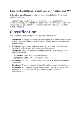
- 1. Important radiological classification of fracture and AVN Schatzker classification system is one method of classifying tibial plateau fractures. Increase in type number denotes increasing severity, reflecting an increase in energy imparted to the bone at the time of injury and also an increasingly worse prognosis 1. The most common fracture of the tibial plateau is type II. Classification This system divides tibial plateau fractures into six types: Schatzker I: wedge-shaped pure cleavage fracture of the lateral tibial plateau, originally defined as having less than 4 mm of depression or displacement Schatzker II: splitting and depression of the lateral tibial plateau; namely, type I fracture with a depressed component Schatzker III: pure depression of the lateral tibial plateau; divided into two subtypes: o Schatzker IIIa: with lateral depression o Schatzker IIIb: with central depression Schatzker IV: medial tibial plateau fracture with a split or depressed component Schatzker V: wedge fracture of both lateral and medial tibial plateau Schatzker VI: transverse tibial metadiaphyseal fracture, along with any type of tibial plateau fracture (metaphyseal-diaphyseal discontinuity)
- 2. Calcaneal fracture Calcaneal fractures are the most common tarsal fracture and can occur in a variety of settings. Epidemiology The calcaneus is the most commonly fractured tarsal bone and accounts for about 2% of all fractures and ~60% of all tarsal fractures 3. Pathology Calcaneal fractures can be divided broadly into two types depending on whether there is articular involvement of the subtalar joint 1. extra-articular: 25-30% o anterior calcaneal process fracture o calcaneal tuberosity avulsion fracture o extra-articular body fracture
- 3. lover's fracture o medial sustentaculum 2. intra-articular: 70-75% o intra-articular body fracture The calcaneus is also a common site of stress fractures, occurring in the posterosuperior aspect. Another method of classification is as type A fractures: the anterior process of the calcaneus is fractured type B: fracture of the mid calcaneus, trochlear process, and sustentaculum tali type C: fracture of the posterior tuberosity Radiographic features Plain radiograph Böhler's angle is the angle between two tangent lines drawn across the anterior and posterior borders of the calcaneus in the lateral view. When Böhler's angle becomes <20º it indicates a calcaneal fracture. On a lateral radiograph, an angle of Gissane of >130° suggests depression of the posterior facet of the subtalar joint. normal Böhler's angle does not exclude a fracture and is unreliable in children 9 calcaneal stress fracture shows vertical linear sclerotic appearance 9 CT CT is the modality of choice to evaluate calcaneal fracture. It can show the extent and extra- or intra-articular components of the fracture and haematoma along the sole of the foot (Mondor sign). Intra-articular fractures are often classified using the Sander classification system, which is one of the only systems that correlate well with patient outcome. Practical points
- 4. If bilateral calcaneal fractures are seen, then the spine should also be evaluated for fracture as the mechanism of injury is often a large load to the axial skeleton, such as jumping from a second-story window. If an intraarticular calcaneal fracture is seen, the images should be scrutinized for a lateral malleolar fleck sign (ankle), which raises the likelihood of peroneal tendon instability
- 5. type 1: includes all intraarticular fractures that have less than 2 mm of articular displacement, regardless of the number of fracture lines/fragments present type 2a: involves one primary fracture line that courses through the lateral aspect of the posterior facet; the primary fracture usually assumes a "y" shaped configuration as it exits medially and laterally out of the calcaneal body; this fracture is often accompanied by one or more accessory fracture lines that do not involve the posterior articular facet type 2b: involves one primary fracture line that courses through the central aspect of the posterior facet; the primary fracture usually assumes a "y" shaped configuration as it exits medially and laterally out of the calcaneal body; this fracture is often accompanied by one or more accessory fracture lines that do not involve the posterior articular facet
- 6. type 2c: involves one primary fracture line that courses through the medial aspect of the posterior facet and is accompanied by a transverse fracture through the body of the calcaneus; this fracture is often accompanied by one or more accessory fracture lines that do not involve the posterior articular facet type 3ab: involves two primary fracture lines, one coursing through the lateral aspect of the posterior facet and the second through the central aspect; this subtype usually presents with depression of the central fragment; the two primary fracture lines may be accompanied by additional accessory fracture lines that do not involve the posterior articular facet type 3ac: involves two primary fracture lines, one coursing through the lateral aspect of the posterior facet and the second through the medial aspect; this subtype usually presents with depression of the central fragment. The two primary fracture lines may be accompanied by additional accessory fracture lines that do not involve the posterior articular facet type 3bc: involves two primary fracture lines, one coursing through the central aspect of the posterior facet and the second through the medial aspect; this subtype usually presents with depression of the central fragment; the two primary fracture lines may be accompanied by additional accessory fracture lines that do not involve the posterior articular facet type 4: involves three or more primary fracture lines with greater than 2 mm of articular displacement, and are therefore severely comminuted Acetabular fracture Classification The Judet and Letournel system for acetabular fractures is the most widely used classification system in clinical practice. It classifies fracture based on oblique pelvic view on plain radiographs. Additional classification systems include: Orthopaedic Trauma Association classification (primarily for research) Harris system (CT imaging based) Radiographic features
- 7. Plain radiograph Initial assessment is often with a portable AP radiograph of the pelvis in the emergency department. Assess the following lines: 1. anterior acetabular wall 2. posterior acetabular wall 3. acetabular roof 4. iliopectineal line: disrupted in fractures involving the anterior column 5. ilioischial line: disrupted in fractures involving the posterior column 6. radiographic U (teardrop) After diagnosis, oblique pelvic views (Judet views) may be used for follow up. These include: 1. iliac oblique view for the posterior pelvic column and anterior acetabular wall 2. obturator oblique view for the anterior pelvic column and posterior acetabular wall CT CT has revolutionised the diagnosis, enabling precise delineation of the fracture configuration and assessment of any articular surface disruption. Many patients with high-energy trauma will have a whole body CT, allowing initial assessment of the femoroacetabular joint as well as any other injuries that are likely to be present, given the typically high energy mechanism of injury 2. For those patients with pelvic insufficiency fractures involving the acetabulum, a standard CT with a bony algorithm may be useful, especially if operative management is under consideration. A repeat CT after traction is sometimes used to assess response to treatment. Treatment and prognosis Treatment analgesia venous thromboembolism prophylaxis traction
- 8. o skin traction o skeletal traction non-operative management o may be indicated in the setting of minimally displaced fracture o more common in developing countries open reduction and internal fixation (ORIF) o articular incongruence/displaced fracture o significantly distorted acetabular roof arc o entrapped intra-articular fragment o subluxation of the femoral head
- 10. The Judet and Letournel classification is more widely used and is applicable to both CT and plain radiographs, whereas, the Harris classification is applicable to CT only . Classification The Harris classification defines acetabular fractures in four categories : category 0: anterior wall or posterior wall only category 1: single-column fractures category 2: both columns (i.e. transverse) o 2a: no extension o 2b: superior extension o 2c: inferior extension o 2d: superior and inferior extension category 3: acetabular dissociated from the axial skeleton Avascular necrosis (AVN), or more correctly osteonecrosis, is a generic term referring to the ischaemic death of the constituents of bone. AVN has a wide variety of causes and can affect nearly any bone in the body. Most sites of involvement have an eponym associated with avascular necrosis of that area, and these sites are discussed individually as each site has unique clinical, aetiologic and prognostic features. Eponymous names for specific sites of avascular necrosis Ahlback disease: medial femoral condyle, i.e. SONK Brailsford disease: head of the radius Buchman disease: iliac crest Burns disease: distal ulna Caffey disease: entire carpus or intercondylar spines of the tibia Dias disease: trochlea of the talus Dietrich disease: head of metacarpals Freiberg infraction: head of the second metatarsal Friedrich disease: medial clavicle Hass disease: humeral head Iselin disease: base of 5th metatarsal Kienböck disease: lunate
- 11. Köhler disease: patella or navicular (children) Kümmell disease: vertebral body Legg-Calvé-Perthes disease: femoral head Mandl disease: greater trochanter Mauclaire disease: metacarpal heads Milch disease: ischial apophysis Mueller-Weiss disease: navicular (adult) Panner disease: capitellum of the humerus Pierson disease: symphysis pubis Preiser disease: scaphoid Sever disease: calcaneal epiphysis Siffert-Arkin disease: distal tibia Thiemann disease: base of phalanges van Neck-Odelberg disease: ischiopubic synchondrosis Radiographic features Radiographic changes alter with the stage of AVN - Ficat staging, Steinberg classification. Radiograph In general, there is initial minor osteopenia, followed by variable density. Gradually microfractures of the subchondral bone accumulate in the dead bone, which is unable to repair leading to the collapse of the articular surface and the crescent sign of AVN. Eventually the cortex collapses and fragments, with superimposed secondary degenerative change. MRI MRI is the most sensitive (~95%) modality and demonstrates changes well before plain films changes are visible. reactive interface line: focal serpentine low signal line with fatty centre (most common appearance and first sign on MRI) double line sign: serpentine peripheral/outer dark (sclerosis) and inner bright (granulation tissue) on T2WI is diagnostic diffuse oedema: oedema is not an early sign; instead, studies show that oedema occurs in advanced stages and is directly correlated with pain rim sign: osteochondral fragmentation secondary degenerative change (i.e. osteoarthritis)
- 12. Nuclear medicine Bone scintigraphy is also quite sensitive (~85%) and is the second option after MRI. It is a choice when multiple sites of involvement must be assessed in patients with risk factors, such as sickle cell disease. The findings are different accordingly to the time of the scan: early disease: often represented by a cold area likely representing the vascular interruption late disease: may show a "doughnut sign": a cold spot with surrounding high uptake ring (surrounding hyperaemia and adjacent synovitis)
- 13. Steinbergclassification Modic type I o T1: low signal o T2: high signal o represents bone marrow oedema and inflammation o T1+C: enhancement
- 14. Modic type II o T1: high signal o T2: iso to high signal o represents normal red haemopoietic bone marrow conversion into yellow fatty marrow as a result of marrow ischaemia Modic type III o T1: low signal o T2: low signal o represents subchondral bony sclerosis Acromioclavicularinjury Acromioclavicular joint injuries are characterised by damage to the acromioclavicular joint and surrounding structures. Almost invariably traumatic in aetiology, they range in severity from a mild sprain to complete disruption. Clinical presentation Acromioclavicular joint injuries usually occur from a direct blow or following a fall onto the shoulder with an adducted arm. This pushes the acromion forcibly inferiorly and medially with respect to the clavicle 7. Radiographic features Imaging can be used to classify acromioclavicular injuries, with the Rockwood system most commonly used to classify injuries into
- 15. six types. Other described systems include the Tossy and the Allman classification systems. In most cases, plain films (including an axillary view) are sufficient for accurate grading although CT or MRI may be useful in cases where plain films are thought to underrepresent the degree of injury. Plain radiograph Standard acromioclavicular joint radiographs consist of a clavicle series including an AP and cephalic angled oblique (10-15º) views. Additional weight-bearing stress views may be of benefit if: initial radiographs are normal, but an injury is suspected surgical intervention on a type III injury would be contemplated (see below) These are performed with the patient erect and holding a weight in the arm. If the joint is normal, then acromioclavicular alignment should remain normal and symmetric. Features of acromioclavicular joint injury include 6: soft tissue swelling/stranding o may be the only finding in type I injuries widening of the acromioclavicular joint o normal: 5-8 mm (narrower in the elderly) o greater than 2-4 mm asymmetry (compared to radiographs of the contralateral side) increased coracoclavicular distance o normal: 10-13 mm o greater than 5 mm asymmetry (compared to the contralateral side) superior displacement of the distal clavicle o the inferior edge of the acromion should be level with the inferior edge of the clavicle
- 16. Slipped upper femoral epiphysis Slipped upper femoral epiphysis (SUFE), also known as a slipped capital femoral epiphysis (SCFE), (plural: epiphyses) is a relatively common condition affecting the physis of the proximal femur in adolescents. It is one of the commonest hip abnormalities in adolescence and is bilateral in ~20% of cases. Epidemiology Slipped upper femoral epiphysis is more common in boys than girls and more common in Afro-Caribbeans than Caucasians. The age of presentation is somewhat dependant on gender with boys presenting later (10-17 years) than girls (8-15 years) 2. Obesity is a significant risk factor.
- 17. Clinical presentation Patients present with hip pain progressing to a limp and even leg length discrepancy. If slipped upper femoral epiphysis is confirmed on imaging, the child should be rested, admitted, and not allowed to weight-bear. Pathology Slipped upper femoral epiphysis is a type I Salter-Harris growth plate injury due to repeated trauma on a background of mechanical and probably hormonal predisposing factors. Radiographic features In all situations, especially when imaging children, the fewest number of radiographs, with the smallest exposed area is performed. Gonad protection is usually used in pelvic x-rays of children. However, there should always be one radiograph without the lead protection so that the entire pelvis is visualised. Plain radiograph The radiographs that are used will depend on the institution where you work: AP and frog-leg: the commonest situation allows assessment of two views AP only: a perceived concern about a risk of worsening a slip means that a frog-leg lateral is only performed if the orthopaedic surgeon or radiologist agrees frog-leg only: to reduce the dose, only frog-leg lateral is performed because it is this view that is most sensitive and the AP rarely adds diagnostic information The slip that occurs is posterior and, to a lesser extent, medial. It is therefore is more easily seen on the frog-leg lateral view rather than the AP hip view. Because the epiphysis moves posteriorly, it appears smaller because of projectional factors. On the AP, a line drawn up the lateral edge of the femoral neck (line of Klein) fails to intersect the epiphysis during the acute phase (Trethowan sign). The metaphysis is displaced laterally and therefore may not overlap posterior lip of the acetabulum as it should normally (loss of triangular sign of Capener) .
- 18. The metaphyseal blanch sign, a sign seen on AP views, involves increases in the density of the proximal metaphysis. It represents the superposition of the femoral neck and the posteriorly displaced capital epiphysis. Alignment of the epiphysis with respect to the femoral metaphysis can be used to grade the degree of slippage:. MRI In the acute stage, marrow oedema results in an increased signal on T2- weighted sequences, e.g. STIR. Marrow oedema is non-specific, and while it may indicate early bone changes in SUFE, there are numerous other causes, e.g. infection or a tumour. MRI can be used to examine the contralateral hip which is important because of the high incidence of bilateral slip. STIR o high signal in epiphysis and metaphysis o joint effusion T1 o low signal in oedematous regions o metaphyseal displacement n an AP radiograph a line along the superior margin of the femoral neck (line of Klein) should intersect the lateral corner of the epiphysis. As the epiphysis slips, the metaphysis can be divided into thirds. mild: lateral edge of epiphysis is within the lateral third of the metaphysis
- 19. moderate: middle third severe: medial third On a true lateral radiograph, the angle (slip angle) which the epiphysis makes with the metaphysis may also be employed (sometimes known as the Southwick head shaft angle). normal: ~0 degrees mild: 0-30 degrees moderate: 30-60 degrees severe: >60 degrees Perthes disease Perthes disease, also known as Legg-Calvé-Perthes disease, refers to idiopathic osteonecrosis of the femoral epiphysis seen in children. It should not be confused with Perthes lesion of the shoulder. It is a diagnosis of exclusion and other causes of osteonecrosis (including sickle cell disease, leukaemia, corticosteroid administration, Gaucher disease) must be ruled out . Epidemiology Perthes disease is relatively uncommon and in Western populations has an incidence approaching 5 to 15:100,000. Boys are five times more likely to be affected than girls. Presentation is typically at a younger age than slipped upper femoral epiphysis (SUFE) with peak presentation at 5-6 years, but confidence intervals are as wide as 2-14 years . Perthes is considered an idiopathic condition, and there are no clear predisposing factors. Clinical presentation Most children present with atraumatic hip pain or limp. Some children have a coincidental history of trauma. This may precipitate the presentation or the realisation of symptoms that in fact had been long-standing.
- 20. Blood tests are typically normal in Perthes. It is important to be certain that there is no other cause of osteonecrosis (e.g. sickle cell disease) during the workup. Pathology The specific cause of osteonecrosis in Perthes disease is unclear. Osteonecrosis generally occurs secondary to the abnormal or damaged blood supply to the femoral epiphysis, leading to fragmentation, bone loss, and eventual structural collapse of the femoral head. In approximately 15% of cases, osteonecrosis occurs bilaterally. Radiographic features The best initial test for the diagnosis of Perthes is a pelvic radiograph. In a small number of patients with Perthes, the radiograph will be normal and persistent symptoms will trigger further imaging, e.g. MRI. The investigation of atraumatic limp will often include a hip ultrasound to look for effusion, but ultrasound is unlikely to pick up osteonecrosis. The radiographic findings are those of osteonecrosis. There are separate systems for staging of Perthes disease: temporal evolution o Waldenström classification o modified Elizabethtown classification severity and prognosis o Catterall classification: extent of femoral head involvement o Salter-Thompson classification: extent of femoral head involvement o Herring classification: lateral pillar involvement healed stage deformity: osteoarthritis risk o Stulberg classification
- 21. Plain radiograph The radiographic changes to the femoral epiphyses depend on the severity of osteonecrosis and the amount of time that there has been an alteration of blood supply: early: there may be no appreciable change established: reduction in epiphysis size, lucency late: fragmentation, destruction As changes progress, the width of the femoral neck increases (coxa magna) in order to increase weight-bearing support. Early signs joint effusion: widening of the medial joint space asymmetrical femoral epiphyseal size (smaller on the affected side) apparent increased density of the femoral head epiphysis blurring of the physeal plate (stage 1) radiolucency of the proximal metaphysis Late signs Eventually, the femoral head begins to fragment (stage 2), with subchondral lucency (crescent sign) and redistribution of weight- bearing stresses leading to thickening of some trabeculae which become more prominent. The typical findings of advanced burnt out (stage 4) Perthes disease are: femoral head deformity with widening and flattening (coxa plana) proximal femoral neck deformity: coxa magna "sagging rope sign" (thin sclerotic line running across the femoral neck) Additionally, tongues of cartilage sometimes extend inferolaterally into the femoral neck, creating lucencies, which must be distinguished from infection or neoplastic lesions 4. The presence of metaphyseal involvement not only increases the likelihood of femoral neck deformity but also make early physeal closure with resulting leg length disparity more likely.
- 22. Arthrography Traditionally arthrography performed under general anaesthesia with conventional fluoroscopy is performed to assess congruence between the femoral head and the acetabulum in a variety of positions . MRI is increasingly replacing this, in an effort to eliminate pelvic irradiation. MRI MRI is gaining an increasing role in a number of scenarios: early diagnosis, before the onset of x-ray findings assessing the extent of cartilaginous involvement, important in prognosis assessing joint congruence in a variety of joint positions (requires open magnet and dynamic imaging) 2 Both arthrography and dynamic MRI assess three main features : deformity of the femoral head (also assessed on static x-rays and MRI) congruence: how well the femoral head contour matches that of the acetabulum containment: the amount of lateral subluxation of the flattened femoral head out of the acetabulum o when severe this may lead to hinge abduction, whereby rather than rotation and medial movement of the femoral head during hip abduction, the flattened head 'hinges' on the lateral lip of the acetabulum, widening the medial joint space 2,3 here are several classification systems for sacral fractures, but the most commonly employed are the Denis classification and subclassification systems, and the Isler classification system. These classification systems are important to understand as proper classification can impact management. AO classification of sacral injuries type A: lower sacrococcygeal injuries
- 23. type B: posterior pelvic injuries type C: spinopelvic injuries The AO sacral injury classification further subdivides these three injury types and also involves neurological and other modifiers that can be added. Denis classification zone 1: fracture involves the sacral ala lateral to the neural foramina zone 2: fracture involves the neural foramina, but does not involve the spinal canal zone 3: fracture is medial to the neural foramen, involving the spinal canal; these may be transverse or longitudinal, and can be sub- classified into 4 types: o type 1: only kyphotic angulation at the fracture site (no translation) o type 2: kyphotic angulation with anterior translation of the distal sacrum o type 3: kyphotic angulation with complete offset of the fracture fragments o type 4: comminuted S1 segment, usually due to axial compression Morphologic injury patterns of zone 3 fractures “H” shaped fracture “U” shaped fracture “ʎ” (lambda) shaped fracture “T” shaped fracture Isler classification Used for fractures that involve the lumbosacral articulation: Isler 1: fracture occurs lateral to the L5/S1 facet Isler 2: fractures line involves the L5/S1 facet Isler 3: fracture line extends medially to the L5/S1 facet