SlideShare una empresa de Scribd logo
1 de 152
Avascular Necrosis of Femoral Head 1   Dr Zameer Ali  St Stephen’s hospital
Definition 2 Osteonecrosis of femoral head refers to the death of osteocytes with subsequent structural changes leading to femoral head collapse and secondary osteoarthritis of hip joint
Definition. 3    Osteonecrosis is defined as an “end stage” condition of the femoral head in which there is  necrosis of the bone, secondary to disruption of the blood supply and causes which are still unknown.
4     The death of cell components of bone & bone marrow from repeated interruptions or a single massive interruption of the blood supply to the bone.
5 AVN may involve all or part of head
6
The Problem. Commonly affects young patients. Need to alter the life style and leisure activity. Accounts for 18 % of all  total hip replacements.  7
8 75% of cases are between 30 to 70 years of age Male: female ratio 4:1 Bilateral involvement is seen in 50 % of cases
History 9 Freund in 1926 gave detailed description of Osteonecrosis  Chandler in 1948 termed disorder as "coronary disease of hip” which accounts for eponym Chandlers disease
. 10 Chandler in 1948 referred this condition as coronary artery disease of hip which even after 50 years correctly describes this condition  Aseptic necrosis was initially used  to distinguish this condition from infections
11 3)Glimser and Kenzora in 1979 analyzed the radiographic changes which accompany AVN Ficat in 1985 states that this condition resulted from blockage of the osseus microcirculation with intramedullary stasis and increased pressure .
Anatomy 12 Head of femur forms about 2/3rd  of sphere and articulates with acetabulum of hip joint  Connecting two trochanters anteriorly forms intertrochanteric line and connecting two trochanters posteriorly forms intertrochanteric crest.
13 ,[object Object]
Ball
Femoral neck
Smooth weight-bearing surfaces
Smooth cartilage
Femur,[object Object]
Blood supply of the Head. 3 main sources.          Ascending       cervical branches          Metaphyseal          blood vessels              Artery of       ligamentum teres. 15
Vascular supply of femoral head  16 Crook described Extra capsular arterial ring located at base of femoral neck  Ascending cervical branches or retinacular arteries run on surface of femoral neck  Arteries of round ligament or ligament teres.
17
18 The proximity of retinacular arteries to bone put them at risk of injury in any fracture of femoral neck. The ascending cervical arteries puts them at risk for injury in any fracture of femoral neck
19 4 groups of ascending cervical arteries 1. Anterior 2. Posterior  3. Medial  4. Lateral  Of these lateral group provides most of blood supply to femoral head and neck
20 The adult pattern of femoral head vascularity usually becomes established with closure of growth plate at approximately 18 years of age
In neonate 21 Three groups of vessels are identified   superior retinacular group or lateral epiphyseal group  inferior metaphyseal group   foveal or medial epiphyseal group All 3 groups anastomose with each other
Between 4 years to  7 years 22 The importance of lateral epiphyseal is established  as metaphyseal and foveal vessels decrease in extent
After 8 years of age  23 Open growth plate represents an effective barrier preventing anastomoses between vessels of head and neck. An increased contribution of foveal vessels
24 During adolescence increased number of inferior metaphyseal vessel is recognized As the growth plate closes the adult pattern appears with anastomosis between 3 arterial systems
Causes. 25 Traumatic causes Non Traumatic causes
Traumatic 26 Fractures of femoral neck. 15% - 50% Dislocation of the hip. 10% - 25%
27 Currently accepted term is Osteonecrosis  which describes main common feature of this condition THE BONE DEATH
Non Traumatic 28 Idiopathic. Chronic alcoholism. Steroid intake. Sickle cell disease. Gauchers disease. Caissons disease. Malignancies. Drug induced
Risk factors 29 Gauchers disease Radiation induced  Liver disease abnormal coagulation factor synthesis
Risk factors 30 Traumatic Osteonecrosis usually involves dislocation of hip or fracture femoral neck . 52% of hips unreduced for more than 12 hours developed AVN 22 % of hips developed AVN  if reduced within 12 hours Femoral neck fractures associated with 15 – 50 % incidence of AVN
Risk factors 31 Alcoholism: Accounts for 10 – 40 % of incidence of AVN. Risk of development of AVN is increased with cumulative doses of alcohol Drug induced: the association between corticosteroid therapy,cushings syndrome, and Osteonecrosis is well established
Risk factors 32 Collagen diseases rheumatoid arthritis,SLE have been associated with AVN Radiation causes obliterative endarteritis and cellular death. Gout sodium urate crystals enhance clotting by activating haegmen factor in intrinsic coagulation system
Pathogenesis. 33 Other than traumatic causes, the mechanism of necrosis is still “obscure”. Coaugulation defect. Genetic predisposition. Emboli formation. “ IDIOPATHIC ”
Pathogenesis 34 The bony compartment function essentially as closed compartment within which one element can expand only at expense of others A unifying concept of pathogenesis of AVN emphasizes the central role of vascular occlusion and ischaemia leading to osteocyte necrosis
35 Pathogenesis of steroid or alcohol induced AVN is not well understood ,but it is suggested that embolic fat  and attendant thrombi occlude microcirculation. Lipocyte hypertrophy and subchondral lipid accumulation may cause  extra vascular intraosseus compression.
36 It matters little whether the initiating factor was capillary occlusion (as in sickle cell disease) venous occlusion (as suggested for perthes disease) or intramedullary tamponade  in Gauchers disease, the end result is diffuse ischaemia involving all the elements
Biological sequence of repair 37
Biological sequence of repair 38  loss of cell viability (cell necrosis) Invasion of marrow spaces of dead bone by proliferating capillaries and cells Differentiation of mesenchymal cells to osteoblast synthesis of new bone. Early remodelling of repaired cancellous bone
Biological sequence of repair 39 Late internal remodeling Resorption of subchondral bone and invasion of articular cartilage
Biological sequence of repair 40 To conclude this reparative process is self limiting and incompletely replaces dead bone with living bone.  In subchondral bone ,bone formation  occurs at slower rate than does resorption resulting in net removal of bone,  loss of structural integerity, subchondral fractures and collapse
Clinical features. Pain.     - Dull boring .     - Progressive.     - Worse at night      -Limp while walking.     - Restricted hip             motion.     - Unable to sit cross               legged. 41
symptoms 42 Clinical manifestation of bone ischaemia and infarction are minimal and non specific  and depend on etiology.
symptoms 43 Initially vague and non specific. Localized or referred  pain in buttocks,thighor knee Gradual increase in intensity of pain and decreased motion especially rotation and abduction Over several years results in limping gait
symptoms 44 Initial pain is commonly misinterpreted as radiating pain from lumbar spine.
Diagnosis 45 Asymptomatic during the early stages. Become symptomatic when significant collapse of the head has occurred.
Management protocol 46 Early diagnosis Radiological evaluation Rule out other causes MRI Quantification Treatment algorithm
Diagnosis 47 Imaging  Routine radiographs Scintigraphy CT scan. MRI. LASER Doppler flometry.
Early Diagnosis – suspicion ? 48 High degree of suspicion in a patient C/o anterior HIP pain, 	Especially with:- 	H/o Cortisone – Skin, Eye, Liver, Asthma, 	RA, Weight gain 	H/o Alcohol abuse 	Traumatic - # N/F, D/ of F, # Acetabulum Hemoglobinopathy – Sickle / Myelo-infiltrating
Other causes 49 Pregnancy Renal Diseases Radiation Gout / Collagen disorder Gaucher’disease Dysbarism  Idiopathic
Imaging 50 Routine radiographs are usually first step in trying to make diagnosis. High quality films taken at least two views 90 degree apart are critical to initial evaluation
Radiology- sequential Changes 51 Crescent Sign Osteoporosis Sclerosis Cystic changes Loss of spherical bearing dome  Partial collapse of head Secondary Osteoarthritis
Xrays. 52 ,[object Object],Early stages : normal film. Subsequently there occurs increased           “ DENSITY “ of the femoral head. Crescent sign. Femoral head collapse. Osteoarthritis of the hip. B/l involvement of femoral head with cystic changes/sclerosis seen
X RAY changes 53 Sclerosis /subchondral cysts
X RAY changes 54 Crescent changes  Flattening of head of femur
Bilateral Cystic changes    With patchy sclerosis 55
Scintigraphy  56 Radionuclide scintigraphy is more sensitive for osteonecrosis than standard radiographs and will reveal changes when standardd radiographs are normal
scintigram 57 The hall mark of vascularity is photopenic effect on scintigram                                RESULTS Decreased uptake by necrotic bone  Increased uptake by remodeling bone Normal uptake by normal bone
Bone scan. Technetium 99 bone scan reveals decreased uptake. It is effective only if done in early stages. During late phase there are very variable resuts. No relationship b/w scan appearance and the function of the hip. 58
MRI 59 MRI is most sensitive technique  for early diagnosis in Osteonecrosis   Can diagnose AVN as early as 48 hours  The classical finding of AVN is decrease in the normally high intensity  signal of marrow of femoral head
MRI. 60 Can detect early stages. Allows to determine exact stage of disease. Tells exactly the extent of damage. Useful in determining the efficacy of treatment.
MRI Geographical area of decreased marrow signal. Necrotic area Surrounded by zone of “low signal” band. Ischaemic bone. 61
MRI Findings. 62
63
MRI - Findings 64 Bone Marrow edema Double Line – Head in Head sign  Crescent sign Collapse Joint effusion Involvement of actabulum Status of other hip Marrow infiltrating disease
LASER Doppler Flometry 65 Laser Doppler flometry is technique at measuring blood cell influx in a capillary bed  The magnitude and  frequency of Doppler shift is proportionate to the velocity and concentration of red cells under probe head
Sequence of radiological  events  in AVN 66 Fragmentation : radiolucent clefts may be seen due to necrosis of involved bone  The entire epiphysis may be absent Mottled trabecular : pattern: scrutiny of trabecular traversing the ischaemic bone demonstrates thickened irregular pattern
67 Sclerosis : with revascularisation new bone is deposited around dead bone resulting in increased bone  density  Subchondral cysts : patchy well circumscribed rarefactions immediately beneath the articular cortex are frequent
68 These cysts are usually seen in region of greatest articular stress and are identical to those found in degenerative joint disease Collapse of articular cortex this generally occurs at the region of maximal stress of involved cortex and represents a localised impaction fracture of weakened bone
Fragmentation 69 Radiolucent clefts seen traversing bone
Sclerosis/subchondral cysts 70 Sclerosis   subchondral cysts
Collapse of articular surface 71 Collapse of articular  surface
72
Staging 73 Several staging systems have been described. The staging system reported by sternbergs and colleagues is similar to that outlined by  Marcus,Ficat and Arlet Sternbergs classification allows physician to quantify extent of involvement of femoral head in both early and late stages
Staging / Grading --- too many 74 Ficat				Radiological Steinberg			Quantification Enneking's         		Stages of Osteonecrosis Marcus and Enneking 	System Japanese criteria		Location Sugioka				Radiological University Of Pennsylvania System Association Research Classification Osseous Committee (ARCO)--	Combination
Fi Stages of Bone Necrosis 75 Ficats radiological staging of osteonecrosis of femoral head
76 Ficat Stages of Bone Necrosis Stage 		Clinical Features 	Radiographs 0 Preclinical		          0			 0 1Preradiographic	          +  2     mild		density changes in femoral head 2aPrecollapsemild		 Diffuse Porosis, Sclerosis,or                      							cyst 2bTransition: 		          		 Flattening, Crescent Sign  3  Collapse	mild/moder		 Broken Contour of Head 					            Certain Sequestrum, 						Joint Space Normal  4 Osteoarthritismod/severe	 	Flattened Contour     							Decreased Joint Space				                                     Collapse of Head
Stage 1 77 Symptoms – none / mild Xrays are normal. Bone scan reveals a “cold spot”.
Stage 2 2A Symptoms are mild. Xray shows increased density.      subchondral cysts       joint line maintained.       normal head contour.     Bone scan reveals increased uptake. 78
2B Crescent sign. Flattening of the head. 79
Crescent sign. 80
Stage 3 Moderate symptoms. Loss of shape Subchondral collapse 81
Stage 4 Severe symptoms. Joint space narrowing. OA changes in acetabulum. 82
Marcus  radiological staging of AVN 83 Stage I    normal or equivocal radiograph Stage II  sclerotic or cystic lesion Stage III  crescent sign Stage IV step off in outline of bone  Stage V  narrowing of joint space with       			degenerative changes
Shimizu’s classification.1995. 84                          Grade 1 lesion. A lesion involving medial 1/3 rd of weight bearing surface of the head. In coronal plane ,these lesions occupy < 1/3 of the head. These lesions rarely go into collapse.
Grade 2. 85 Lesions involve 1/3 – 2/3 of the weight bearing surface of the head. Involve ½ of the head in coronal plane. Such lesions collapse in 30% of patients.
Grade 3. 86 Lesions involving 2/3 of the weight bearing surface of the femoral head. Such lesions collapse in 70% of patients in around 3 years.
Treatment 87 Conservative /non surgical treatment Core decompression Bone grafting Cancellous autogenic/allogenic bone graft Osteochondral graft Muscle pedicle bone graft Free vascularized graft
Treatment 88 Electric stimulation Osteotomy Joint reconstruction  Cup arthoplasty Surface arthoplasty Hemiarthoplasty Total joint arthoplasty
89 The natural history of osteonecrosis in its early stage, before subchondral collapse, is still unclear, but evidence suggests that the rate of progression is high, especially in symptomatic patients.
90 Once subchondral collapse occurs and joint space is lost, progressive osteoarthritis generally is considered inevitable.
91  Many studies have reported an extremely poor prognosis, with a rate of femoral head collapse of more than 85% at 2 years in symptomatic patients (stage I or II disease)
92 No treatment method has proved to be completely effective in arresting the disease process before subchondral collapse or in slowing the progression of femoral head destruction and osteoarthritis after subchondral collapse
93     The rate and course of progression of the disease are unpredictable, and the radiographic picture may not correlate with the clinical symptoms; some patients maintain tolerable function for an extended period after femoral head collapse.
94 Conservative treatment, such as crutch ambulation or bed rest, generally is ineffective. However, symptomatic patients that may benefit from a head-preserving technique should be placed on crutches until surgical treatment is carried out to prevent collapse in the interim
Management. 95 Grade 1 lesions        - Continuous supervision  to detect any                   changes.        - Symptomatic treatment of pain.
Grade 1 lesions 96 Conservative treatment such as observation , Analgesics Limited weight bearing may be successful in minimal affected cases
Core decompression Grade 2 lesions 97 Ficart and Hungerford have popularized the technique of core decompression of femoral head Rationale is that removing necrotic bone decompresses the rigid osseous  chamber, thereby improving blood flow and preventing additional ischaemic events
98 The theoretical advantage of core decompression is based on the belief that the procedure relieves intraosseous pressure caused by venous congestion, thereby allowing improved vascularity and possibly slowing the progression of the disease
99 several authors noted that the results of core decompression are better than those of nonoperative treatment.
100 Several reports noted that the earlier the stage of the disease, the better the results with core decompression. . For more advanced Ficat stages (IIB or III) the results of core decompression are much less predictable, so alternative treatment methods should be explored.
101 Review of the literature currently supports the use of core decompression for the treatment of Ficat stages I and IIA small central lesions in young, nonobese patients who are not taking steroids. This surgery is relatively simple to perform and has a very low complication rate
102  The surgical field for subsequent total hip arthroplasty, if needed, is not substantially altered
Bone grafting  103 Phemister introduced concept of using cortical strut graft in core decompression channel The accurate placement of graft within lesion and under subchondral bone is important
104  Structural bone grafting techniques after core decompression have been described using cortical bone, cancellous bone, vascularized bone graft, and debridement of necrotic bone from the femoral head.
105 Insertion of cancellous bone into channel speeds up reossification by osteoinductive and osteoconductive  properties of bone graft. Meyers procedure  used muscle pedicle bone graft  based on quardratus femoris muscle with cancellous bone chips
106 Baksi employed multiple drilling and muscle pedicle  grafting using tensor fascia lata muscle anteriorly
107  Advances in microsurgical techniques made it possible to preserve the intrinsic vascularity of bone graft, several authors independently proposed implanting a vascularised bone graft into the core of the femoral head. 
Vascular fibular strut grafting 108
109  The rationale for vascularized bone grafting is based on four aspects of the operation and postoperative care: (1) decompression of the femoral head, which may interrupt the cycle of ischemia and intraosseous hypertension that is believed to contribute to the disease;  (2) excision of the sequestrum, which might inhibit revascularization of the femoral head
110 (3) filling of the defect that is created with osteoinductive cancellous graft and a viable cortical strut to support the subchondral surface and to enhance the revascularization process  (4) protection of the healing construct by a period of limited weight-bearing. 
Advantages 111 Advantages of free vascularized fibular grafting compared with total hip arthroplasty:  (1) the presence of a healed femoral head may allow more activity,  (2) there is no increased risk associated with the presence of a foreign body,
112 (3) if performed before the development of a subchondral fracture, the procedure offers the possibility of survival of a viable femoral head for the life of the patient, and  (4) if total hip arthroplasty is ultimately needed, it is much simpler to perform than is a revision arthroplasty after a failed total hip arthroplasty.
Disadvantages 113 Disadvantages include a longer recovery period and less uniform and less complete relief of pain than after total hip arthroplasty.
Strut Fibular Grafting 114 ,[object Object]
Removal of Necrotic Bone
Grafting of defect with cancellous graft
Viable cortical Bone strut to support subchondral bone.
Age 20 – 50, stage 2,[object Object]
Grade 2. If detected before the collapse    Core decompression and fibular grafting.     Realignment osteotomies. 116
Fibular strut grafting  117
118
Pre OP Post OP 119
Electrical stimulation 120 Electrical stimulation has been advocated for AVN  because its histological appearance is similar to that of non union Currently used in combination with head  salvage procedures
osteotomies 121  Various proximal femoral osteotomies have been developed for the treatment of osteonecrosis with the intent to move the involved necrotic segment of the femoral head from the principal weight-bearing area.  These procedures have achieved best results for small- or medium-sized lesions (less than 30% femoral head involvement) in young patients in whom it is optimal to delay a total hip arthroplasty.
osteotomies 122 Various osteotomies have been described  Varus osteotomy Valgus  derotation osteotomy  Rotation osteotomy aid the loss of structural integrity and collapse by redirecting the forces on femoral head
123 Intertrochanteric osteotomy may be considered for the treatment of stage II or III osteonecrosis of the femoral head in which less than 30% of the femoral head is involved.  Plain films and MRI can establish the extent of femoral head involvement and can determine if a satisfactory area of live bone is present under unaffected cartilage in the femoral head and whether this area can be rotated into a position of weight-bearing.
124 Valgus flexion osteotomy  is described by wagner when lesion is anterolateral  and total angle of necrosis is 200 degree and patient is  young and active If necrotic lesion is central varus extension osteotomy is recommended
Transtrochanteric Rotational Osteotomy 125 In 1978 Sugioka described a transtrochanteric rotational osteotomy of the femoral head for idiopathic osteonecrosis
126 The rationale of the procedure is to reposition the necrotic anterosuperior part of the femoral head to a non-weight-bearing locale.  The femoral head and neck segment is rotated anteriorly around its longitudinal axis so that the weight-bearing force is transmitted to what was previously the posterior articular surface of the femoral head, which is not involved in the ischemic process 
127 Sugioka emphasized the need for a preoperative lateral roentgenogram of the femoral head while the patient is supine and the hip is flexed exactly 90 degrees, abducted 45 degrees, and in neutral rotation. The intact area of the posterior part of the femoral head on this lateral view should be greater than one third of the total articular surface of the head to ensure the best result after his osteotomy
Post op regimen 128 Skin traction of 2 kg is applied continuously for the first week and for an additional 2 weeks at night only. As soon as pain tolerance allows, quadriceps setting is begun. Active range-of-motion exercises of the hip are begun at 10 to 14 days.
129 Walking exercises in a pool generally are allowed at 5 to 6 weeks.  Partial weight-bearing with crutches is begun at 8 weeks, and the use of crutches is recommended for 6 months after surgery.  If the necrotic area of the femoral head is extensive or if involvement is bilateral, crutch use is encouraged for up to 1 year postoperatively.
Replacement - options 130 Hemiarthroplasty Bipolar arthroplasty Surface replacement arthroplasty. Newer material for THR ceramic on ceramic Non cemented / cemented THR
Birmingham Surface replacement 131 Surface replacement has some advantages over THR because it preserves femoral head and neck and allows future THR if necessary Effective in cases when femoral head is not involved entirely
The BHR*System Healthy hip Cuts Implant components Implanted
Overview Named for Birmingham, England, where the device’s creators practice medicine Used globally since 1997; More than 65,000 implanted In an international study of 1,626 hips, 99.5% of patients were “Pleased” or “Extremely Pleased” with the results of the BIRMINGHAM HIP Resurfacing (BHR) System.
134 Many failures of resurfacing hemiarthroplasty have been attributed to acetabular cartilage wear. Attention should be given to the quality of the acetabular cartilage on preoperative roentgenographic studies. Intraoperative assessment of the acetabular cartilage is mandatory before implanting a resurfacing hemiarthroplasty prosthesis. If the quality of the acetabular cartilage is in question, a total hip arthroplasty should be performed.
135 Resurfacing hemiarthroplasty is an attractive alternative for young patients with advanced osteonecrosis because very little bone is sacrificed. Should failure occur, conversion to total joint arthroplasty is nearly as simple as primary total hip surgery.  Clearly, the results of primary total hip arthroplasty for osteonecrosis are better than resurfacing hemiarthroplasty. However, this procedure can delay total joint arthroplasty and buy valuable time in a young patient.
Head size Closely matches the size of natural femoral head Larger than the head of a total hip replacement Larger head means a reduced chance of dislocation after surgery—a leading cause of revision surgery 1-3% of total hips dislocate over the lifetime of the implant 0.3% of BHR* implants dislocated in the first 5 years after surgery (in a study of 2,385 hips) Healthy head BHR head Total hip head
Who is the typical candidate for BHR*System? Adults under age 60 for whom total hip replacement may not be appropriate due to an increased level of physical activity Active adults over age 60 may be candidates, depending on their bone quality
Conventional hip replacement Healthy hip Cuts Implant components Implanted
The key benefits Head size Advanced bearing surface Bone conservation
Hip with osteoarthritis Bone cuts fora traditionalhip replacement Bone conservation Preserves your natural femoral neck Neck length and angle determine accurate leg length With the BHR*System, you retain  original equipment; with a total hip, your femoral neck is replaced by the implant Bone cuts forBHR System
“Minimally Invasive.” Soft Tissue No. Incision length of 6 to 8 inches Bone Yes. Preserves your body’s natural bone structure; It resurfaces rather than replaces Conserved bone
Conventional vs. the BHR*System Total hip cuts BHR System cuts
Bone conservation (cont.) Revises to a primary If you need “revision” surgery, you don’t get a revision implant The follow-up procedure would be the same total hip replacement you would otherwise have received
Total Hip Arthroplasty and Bipolar Hemiarthroplasty.  144 Most series that have examined both unipolar and bipolar hemiarthroplasty for the treatment of osteonecrosis have reported uniformly poor results.
THR 145 Patient aged 50 & more  Advance osteoarthritis and reduction of joint space. Radiation necrosis  Result less than Ideal. – necrotic bone Poor in Sickle cell disease. Cementless are superior over cemented THR

Más contenido relacionado

La actualidad más candente

Avascular necrosis of hip
Avascular necrosis of hipAvascular necrosis of hip
Avascular necrosis of hip
vinod naneria
 
Avascular Necrosis of the Femoral Head
Avascular Necrosis of the Femoral HeadAvascular Necrosis of the Femoral Head
Avascular Necrosis of the Femoral Head
Qazi Manaan
 

La actualidad más candente (20)

CORA (center of rotation of angulation)
CORA (center of rotation of angulation)CORA (center of rotation of angulation)
CORA (center of rotation of angulation)
 
AVASCULAR NECROSIS OF HIP (AVN HIP)
AVASCULAR NECROSIS OF HIP (AVN HIP)AVASCULAR NECROSIS OF HIP (AVN HIP)
AVASCULAR NECROSIS OF HIP (AVN HIP)
 
Anterior shoulder instability st4-6
Anterior shoulder instability   st4-6Anterior shoulder instability   st4-6
Anterior shoulder instability st4-6
 
Capitellum fractures
Capitellum fracturesCapitellum fractures
Capitellum fractures
 
Avascular necrosis hip
Avascular necrosis hipAvascular necrosis hip
Avascular necrosis hip
 
Congenital pseudoarthrosis tibia
Congenital pseudoarthrosis tibiaCongenital pseudoarthrosis tibia
Congenital pseudoarthrosis tibia
 
Avascular Necrosis
Avascular NecrosisAvascular Necrosis
Avascular Necrosis
 
Leg Calve Perthes disease
Leg Calve Perthes disease Leg Calve Perthes disease
Leg Calve Perthes disease
 
Perilunate dislocations
Perilunate dislocationsPerilunate dislocations
Perilunate dislocations
 
Operative treatment of osteoporotic spinal fractures
Operative treatment of osteoporotic spinal fracturesOperative treatment of osteoporotic spinal fractures
Operative treatment of osteoporotic spinal fractures
 
Myositis ossificans
Myositis ossificansMyositis ossificans
Myositis ossificans
 
Avn hip
Avn hipAvn hip
Avn hip
 
SCFE
SCFESCFE
SCFE
 
Avascular necrosis
Avascular necrosisAvascular necrosis
Avascular necrosis
 
Recent advances in genetic orthopaedics
Recent advances in genetic orthopaedicsRecent advances in genetic orthopaedics
Recent advances in genetic orthopaedics
 
DISH: Diffuse Idiopathic Skeletal Hyperostosis of the spine
DISH: Diffuse Idiopathic Skeletal Hyperostosis of the spineDISH: Diffuse Idiopathic Skeletal Hyperostosis of the spine
DISH: Diffuse Idiopathic Skeletal Hyperostosis of the spine
 
Avascular necrosis of hip
Avascular necrosis of hipAvascular necrosis of hip
Avascular necrosis of hip
 
Avascular Necrosis of the Femoral Head
Avascular Necrosis of the Femoral HeadAvascular Necrosis of the Femoral Head
Avascular Necrosis of the Femoral Head
 
Current Concepts in Treatment of Proximal Humerus Fractures
Current Concepts in Treatment of Proximal Humerus Fractures Current Concepts in Treatment of Proximal Humerus Fractures
Current Concepts in Treatment of Proximal Humerus Fractures
 
Lumbar interbody fusion indications techniques and complications
Lumbar interbody fusion indications techniques and complicationsLumbar interbody fusion indications techniques and complications
Lumbar interbody fusion indications techniques and complications
 

Similar a AVN

Avascular necrosis femoral head by DR RAJAT MALOT (MS,DNB, MNAMS,FELLLOWSHIP ...
Avascular necrosis femoral head by DR RAJAT MALOT (MS,DNB, MNAMS,FELLLOWSHIP ...Avascular necrosis femoral head by DR RAJAT MALOT (MS,DNB, MNAMS,FELLLOWSHIP ...
Avascular necrosis femoral head by DR RAJAT MALOT (MS,DNB, MNAMS,FELLLOWSHIP ...
chitrapandey
 
Cervical myelopathy cme
Cervical myelopathy cmeCervical myelopathy cme
Cervical myelopathy cme
group7usmkk
 
Biological options in avn
Biological options in avnBiological options in avn
Biological options in avn
Paudel Sushil
 
Pagests disease,eosinophilic granuloma,heterotopic ossification
Pagests disease,eosinophilic granuloma,heterotopic ossificationPagests disease,eosinophilic granuloma,heterotopic ossification
Pagests disease,eosinophilic granuloma,heterotopic ossification
luay hassan
 

Similar a AVN (20)

Avascular necross
Avascular necrossAvascular necross
Avascular necross
 
Avascular necross
Avascular necrossAvascular necross
Avascular necross
 
Avascular necrosis femoral head by DR RAJAT MALOT (MS,DNB, MNAMS,FELLLOWSHIP ...
Avascular necrosis femoral head by DR RAJAT MALOT (MS,DNB, MNAMS,FELLLOWSHIP ...Avascular necrosis femoral head by DR RAJAT MALOT (MS,DNB, MNAMS,FELLLOWSHIP ...
Avascular necrosis femoral head by DR RAJAT MALOT (MS,DNB, MNAMS,FELLLOWSHIP ...
 
Avascular necrosis femoral head by DR RAJAT MALOT (MS,DNB,Fellowship paediatr...
Avascular necrosis femoral head by DR RAJAT MALOT (MS,DNB,Fellowship paediatr...Avascular necrosis femoral head by DR RAJAT MALOT (MS,DNB,Fellowship paediatr...
Avascular necrosis femoral head by DR RAJAT MALOT (MS,DNB,Fellowship paediatr...
 
Case record...Cervical vascular spondylotic myelopathy
Case record...Cervical vascular spondylotic myelopathyCase record...Cervical vascular spondylotic myelopathy
Case record...Cervical vascular spondylotic myelopathy
 
Ankylos ing spondylitis
Ankylos ing spondylitisAnkylos ing spondylitis
Ankylos ing spondylitis
 
Cervical spondylosis
Cervical spondylosisCervical spondylosis
Cervical spondylosis
 
Perthes disease in children
Perthes disease in childrenPerthes disease in children
Perthes disease in children
 
avn-131221141653-phpapp02 (1).pdf
avn-131221141653-phpapp02 (1).pdfavn-131221141653-phpapp02 (1).pdf
avn-131221141653-phpapp02 (1).pdf
 
Avn
AvnAvn
Avn
 
Avascular necrosis of femur
Avascular necrosis of femurAvascular necrosis of femur
Avascular necrosis of femur
 
Final case presentation sci (kimberly walsh)
Final case presentation sci (kimberly walsh)Final case presentation sci (kimberly walsh)
Final case presentation sci (kimberly walsh)
 
Cervical myelopathy cme
Cervical myelopathy cmeCervical myelopathy cme
Cervical myelopathy cme
 
Ddd rem rai2
Ddd rem rai2Ddd rem rai2
Ddd rem rai2
 
Primary vertebral body...........
Primary vertebral body...........Primary vertebral body...........
Primary vertebral body...........
 
Biological options in avn
Biological options in avnBiological options in avn
Biological options in avn
 
Mm and pagets
Mm and pagetsMm and pagets
Mm and pagets
 
Lumbar canal stenosis
Lumbar canal stenosisLumbar canal stenosis
Lumbar canal stenosis
 
Pagests disease,eosinophilic granuloma,heterotopic ossification
Pagests disease,eosinophilic granuloma,heterotopic ossificationPagests disease,eosinophilic granuloma,heterotopic ossification
Pagests disease,eosinophilic granuloma,heterotopic ossification
 
Pott Disease
Pott DiseasePott Disease
Pott Disease
 

Último

Spellings Wk 3 English CAPS CARES Please Practise
Spellings Wk 3 English CAPS CARES Please PractiseSpellings Wk 3 English CAPS CARES Please Practise
Spellings Wk 3 English CAPS CARES Please Practise
AnaAcapella
 

Último (20)

Python Notes for mca i year students osmania university.docx
Python Notes for mca i year students osmania university.docxPython Notes for mca i year students osmania university.docx
Python Notes for mca i year students osmania university.docx
 
Google Gemini An AI Revolution in Education.pptx
Google Gemini An AI Revolution in Education.pptxGoogle Gemini An AI Revolution in Education.pptx
Google Gemini An AI Revolution in Education.pptx
 
SOC 101 Demonstration of Learning Presentation
SOC 101 Demonstration of Learning PresentationSOC 101 Demonstration of Learning Presentation
SOC 101 Demonstration of Learning Presentation
 
2024-NATIONAL-LEARNING-CAMP-AND-OTHER.pptx
2024-NATIONAL-LEARNING-CAMP-AND-OTHER.pptx2024-NATIONAL-LEARNING-CAMP-AND-OTHER.pptx
2024-NATIONAL-LEARNING-CAMP-AND-OTHER.pptx
 
Unit 3 Emotional Intelligence and Spiritual Intelligence.pdf
Unit 3 Emotional Intelligence and Spiritual Intelligence.pdfUnit 3 Emotional Intelligence and Spiritual Intelligence.pdf
Unit 3 Emotional Intelligence and Spiritual Intelligence.pdf
 
Unit-V; Pricing (Pharma Marketing Management).pptx
Unit-V; Pricing (Pharma Marketing Management).pptxUnit-V; Pricing (Pharma Marketing Management).pptx
Unit-V; Pricing (Pharma Marketing Management).pptx
 
Application orientated numerical on hev.ppt
Application orientated numerical on hev.pptApplication orientated numerical on hev.ppt
Application orientated numerical on hev.ppt
 
How to Create and Manage Wizard in Odoo 17
How to Create and Manage Wizard in Odoo 17How to Create and Manage Wizard in Odoo 17
How to Create and Manage Wizard in Odoo 17
 
How to setup Pycharm environment for Odoo 17.pptx
How to setup Pycharm environment for Odoo 17.pptxHow to setup Pycharm environment for Odoo 17.pptx
How to setup Pycharm environment for Odoo 17.pptx
 
Sociology 101 Demonstration of Learning Exhibit
Sociology 101 Demonstration of Learning ExhibitSociology 101 Demonstration of Learning Exhibit
Sociology 101 Demonstration of Learning Exhibit
 
Introduction to Nonprofit Accounting: The Basics
Introduction to Nonprofit Accounting: The BasicsIntroduction to Nonprofit Accounting: The Basics
Introduction to Nonprofit Accounting: The Basics
 
Micro-Scholarship, What it is, How can it help me.pdf
Micro-Scholarship, What it is, How can it help me.pdfMicro-Scholarship, What it is, How can it help me.pdf
Micro-Scholarship, What it is, How can it help me.pdf
 
Graduate Outcomes Presentation Slides - English
Graduate Outcomes Presentation Slides - EnglishGraduate Outcomes Presentation Slides - English
Graduate Outcomes Presentation Slides - English
 
TỔNG ÔN TẬP THI VÀO LỚP 10 MÔN TIẾNG ANH NĂM HỌC 2023 - 2024 CÓ ĐÁP ÁN (NGỮ Â...
TỔNG ÔN TẬP THI VÀO LỚP 10 MÔN TIẾNG ANH NĂM HỌC 2023 - 2024 CÓ ĐÁP ÁN (NGỮ Â...TỔNG ÔN TẬP THI VÀO LỚP 10 MÔN TIẾNG ANH NĂM HỌC 2023 - 2024 CÓ ĐÁP ÁN (NGỮ Â...
TỔNG ÔN TẬP THI VÀO LỚP 10 MÔN TIẾNG ANH NĂM HỌC 2023 - 2024 CÓ ĐÁP ÁN (NGỮ Â...
 
Sensory_Experience_and_Emotional_Resonance_in_Gabriel_Okaras_The_Piano_and_Th...
Sensory_Experience_and_Emotional_Resonance_in_Gabriel_Okaras_The_Piano_and_Th...Sensory_Experience_and_Emotional_Resonance_in_Gabriel_Okaras_The_Piano_and_Th...
Sensory_Experience_and_Emotional_Resonance_in_Gabriel_Okaras_The_Piano_and_Th...
 
Fostering Friendships - Enhancing Social Bonds in the Classroom
Fostering Friendships - Enhancing Social Bonds  in the ClassroomFostering Friendships - Enhancing Social Bonds  in the Classroom
Fostering Friendships - Enhancing Social Bonds in the Classroom
 
Kodo Millet PPT made by Ghanshyam bairwa college of Agriculture kumher bhara...
Kodo Millet  PPT made by Ghanshyam bairwa college of Agriculture kumher bhara...Kodo Millet  PPT made by Ghanshyam bairwa college of Agriculture kumher bhara...
Kodo Millet PPT made by Ghanshyam bairwa college of Agriculture kumher bhara...
 
Spellings Wk 3 English CAPS CARES Please Practise
Spellings Wk 3 English CAPS CARES Please PractiseSpellings Wk 3 English CAPS CARES Please Practise
Spellings Wk 3 English CAPS CARES Please Practise
 
Wellbeing inclusion and digital dystopias.pptx
Wellbeing inclusion and digital dystopias.pptxWellbeing inclusion and digital dystopias.pptx
Wellbeing inclusion and digital dystopias.pptx
 
HMCS Vancouver Pre-Deployment Brief - May 2024 (Web Version).pptx
HMCS Vancouver Pre-Deployment Brief - May 2024 (Web Version).pptxHMCS Vancouver Pre-Deployment Brief - May 2024 (Web Version).pptx
HMCS Vancouver Pre-Deployment Brief - May 2024 (Web Version).pptx
 

AVN

  • 1. Avascular Necrosis of Femoral Head 1 Dr Zameer Ali St Stephen’s hospital
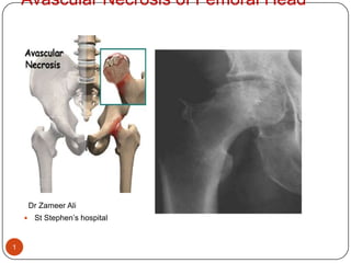
  • 2. Definition 2 Osteonecrosis of femoral head refers to the death of osteocytes with subsequent structural changes leading to femoral head collapse and secondary osteoarthritis of hip joint
  • 3. Definition. 3 Osteonecrosis is defined as an “end stage” condition of the femoral head in which there is necrosis of the bone, secondary to disruption of the blood supply and causes which are still unknown.
  • 4. 4 The death of cell components of bone & bone marrow from repeated interruptions or a single massive interruption of the blood supply to the bone.
  • 5. 5 AVN may involve all or part of head
  • 6. 6
  • 7. The Problem. Commonly affects young patients. Need to alter the life style and leisure activity. Accounts for 18 % of all total hip replacements. 7
  • 8. 8 75% of cases are between 30 to 70 years of age Male: female ratio 4:1 Bilateral involvement is seen in 50 % of cases
  • 9. History 9 Freund in 1926 gave detailed description of Osteonecrosis Chandler in 1948 termed disorder as "coronary disease of hip” which accounts for eponym Chandlers disease
  • 10. . 10 Chandler in 1948 referred this condition as coronary artery disease of hip which even after 50 years correctly describes this condition Aseptic necrosis was initially used to distinguish this condition from infections
  • 11. 11 3)Glimser and Kenzora in 1979 analyzed the radiographic changes which accompany AVN Ficat in 1985 states that this condition resulted from blockage of the osseus microcirculation with intramedullary stasis and increased pressure .
  • 12. Anatomy 12 Head of femur forms about 2/3rd of sphere and articulates with acetabulum of hip joint Connecting two trochanters anteriorly forms intertrochanteric line and connecting two trochanters posteriorly forms intertrochanteric crest.
  • 13.
  • 14. Ball
  • 18.
  • 19. Blood supply of the Head. 3 main sources. Ascending cervical branches Metaphyseal blood vessels Artery of ligamentum teres. 15
  • 20. Vascular supply of femoral head 16 Crook described Extra capsular arterial ring located at base of femoral neck Ascending cervical branches or retinacular arteries run on surface of femoral neck Arteries of round ligament or ligament teres.
  • 21. 17
  • 22. 18 The proximity of retinacular arteries to bone put them at risk of injury in any fracture of femoral neck. The ascending cervical arteries puts them at risk for injury in any fracture of femoral neck
  • 23. 19 4 groups of ascending cervical arteries 1. Anterior 2. Posterior 3. Medial 4. Lateral Of these lateral group provides most of blood supply to femoral head and neck
  • 24. 20 The adult pattern of femoral head vascularity usually becomes established with closure of growth plate at approximately 18 years of age
  • 25. In neonate 21 Three groups of vessels are identified superior retinacular group or lateral epiphyseal group inferior metaphyseal group foveal or medial epiphyseal group All 3 groups anastomose with each other
  • 26. Between 4 years to 7 years 22 The importance of lateral epiphyseal is established as metaphyseal and foveal vessels decrease in extent
  • 27. After 8 years of age 23 Open growth plate represents an effective barrier preventing anastomoses between vessels of head and neck. An increased contribution of foveal vessels
  • 28. 24 During adolescence increased number of inferior metaphyseal vessel is recognized As the growth plate closes the adult pattern appears with anastomosis between 3 arterial systems
  • 29. Causes. 25 Traumatic causes Non Traumatic causes
  • 30. Traumatic 26 Fractures of femoral neck. 15% - 50% Dislocation of the hip. 10% - 25%
  • 31. 27 Currently accepted term is Osteonecrosis which describes main common feature of this condition THE BONE DEATH
  • 32. Non Traumatic 28 Idiopathic. Chronic alcoholism. Steroid intake. Sickle cell disease. Gauchers disease. Caissons disease. Malignancies. Drug induced
  • 33. Risk factors 29 Gauchers disease Radiation induced Liver disease abnormal coagulation factor synthesis
  • 34. Risk factors 30 Traumatic Osteonecrosis usually involves dislocation of hip or fracture femoral neck . 52% of hips unreduced for more than 12 hours developed AVN 22 % of hips developed AVN if reduced within 12 hours Femoral neck fractures associated with 15 – 50 % incidence of AVN
  • 35. Risk factors 31 Alcoholism: Accounts for 10 – 40 % of incidence of AVN. Risk of development of AVN is increased with cumulative doses of alcohol Drug induced: the association between corticosteroid therapy,cushings syndrome, and Osteonecrosis is well established
  • 36. Risk factors 32 Collagen diseases rheumatoid arthritis,SLE have been associated with AVN Radiation causes obliterative endarteritis and cellular death. Gout sodium urate crystals enhance clotting by activating haegmen factor in intrinsic coagulation system
  • 37. Pathogenesis. 33 Other than traumatic causes, the mechanism of necrosis is still “obscure”. Coaugulation defect. Genetic predisposition. Emboli formation. “ IDIOPATHIC ”
  • 38. Pathogenesis 34 The bony compartment function essentially as closed compartment within which one element can expand only at expense of others A unifying concept of pathogenesis of AVN emphasizes the central role of vascular occlusion and ischaemia leading to osteocyte necrosis
  • 39. 35 Pathogenesis of steroid or alcohol induced AVN is not well understood ,but it is suggested that embolic fat and attendant thrombi occlude microcirculation. Lipocyte hypertrophy and subchondral lipid accumulation may cause extra vascular intraosseus compression.
  • 40. 36 It matters little whether the initiating factor was capillary occlusion (as in sickle cell disease) venous occlusion (as suggested for perthes disease) or intramedullary tamponade in Gauchers disease, the end result is diffuse ischaemia involving all the elements
  • 42. Biological sequence of repair 38 loss of cell viability (cell necrosis) Invasion of marrow spaces of dead bone by proliferating capillaries and cells Differentiation of mesenchymal cells to osteoblast synthesis of new bone. Early remodelling of repaired cancellous bone
  • 43. Biological sequence of repair 39 Late internal remodeling Resorption of subchondral bone and invasion of articular cartilage
  • 44. Biological sequence of repair 40 To conclude this reparative process is self limiting and incompletely replaces dead bone with living bone. In subchondral bone ,bone formation occurs at slower rate than does resorption resulting in net removal of bone, loss of structural integerity, subchondral fractures and collapse
  • 45. Clinical features. Pain. - Dull boring . - Progressive. - Worse at night -Limp while walking. - Restricted hip motion. - Unable to sit cross legged. 41
  • 46. symptoms 42 Clinical manifestation of bone ischaemia and infarction are minimal and non specific and depend on etiology.
  • 47. symptoms 43 Initially vague and non specific. Localized or referred pain in buttocks,thighor knee Gradual increase in intensity of pain and decreased motion especially rotation and abduction Over several years results in limping gait
  • 48. symptoms 44 Initial pain is commonly misinterpreted as radiating pain from lumbar spine.
  • 49. Diagnosis 45 Asymptomatic during the early stages. Become symptomatic when significant collapse of the head has occurred.
  • 50. Management protocol 46 Early diagnosis Radiological evaluation Rule out other causes MRI Quantification Treatment algorithm
  • 51. Diagnosis 47 Imaging Routine radiographs Scintigraphy CT scan. MRI. LASER Doppler flometry.
  • 52. Early Diagnosis – suspicion ? 48 High degree of suspicion in a patient C/o anterior HIP pain, Especially with:- H/o Cortisone – Skin, Eye, Liver, Asthma, RA, Weight gain H/o Alcohol abuse Traumatic - # N/F, D/ of F, # Acetabulum Hemoglobinopathy – Sickle / Myelo-infiltrating
  • 53. Other causes 49 Pregnancy Renal Diseases Radiation Gout / Collagen disorder Gaucher’disease Dysbarism Idiopathic
  • 54. Imaging 50 Routine radiographs are usually first step in trying to make diagnosis. High quality films taken at least two views 90 degree apart are critical to initial evaluation
  • 55. Radiology- sequential Changes 51 Crescent Sign Osteoporosis Sclerosis Cystic changes Loss of spherical bearing dome Partial collapse of head Secondary Osteoarthritis
  • 56.
  • 57. X RAY changes 53 Sclerosis /subchondral cysts
  • 58. X RAY changes 54 Crescent changes Flattening of head of femur
  • 59. Bilateral Cystic changes With patchy sclerosis 55
  • 60. Scintigraphy 56 Radionuclide scintigraphy is more sensitive for osteonecrosis than standard radiographs and will reveal changes when standardd radiographs are normal
  • 61. scintigram 57 The hall mark of vascularity is photopenic effect on scintigram RESULTS Decreased uptake by necrotic bone Increased uptake by remodeling bone Normal uptake by normal bone
  • 62. Bone scan. Technetium 99 bone scan reveals decreased uptake. It is effective only if done in early stages. During late phase there are very variable resuts. No relationship b/w scan appearance and the function of the hip. 58
  • 63. MRI 59 MRI is most sensitive technique for early diagnosis in Osteonecrosis Can diagnose AVN as early as 48 hours The classical finding of AVN is decrease in the normally high intensity signal of marrow of femoral head
  • 64. MRI. 60 Can detect early stages. Allows to determine exact stage of disease. Tells exactly the extent of damage. Useful in determining the efficacy of treatment.
  • 65. MRI Geographical area of decreased marrow signal. Necrotic area Surrounded by zone of “low signal” band. Ischaemic bone. 61
  • 67. 63
  • 68. MRI - Findings 64 Bone Marrow edema Double Line – Head in Head sign Crescent sign Collapse Joint effusion Involvement of actabulum Status of other hip Marrow infiltrating disease
  • 69. LASER Doppler Flometry 65 Laser Doppler flometry is technique at measuring blood cell influx in a capillary bed The magnitude and frequency of Doppler shift is proportionate to the velocity and concentration of red cells under probe head
  • 70. Sequence of radiological events in AVN 66 Fragmentation : radiolucent clefts may be seen due to necrosis of involved bone The entire epiphysis may be absent Mottled trabecular : pattern: scrutiny of trabecular traversing the ischaemic bone demonstrates thickened irregular pattern
  • 71. 67 Sclerosis : with revascularisation new bone is deposited around dead bone resulting in increased bone density Subchondral cysts : patchy well circumscribed rarefactions immediately beneath the articular cortex are frequent
  • 72. 68 These cysts are usually seen in region of greatest articular stress and are identical to those found in degenerative joint disease Collapse of articular cortex this generally occurs at the region of maximal stress of involved cortex and represents a localised impaction fracture of weakened bone
  • 73. Fragmentation 69 Radiolucent clefts seen traversing bone
  • 74. Sclerosis/subchondral cysts 70 Sclerosis subchondral cysts
  • 75. Collapse of articular surface 71 Collapse of articular surface
  • 76. 72
  • 77. Staging 73 Several staging systems have been described. The staging system reported by sternbergs and colleagues is similar to that outlined by Marcus,Ficat and Arlet Sternbergs classification allows physician to quantify extent of involvement of femoral head in both early and late stages
  • 78. Staging / Grading --- too many 74 Ficat Radiological Steinberg Quantification Enneking's Stages of Osteonecrosis Marcus and Enneking System Japanese criteria Location Sugioka Radiological University Of Pennsylvania System Association Research Classification Osseous Committee (ARCO)-- Combination
  • 79. Fi Stages of Bone Necrosis 75 Ficats radiological staging of osteonecrosis of femoral head
  • 80. 76 Ficat Stages of Bone Necrosis Stage Clinical Features Radiographs 0 Preclinical 0 0 1Preradiographic + 2 mild density changes in femoral head 2aPrecollapsemild Diffuse Porosis, Sclerosis,or cyst 2bTransition: Flattening, Crescent Sign 3 Collapse mild/moder Broken Contour of Head Certain Sequestrum, Joint Space Normal 4 Osteoarthritismod/severe Flattened Contour Decreased Joint Space Collapse of Head
  • 81. Stage 1 77 Symptoms – none / mild Xrays are normal. Bone scan reveals a “cold spot”.
  • 82. Stage 2 2A Symptoms are mild. Xray shows increased density. subchondral cysts joint line maintained. normal head contour. Bone scan reveals increased uptake. 78
  • 83. 2B Crescent sign. Flattening of the head. 79
  • 85. Stage 3 Moderate symptoms. Loss of shape Subchondral collapse 81
  • 86. Stage 4 Severe symptoms. Joint space narrowing. OA changes in acetabulum. 82
  • 87. Marcus radiological staging of AVN 83 Stage I normal or equivocal radiograph Stage II sclerotic or cystic lesion Stage III crescent sign Stage IV step off in outline of bone Stage V narrowing of joint space with degenerative changes
  • 88. Shimizu’s classification.1995. 84 Grade 1 lesion. A lesion involving medial 1/3 rd of weight bearing surface of the head. In coronal plane ,these lesions occupy < 1/3 of the head. These lesions rarely go into collapse.
  • 89. Grade 2. 85 Lesions involve 1/3 – 2/3 of the weight bearing surface of the head. Involve ½ of the head in coronal plane. Such lesions collapse in 30% of patients.
  • 90. Grade 3. 86 Lesions involving 2/3 of the weight bearing surface of the femoral head. Such lesions collapse in 70% of patients in around 3 years.
  • 91. Treatment 87 Conservative /non surgical treatment Core decompression Bone grafting Cancellous autogenic/allogenic bone graft Osteochondral graft Muscle pedicle bone graft Free vascularized graft
  • 92. Treatment 88 Electric stimulation Osteotomy Joint reconstruction Cup arthoplasty Surface arthoplasty Hemiarthoplasty Total joint arthoplasty
  • 93. 89 The natural history of osteonecrosis in its early stage, before subchondral collapse, is still unclear, but evidence suggests that the rate of progression is high, especially in symptomatic patients.
  • 94. 90 Once subchondral collapse occurs and joint space is lost, progressive osteoarthritis generally is considered inevitable.
  • 95. 91  Many studies have reported an extremely poor prognosis, with a rate of femoral head collapse of more than 85% at 2 years in symptomatic patients (stage I or II disease)
  • 96. 92 No treatment method has proved to be completely effective in arresting the disease process before subchondral collapse or in slowing the progression of femoral head destruction and osteoarthritis after subchondral collapse
  • 97. 93 The rate and course of progression of the disease are unpredictable, and the radiographic picture may not correlate with the clinical symptoms; some patients maintain tolerable function for an extended period after femoral head collapse.
  • 98. 94 Conservative treatment, such as crutch ambulation or bed rest, generally is ineffective. However, symptomatic patients that may benefit from a head-preserving technique should be placed on crutches until surgical treatment is carried out to prevent collapse in the interim
  • 99. Management. 95 Grade 1 lesions - Continuous supervision to detect any changes. - Symptomatic treatment of pain.
  • 100. Grade 1 lesions 96 Conservative treatment such as observation , Analgesics Limited weight bearing may be successful in minimal affected cases
  • 101. Core decompression Grade 2 lesions 97 Ficart and Hungerford have popularized the technique of core decompression of femoral head Rationale is that removing necrotic bone decompresses the rigid osseous chamber, thereby improving blood flow and preventing additional ischaemic events
  • 102. 98 The theoretical advantage of core decompression is based on the belief that the procedure relieves intraosseous pressure caused by venous congestion, thereby allowing improved vascularity and possibly slowing the progression of the disease
  • 103. 99 several authors noted that the results of core decompression are better than those of nonoperative treatment.
  • 104. 100 Several reports noted that the earlier the stage of the disease, the better the results with core decompression. . For more advanced Ficat stages (IIB or III) the results of core decompression are much less predictable, so alternative treatment methods should be explored.
  • 105. 101 Review of the literature currently supports the use of core decompression for the treatment of Ficat stages I and IIA small central lesions in young, nonobese patients who are not taking steroids. This surgery is relatively simple to perform and has a very low complication rate
  • 106. 102 The surgical field for subsequent total hip arthroplasty, if needed, is not substantially altered
  • 107. Bone grafting 103 Phemister introduced concept of using cortical strut graft in core decompression channel The accurate placement of graft within lesion and under subchondral bone is important
  • 108. 104  Structural bone grafting techniques after core decompression have been described using cortical bone, cancellous bone, vascularized bone graft, and debridement of necrotic bone from the femoral head.
  • 109. 105 Insertion of cancellous bone into channel speeds up reossification by osteoinductive and osteoconductive properties of bone graft. Meyers procedure used muscle pedicle bone graft based on quardratus femoris muscle with cancellous bone chips
  • 110. 106 Baksi employed multiple drilling and muscle pedicle grafting using tensor fascia lata muscle anteriorly
  • 111. 107  Advances in microsurgical techniques made it possible to preserve the intrinsic vascularity of bone graft, several authors independently proposed implanting a vascularised bone graft into the core of the femoral head. 
  • 112. Vascular fibular strut grafting 108
  • 113. 109  The rationale for vascularized bone grafting is based on four aspects of the operation and postoperative care: (1) decompression of the femoral head, which may interrupt the cycle of ischemia and intraosseous hypertension that is believed to contribute to the disease; (2) excision of the sequestrum, which might inhibit revascularization of the femoral head
  • 114. 110 (3) filling of the defect that is created with osteoinductive cancellous graft and a viable cortical strut to support the subchondral surface and to enhance the revascularization process (4) protection of the healing construct by a period of limited weight-bearing. 
  • 115. Advantages 111 Advantages of free vascularized fibular grafting compared with total hip arthroplasty: (1) the presence of a healed femoral head may allow more activity, (2) there is no increased risk associated with the presence of a foreign body,
  • 116. 112 (3) if performed before the development of a subchondral fracture, the procedure offers the possibility of survival of a viable femoral head for the life of the patient, and (4) if total hip arthroplasty is ultimately needed, it is much simpler to perform than is a revision arthroplasty after a failed total hip arthroplasty.
  • 117. Disadvantages 113 Disadvantages include a longer recovery period and less uniform and less complete relief of pain than after total hip arthroplasty.
  • 118.
  • 120. Grafting of defect with cancellous graft
  • 121. Viable cortical Bone strut to support subchondral bone.
  • 122.
  • 123. Grade 2. If detected before the collapse Core decompression and fibular grafting. Realignment osteotomies. 116
  • 125. 118
  • 126. Pre OP Post OP 119
  • 127. Electrical stimulation 120 Electrical stimulation has been advocated for AVN because its histological appearance is similar to that of non union Currently used in combination with head salvage procedures
  • 128. osteotomies 121  Various proximal femoral osteotomies have been developed for the treatment of osteonecrosis with the intent to move the involved necrotic segment of the femoral head from the principal weight-bearing area. These procedures have achieved best results for small- or medium-sized lesions (less than 30% femoral head involvement) in young patients in whom it is optimal to delay a total hip arthroplasty.
  • 129. osteotomies 122 Various osteotomies have been described Varus osteotomy Valgus derotation osteotomy Rotation osteotomy aid the loss of structural integrity and collapse by redirecting the forces on femoral head
  • 130. 123 Intertrochanteric osteotomy may be considered for the treatment of stage II or III osteonecrosis of the femoral head in which less than 30% of the femoral head is involved. Plain films and MRI can establish the extent of femoral head involvement and can determine if a satisfactory area of live bone is present under unaffected cartilage in the femoral head and whether this area can be rotated into a position of weight-bearing.
  • 131. 124 Valgus flexion osteotomy is described by wagner when lesion is anterolateral and total angle of necrosis is 200 degree and patient is young and active If necrotic lesion is central varus extension osteotomy is recommended
  • 132. Transtrochanteric Rotational Osteotomy 125 In 1978 Sugioka described a transtrochanteric rotational osteotomy of the femoral head for idiopathic osteonecrosis
  • 133. 126 The rationale of the procedure is to reposition the necrotic anterosuperior part of the femoral head to a non-weight-bearing locale. The femoral head and neck segment is rotated anteriorly around its longitudinal axis so that the weight-bearing force is transmitted to what was previously the posterior articular surface of the femoral head, which is not involved in the ischemic process 
  • 134. 127 Sugioka emphasized the need for a preoperative lateral roentgenogram of the femoral head while the patient is supine and the hip is flexed exactly 90 degrees, abducted 45 degrees, and in neutral rotation. The intact area of the posterior part of the femoral head on this lateral view should be greater than one third of the total articular surface of the head to ensure the best result after his osteotomy
  • 135. Post op regimen 128 Skin traction of 2 kg is applied continuously for the first week and for an additional 2 weeks at night only. As soon as pain tolerance allows, quadriceps setting is begun. Active range-of-motion exercises of the hip are begun at 10 to 14 days.
  • 136. 129 Walking exercises in a pool generally are allowed at 5 to 6 weeks. Partial weight-bearing with crutches is begun at 8 weeks, and the use of crutches is recommended for 6 months after surgery. If the necrotic area of the femoral head is extensive or if involvement is bilateral, crutch use is encouraged for up to 1 year postoperatively.
  • 137. Replacement - options 130 Hemiarthroplasty Bipolar arthroplasty Surface replacement arthroplasty. Newer material for THR ceramic on ceramic Non cemented / cemented THR
  • 138. Birmingham Surface replacement 131 Surface replacement has some advantages over THR because it preserves femoral head and neck and allows future THR if necessary Effective in cases when femoral head is not involved entirely
  • 139. The BHR*System Healthy hip Cuts Implant components Implanted
  • 140. Overview Named for Birmingham, England, where the device’s creators practice medicine Used globally since 1997; More than 65,000 implanted In an international study of 1,626 hips, 99.5% of patients were “Pleased” or “Extremely Pleased” with the results of the BIRMINGHAM HIP Resurfacing (BHR) System.
  • 141. 134 Many failures of resurfacing hemiarthroplasty have been attributed to acetabular cartilage wear. Attention should be given to the quality of the acetabular cartilage on preoperative roentgenographic studies. Intraoperative assessment of the acetabular cartilage is mandatory before implanting a resurfacing hemiarthroplasty prosthesis. If the quality of the acetabular cartilage is in question, a total hip arthroplasty should be performed.
  • 142. 135 Resurfacing hemiarthroplasty is an attractive alternative for young patients with advanced osteonecrosis because very little bone is sacrificed. Should failure occur, conversion to total joint arthroplasty is nearly as simple as primary total hip surgery. Clearly, the results of primary total hip arthroplasty for osteonecrosis are better than resurfacing hemiarthroplasty. However, this procedure can delay total joint arthroplasty and buy valuable time in a young patient.
  • 143. Head size Closely matches the size of natural femoral head Larger than the head of a total hip replacement Larger head means a reduced chance of dislocation after surgery—a leading cause of revision surgery 1-3% of total hips dislocate over the lifetime of the implant 0.3% of BHR* implants dislocated in the first 5 years after surgery (in a study of 2,385 hips) Healthy head BHR head Total hip head
  • 144. Who is the typical candidate for BHR*System? Adults under age 60 for whom total hip replacement may not be appropriate due to an increased level of physical activity Active adults over age 60 may be candidates, depending on their bone quality
  • 145. Conventional hip replacement Healthy hip Cuts Implant components Implanted
  • 146. The key benefits Head size Advanced bearing surface Bone conservation
  • 147. Hip with osteoarthritis Bone cuts fora traditionalhip replacement Bone conservation Preserves your natural femoral neck Neck length and angle determine accurate leg length With the BHR*System, you retain original equipment; with a total hip, your femoral neck is replaced by the implant Bone cuts forBHR System
  • 148. “Minimally Invasive.” Soft Tissue No. Incision length of 6 to 8 inches Bone Yes. Preserves your body’s natural bone structure; It resurfaces rather than replaces Conserved bone
  • 149. Conventional vs. the BHR*System Total hip cuts BHR System cuts
  • 150. Bone conservation (cont.) Revises to a primary If you need “revision” surgery, you don’t get a revision implant The follow-up procedure would be the same total hip replacement you would otherwise have received
  • 151. Total Hip Arthroplasty and Bipolar Hemiarthroplasty.  144 Most series that have examined both unipolar and bipolar hemiarthroplasty for the treatment of osteonecrosis have reported uniformly poor results.
  • 152. THR 145 Patient aged 50 & more Advance osteoarthritis and reduction of joint space. Radiation necrosis Result less than Ideal. – necrotic bone Poor in Sickle cell disease. Cementless are superior over cemented THR
  • 153. 146 With new bearing surfaces becoming available, such as ceramic on ceramic, metal on metal, and highly cross-linked polyethylene, results may improve even more. The results of primary total joint replacement for osteonecrosis are now approaching those reported for osteoarthritis in aged-matched patients.
  • 154. Malakar post alcohol AVN Bil THR 1991 147
  • 155. THR 148 At the end stage of osteonecrosis, when severe arthritic changes are noted on both sides of the hip joint, total hip arthroplasty is one of the only viable operative options available
  • 156. 149  Given the young age of most patients affected with this disease, if total joint replacement is elected, the patient should be well informed of the almost certain need for one or more revision hip replacements later in life.
  • 157. Girdle stone arthoplasty 150 Used as salvage procedure in special circumstances like painful hip with superimposed sepsis, failed THR with sepsis Femur without good bone stock Conversion to THR can be taken at later stage
  • 158. Hip fusion 151 Not frequently recommended because of high failure rates
  • 159. 152 ….Thank you…