SlideShare una empresa de Scribd logo
1 de 69
Group Discussion
Presenter :
Dr. Durga Singh (JR1)
Moderator :
Prof. Dr. Sunil Kumar
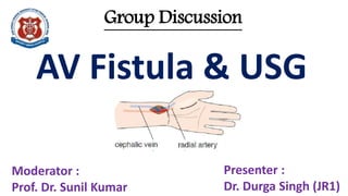
AV Fistula & USG
AV
Fistula
Ultrasound evaluation before and after
hemodialysis access
• USG / Physical examination / conventional venography
• AV Fistula >>> AV graft
• Pre-operative vascular mapping - increased
visualization of veins , AVFs , success , cause of immature
AVF , treatment of failed AVF
AV
Fistula
AV Fistula
A native AVF is a surgically created , direct
anastomosis b/w an artery & a vein , placed in
either the forearm or upper arm
AV
Fistula
Q1. MC type of fistula is
a. Traumatic
b. Iatrogenic
c. Congenital
d. None
Reference – Bailey and love’s short practice of
surgery
Ans - Iatrogenic
AV
Fistula
AV Fistula
Q2. Distal needle placed in the AVF carries blood –
a. From machine to the pt
b. From pt to the machine
Reference - Chapter 17 , Ultrasound evaluation
before and after hemodialyis access ,
Introduction to vascular ultrasonography -
Zwiebel (5th edition)
Ans - B
AV
Fistula
Types of AV Fistula
AV
Fistula
Types of AV Fistula
Q3. MC type of fistula used for dialysis
is where we join
a. end of vein to end of artery
b. end of vein to side of artery
c. side of vein to end of artery
d. side of vein to side of artery
Reference – Bailey and love’s short
practice of surgery
Ans - B
Brescia – Cimino A-V fistula
AV
Fistula
A-V access for hemodialysis in
preferential order
AV Fistula > AV Graft
Forearm > arm
Non dominant > dominant
Non dominant fistula (FA) - dominant fistula (FA) -
non dominant fistula (arm) – dominant fistula (arm)
non dominant graft (FA>A) - dominant graft (FA>A) –
thigh graft – venous catheter
AV
Fistula
A-V access for
hemodialysis
in preferential
order
AV
Fistula
Vascular mapping prior to hemodialysis
access placement
• Terminology used is proximal/distal or cranial/caudal
• General principles :
1. High resolution linear USG transducer (>-7Hz)
2. Transverse plane : identify vessels ( A/V ) – Diameter ,
wall thickness , compressibility , depth , intimal
thickening , stenosis , concentric calcification
3. Longitudinal plane – C/D , S/D
Anatomy
AV
Fistula
Assessment
1. Subclavian , Internal jugular and Central
Vein Assessment
• Stenosis or thrombosis evaluation
• Spectral waveforms – in medial portion of
subclavian vein and caudal portion of IJV for
respiratory phasicity and transmitted cardiac
pulsatility
AV
Fistula
• Absent flow features – Central venous stenosis or obstruction
• Examine C/L subclavian and IJV
• If flow abnormality is U/L , BCV stenosis / occlusion is likely
• If flow abnormality is B/L , SVC stenosis / obstruction is likely
AV
Fistula
AV
Fistula
2. Forearm Assessment ( positioning )
• RA diameter at wrist / FA (>-2mm) ND
• UA diameter at wrist / FA (>-2mm)
• RA diameter at wrist / FA (>-2mm) D
• UA diameter at wrist / FA (>-2mm)
IF NO FA ARTERY IS SATISFACTORY – Pt is not a
candidate for forearm AVF
AV
Fistula
AV
Fistula
• Look for intimal thickening / stenosis /concentric
calcification
• A high RA takeoff from the BA or even AA in the
upper arm is a common anatomic variant. The
presence of this variant can be suspected when two
arteries with accompanying paired veins are seen in the
upper arm. These arteries should be followed into the
forearm, where they assume the respective positions
of the radial and ulnar arteries.
Hemodialysis access surgeons are reluctant to place a
forearm graft or upper arm straight graft in a patient
with a high radial artery takeoff, as the chance for
arterial steal is increased.
Infrequently, a prominent arterial branch that courses
posteriorly toward the elbow can mimic a high radial
artery takeoff. Following the course of the artery more
distally allows differentiation.
AV
Fistula
• It is important to analyze the spectral waveform of the BA
and RA arteries to detect either proximal or distal arterial
obstruction.
AV
Fistula
• It is important to analyze the spectral waveform
of the BA and RA arteries to detect either proximal or
distal arterial obstruction.
With proximal obstruction, the waveforms are
monophasic and dampened.
With distal obstruction, the waveforms have a
normal triphasic pattern, but the velocity may be
reduced because of diminished outflow.
AV
Fistula
• Malovrh noted a higher success rate in FA fistulas in patients
who converted from triphasic to monophasic flow after release
of a clenched fist (Clenched fist maneuver)
Arterial
hyperemic
response – useful
to predict risk of
arterial steal
Clenched fist (3min)
– high resistance flow
(triphasic)
Released fist – low
resistance
(monophasic) and RI
<0.70
Failure of such
response is regarded
as C/I to AVF
AV
Fistula
Modified Allen’s test for patency of palmar arch
AV
Fistula
Q4. Diameter criteria for arteries during fistula mapping for
hemodialysis
a. 5mm
b. 3mm
c. 2.5mm
d. 1.6-1.7mm
Reference - Chapter 17 , Ultrasound evaluation before and after
hemodialyis access , Introduction to vascular ultrasonography -
Zwiebel (5th edition)
Ans - D
Anatomy
AV
Fistula
• If RA / UA meets size criteria at wrist / FA - NEXT
evaluate CV
• Tourniquet / percuss for 3 min ---
AV
Fistula
• If RA / UA meets size criteria at wrist / FA - NEXT
evaluate CV
• Tourniquet / percuss for 3 min ---
>2.5mm / continuous upto tourniquet / patency
/compressibilty / wall thickness / depth / follow in
cephalad direction for continuity / stenosis / branch
points/ follow till axilla or subclavian vein (Muscle or
arm position )
It is possible creating a FA CV AVF , even if the
CV in the upper arm is small or thrombosed. If
the CV in the FA drains into the
brachial or basilic veins via an adequately
sized median cubital or other branch vein, it is
suitable for AVF creation.
Carefully assess vein branch
points, as areas of focal stenosis
may occur at accessory vein
takeoffs. These stenoses may
significantly limit flow in a
subsequently created access.
Depth of the CV from the skin surface should
be measured during the USG mapping
procedure. If the vein is greater than 5mm in
depth, it will likely be difficult to palpate the
vein with sufficient confidence to permit the
insertion of a 15G needle into it for
hemodialysis - SUPERFICIALIZE
AV
Fistula
1. It may be possible to create a FA CV AVF, even if the CV in the upper arm is small or
occluded by thrombus. If the CV in the FA drains into the brachial or basilic veins via
an adequately sized median cubital or other branch vein, it is suitable for AVF
creation.
2. It is important to carefully assess vein branch points, as areas of focal stenosis may
occur at accessory vein takeoffs. These stenoses may significantly limit flow in a
subsequently created access.
3. CV may meet diameter criteria for AVF creation yet may be too deep,to access easily
at hemodialysis once the AVF is mature. Thus, the depth of the CV from the skin
surface should be measured during the USG mapping procedure. If the vein is
greater than 5mm in depth, it will likely be difficult to palpate the vein with sufficient
confidence to permit the insertion of a 15G needle into it for hemodialysis.
Detection of a vein that is too deep but otherwise suitable for an AVF allows the
surgeon to inform the patient preoperatively about the potential need for a second
procedure to "superficialize" the vein in the subcutaneous tissues. This discussion
allows the patient to decide whether to accept the procedure.
AV
Fistula
• If CV is not adeqaute ----
Assess basilic vein ------suitable vein in volar then dosral
surface of FA
>2.5mm / continuous upto tourniquet / patency /
follow in cephalad direction for continuity / stenosis /
branch points/ follow till axilla /drainage point
IF NO VEIN IS SUFFICIENT IN FA – repeat same thing on
dominant side
AV
Fistula
3. Arm Assessment (Nondominant 1st ,
tourniquet at axilla )
• BA diameter at elbow above its bifurcation(>-2mm)
• CV diameter at elbow (>-2.5mm diameter and extend
approx 2 cm below antecubital fossa , evaluate median
cubital branch of CV)
• BV ( 2 cm caudal – transposition or diameter 4mm for
graft / BVs for graft
AV
Fistula
• Suitable vein – Continuity with deep venous system
continuity / adequate diameter / empty point
diameter / continuity / stenosis / branch points/
follow till axilla or subclavian vein
AV
Fistula
Q5. Diameter criteria for veins during graft mapping for hemodialysis
a. 5mm
b. 4mm
c. 3mm
d. 2.5mm
Reference - Chapter 17 , Ultrasound evaluation before and after
hemodialyis access , Introduction to vascular ultrasonography -
Zwiebel (5th edition)
Ans - B
AV
Fistula
Reporting :-
AV
Fistula
AV
Fistula
AV
Fistula
AVF maturity
assessment
AV
Fistula
AVF maturity assessment
Mature AVF in the United
States is a fistula that is usable
for hemodialysis at a flow of
350 cc/min at six dialysis
sessions in one month.
Practitioners in other
countries, particularly in
Europe, accept lower AVF
flows with subsequently
longer dialysis times.
A mature AVF can be
identified clinically as one
that has a large, easily
palpable vein that can
provide access for two 15-
gauge needles.
If AVF maturity is in doubt ,
use USG (Maturity and
Triage)
AV
Fistula
Sonographic evaluation of
AVF Maturity
• >- 7 Hz
• No tourniquet
• Positioning
• Feeding artery , anastomosis , draining vein
• Transverse plane – vessel diameter , wall thickness ,
compressibility , depth
• Longitudinal plane – C/D , S/D , stenosis , functional
stenosis
AV
Fistula
AV
Fistula
AV
Fistula
• The diameter of the draining vein is measured
routinely in the caudal, mid-, and cranial portions of
the forearm, and similarly in the upper arm when a
forearm AVF is evaluated. The entire draining vein
should be scanned, and the minimum diameter
should be measured, even if it occurs at a location
not routinely measured.
• The depth of the anterior wall of the AVF / vein
from the skin surface is also measured (5mm).
AV
Fistula
Blood flow is measured in the AVF in mL/min.
If the blood flow equals or exceeds 500 mL/min, the likelihood of fistula
adequacy is nearly twice as great as with lower flow rates.
Q6. Formula for calculating flow volume in ml/min in a case of A-V fistula is
a. Cross sectional area (cm2) x mean velocity (cm/sec) x 30
b. Cross sectional area (cm2) x mean velocity (cm/sec) x 60
c. Cross sectional area / AT
d. 3.14 x diameter x diameter / 4 (cm2) x mean velocity (cm/sec) x 60
Reference - Chapter 17 , Ultrasound evaluation before and after hemodialyis
access , Introduction to vascular ultrasonography - Zwiebel (5th edition)
Ans – b and d
AV
Fistula
Blood flow is measured in the AVF in mL/min.
If the blood flow equals or exceeds 500 mL/min, the
likelihood of fistula adequacy is nearly twice as great as with
lower flow rates.
AV
Fistula
Combining venous diameter and volume flow
measurement increased our ability to predict
fistula adequacy. A venous diameter of 4 mm or
greater and flow volume equaling or exceeding 500
mL/min confirmed AVF maturity in 95% of cases,
versus a maturity rate of only 33% when neither
criterion is met.
AV
Fistula
KEY ADDITIONAL POINTS REGARDING
AVF EVALUATION :-
1. Look for the presence of large vein branches involving the
first 10cm of the draining vein . These accessory branches
may divert a significant amount of flow from the primary
draining vein with resultant decrease in flow to below
functional levels. Such flow diversion is a frequent reason for
AVF immaturity.These branches can be surgically ligated,
thereby increasing the likelihood that the AVF will mature
AV
Fistula
2. Occasionally, a patient with an AVF may present for evaluation of
arm swelling. Respiratory phasicity and transmitted cardiac
pulsatility should be evaluated in the subclavian and internal
jugular veins, to assess for the possibility of a central venous
stenosis . The brachial veins should also be evaluated for the
presence of deep venous thrombosis.
3. Infrequently, a patient will have symptoms of arterial steal with
an AVF , such as hand pain and numbness, particularly during
dialysis. Flow direction of the distal radial artery is evaluated
using spectral and color Doppler. Arterial steal is diagnosed when
the flow in the radial artery is reversed.
AV
Fistula
Causes of immature fistula –
1. Stenosis at or near the fistula (Angioplasty –
surgical revision)
2. One or more accessory veins (Ligation)
3. Deep draining vein (Fistula surgically placed in
more superficial soft tissues)
Immature fistulas can be converted into usable fistula with
correction of underlying problem
AV
Fistula
Complications of A-V access for
hemodialysis –
1. Stenosis and occlusion
2. Aneurysm and pseudoaneurysm
3. Infected and non infected collections
4. Arterial steal syndrome
5. High output cardiac failure
AV
Fistula
Complications of A-V access for
hemodialysis –
Stenosis and occlusion
B mode (diameter reduction)
C/D (aliasing)
S/D ( PSV ratio)
AV
Fistula
• If the draining vein is visibly narrowed, PSV are measured at
the stenosis and 2 cm caudal to the stenosis.
A PSV ratio is calculated by dividing PSV at the stenosis by the
PSV obtained 2 cm caudal to the stenosis.
If the PSV ratio is 2 or more, it is classified as a ~50%
diameter stenosis.
• Both arteriovenous and draining vein stenoses may be
treated with angioplasty or surgical revision.
AV
Fistula
Q7. The most frequent location of AVF stenoses is
a. Draining vein
b. Feeding artery
c. Central veins
d. Perianastomotic
Reference - Chapter 17 , Ultrasound evaluation before and
after hemodialyis access , Introduction to vascular
ultrasonography - Zwiebel (5th edition)
Ans - D
AV
Fistula
PSV is measured at the anastomosis and 2 cm cephalad to the
anastomosis in the feeding artery.
A PSV ratio is then calculated by dividing the PSV at the
anastomosis by the PSV obtained 2 cm cranial to the anastomosis
We generally begin to be concerned about stenosis at the AV
anastomosis when the PSV ratio reaches 3
Visual confirmation of a stenosis at the AV anastomosis is
useful, as the PSV in the draining vein may be significantly
elevated merely because of the acute angulation of the draining
vein at the anastomosis.
PSV at fistula
PSV 2cm cranial to fistula in artery
AV
Fistula
Complications of A-V access for
hemodialysis –
Aneurysm and pseudoaneurysm
AV
Fistula
Complications of A-V access for
hemodialysis –
Arterial steal syndrome
AV
Fistula
Birectional flow in distal radial artery Restoration of antegrade flow on gentle
compression of fistula
AV
Fistula
Q8. AVF stenosis at the anastomosis site is suspected if the
PSV ratio reaches
a. 1
b. 2
c. 3
d. 4
Reference - Chapter 17 , Ultrasound evaluation before and
after hemodialyis access , Introduction to vascular
ultrasonography - Zwiebel (5th edition)
Ans - c
AV
Fistula
Q9. AVF stenosis in the draining vein is suspected if the PSV
ratio reaches
a. 1
b. 2
c. 3
d. 4
Reference - Chapter 17 , Ultrasound evaluation before and
after hemodialyis access , Introduction to vascular
ultrasonography - Zwiebel (5th edition)
Ans - b
AV
Fistula
Reporting :-
AV
Fistula
AV
Fistula
Thank you
By :- Dr Durga Singh

Más contenido relacionado

La actualidad más candente

Doppler ultrasound in transplant renal artery stenosis
Doppler ultrasound in transplant renal artery stenosisDoppler ultrasound in transplant renal artery stenosis
Doppler ultrasound in transplant renal artery stenosisSamir Haffar
 
Doppler ultrasound of the kidneys
Doppler ultrasound of the kidneysDoppler ultrasound of the kidneys
Doppler ultrasound of the kidneysSamir Haffar
 
Haemodialysis fistula doppler
Haemodialysis fistula dopplerHaemodialysis fistula doppler
Haemodialysis fistula dopplerSahroz Khan
 
Peripheral arterial doppler
Peripheral  arterial dopplerPeripheral  arterial doppler
Peripheral arterial dopplerAnish Choudhary
 
Doppler ultrasound of the portal system - Normal findings
Doppler ultrasound of the portal system - Normal findingsDoppler ultrasound of the portal system - Normal findings
Doppler ultrasound of the portal system - Normal findingsSamir Haffar
 
Hepato portal doppler ultrasound
Hepato portal doppler ultrasoundHepato portal doppler ultrasound
Hepato portal doppler ultrasoundRavi patel
 
4 peripheral venous duplex pt 4 varices dr ahmed esawy
4 peripheral venous duplex pt 4 varices dr ahmed esawy4 peripheral venous duplex pt 4 varices dr ahmed esawy
4 peripheral venous duplex pt 4 varices dr ahmed esawyAHMED ESAWY
 
Venous Doppler upper limb
Venous Doppler upper limb Venous Doppler upper limb
Venous Doppler upper limb Milan Silwal
 
portal doppler ppt .pptx
portal doppler ppt .pptxportal doppler ppt .pptx
portal doppler ppt .pptxdypradio
 
lower limb doppler examination -The essentials
lower limb doppler examination -The essentialslower limb doppler examination -The essentials
lower limb doppler examination -The essentialsAhmed Bahnassy
 
Doppler ultrasound of A-V access for hemodialysis
Doppler ultrasound of A-V access for hemodialysisDoppler ultrasound of A-V access for hemodialysis
Doppler ultrasound of A-V access for hemodialysisSamir Haffar
 
Venous Doppler Lower limb Dr Mukesh Tilgam
Venous Doppler Lower limb Dr Mukesh TilgamVenous Doppler Lower limb Dr Mukesh Tilgam
Venous Doppler Lower limb Dr Mukesh TilgamDrmukesh Tilgam
 

La actualidad más candente (20)

Doppler of the portal system
Doppler of the portal systemDoppler of the portal system
Doppler of the portal system
 
New carotid doppler ultrasound
New carotid doppler ultrasoundNew carotid doppler ultrasound
New carotid doppler ultrasound
 
Carotid doppler ii
Carotid doppler iiCarotid doppler ii
Carotid doppler ii
 
Doppler ultrasound in transplant renal artery stenosis
Doppler ultrasound in transplant renal artery stenosisDoppler ultrasound in transplant renal artery stenosis
Doppler ultrasound in transplant renal artery stenosis
 
Doppler ultrasound of the kidneys
Doppler ultrasound of the kidneysDoppler ultrasound of the kidneys
Doppler ultrasound of the kidneys
 
Haemodialysis fistula doppler
Haemodialysis fistula dopplerHaemodialysis fistula doppler
Haemodialysis fistula doppler
 
Peripheral arterial doppler
Peripheral  arterial dopplerPeripheral  arterial doppler
Peripheral arterial doppler
 
Doppler ultrasound of the portal system - Normal findings
Doppler ultrasound of the portal system - Normal findingsDoppler ultrasound of the portal system - Normal findings
Doppler ultrasound of the portal system - Normal findings
 
Hepato portal doppler ultrasound
Hepato portal doppler ultrasoundHepato portal doppler ultrasound
Hepato portal doppler ultrasound
 
4 peripheral venous duplex pt 4 varices dr ahmed esawy
4 peripheral venous duplex pt 4 varices dr ahmed esawy4 peripheral venous duplex pt 4 varices dr ahmed esawy
4 peripheral venous duplex pt 4 varices dr ahmed esawy
 
USG of Aorta and Coeliac axis
USG of Aorta and Coeliac axisUSG of Aorta and Coeliac axis
USG of Aorta and Coeliac axis
 
Portal venous doppler
Portal venous dopplerPortal venous doppler
Portal venous doppler
 
Venous Doppler upper limb
Venous Doppler upper limb Venous Doppler upper limb
Venous Doppler upper limb
 
portal doppler ppt .pptx
portal doppler ppt .pptxportal doppler ppt .pptx
portal doppler ppt .pptx
 
CAROTID DOPPLER STUDY
CAROTID DOPPLER STUDYCAROTID DOPPLER STUDY
CAROTID DOPPLER STUDY
 
Hepatic doppler
Hepatic dopplerHepatic doppler
Hepatic doppler
 
lower limb doppler examination -The essentials
lower limb doppler examination -The essentialslower limb doppler examination -The essentials
lower limb doppler examination -The essentials
 
Doppler ultrasound of A-V access for hemodialysis
Doppler ultrasound of A-V access for hemodialysisDoppler ultrasound of A-V access for hemodialysis
Doppler ultrasound of A-V access for hemodialysis
 
Mdct in cad
Mdct in cadMdct in cad
Mdct in cad
 
Venous Doppler Lower limb Dr Mukesh Tilgam
Venous Doppler Lower limb Dr Mukesh TilgamVenous Doppler Lower limb Dr Mukesh Tilgam
Venous Doppler Lower limb Dr Mukesh Tilgam
 

Similar a Role of USG in A-V fistula assessment

Vascular access in Haemodialysis (2).pptx
Vascular access in Haemodialysis (2).pptxVascular access in Haemodialysis (2).pptx
Vascular access in Haemodialysis (2).pptxMithunAhmed5
 
Role of medical imaging in management of arteriovenous fistula Dr. Muhammad B...
Role of medical imaging in management of arteriovenous fistula Dr. Muhammad B...Role of medical imaging in management of arteriovenous fistula Dr. Muhammad B...
Role of medical imaging in management of arteriovenous fistula Dr. Muhammad B...Dr. Muhammad Bin Zulfiqar
 
Vascular access surgery by Dr. Ali Mujtaba
Vascular access surgery by Dr. Ali MujtabaVascular access surgery by Dr. Ali Mujtaba
Vascular access surgery by Dr. Ali MujtabaDr Ali MUJTABA
 
Hemodialysis procedure dr. mohamed kamal
Hemodialysis procedure   dr. mohamed kamalHemodialysis procedure   dr. mohamed kamal
Hemodialysis procedure dr. mohamed kamalFarragBahbah
 
arteriovenous fistula AVF Presentation.pptx
arteriovenous fistula AVF Presentation.pptxarteriovenous fistula AVF Presentation.pptx
arteriovenous fistula AVF Presentation.pptxDr Tajamul Hassan
 
VENOUS DRAINAGE OF THE LOWER LIMBS.ppt
VENOUS DRAINAGE OF THE LOWER LIMBS.pptVENOUS DRAINAGE OF THE LOWER LIMBS.ppt
VENOUS DRAINAGE OF THE LOWER LIMBS.pptAbubakar Hammadama
 
vascular access for dialysis access: seminar
vascular access for dialysis access: seminarvascular access for dialysis access: seminar
vascular access for dialysis access: seminarMd Rahman
 
Av fistula 3 al mansoura aldawly - nephrolody day dr aboelfotoh
Av fistula 3 al mansoura aldawly - nephrolody day   dr aboelfotohAv fistula 3 al mansoura aldawly - nephrolody day   dr aboelfotoh
Av fistula 3 al mansoura aldawly - nephrolody day dr aboelfotohFarragBahbah
 
Long term Hemodialysis Vascular Access (AVFs and AVGs)
Long term Hemodialysis Vascular Access (AVFs and AVGs)Long term Hemodialysis Vascular Access (AVFs and AVGs)
Long term Hemodialysis Vascular Access (AVFs and AVGs)SAMEH ATTIA ALI ABDELHAMID
 
centralvenouscatheter-1.pdf
centralvenouscatheter-1.pdfcentralvenouscatheter-1.pdf
centralvenouscatheter-1.pdfisha sharma
 
CENTRAL VENOUS CATHETER
CENTRAL VENOUS CATHETERCENTRAL VENOUS CATHETER
CENTRAL VENOUS CATHETERAvijit Prusty
 
Acs0626 Medical Management Of Vascular Disease
Acs0626 Medical Management Of Vascular DiseaseAcs0626 Medical Management Of Vascular Disease
Acs0626 Medical Management Of Vascular Diseasemedbookonline
 
Central venous pressure
Central venous pressureCentral venous pressure
Central venous pressureDavis Kurian
 
Fistula arterio-venoasa la pacientii dializati
Fistula arterio-venoasa la pacientii dializatiFistula arterio-venoasa la pacientii dializati
Fistula arterio-venoasa la pacientii dializatiALEXANDRU ANDRITOIU
 
Tamer elsaid mansouraoct2019
Tamer elsaid mansouraoct2019Tamer elsaid mansouraoct2019
Tamer elsaid mansouraoct2019FAARRAG
 
Interventional Radiology in General Surgery.pptx
Interventional Radiology in General Surgery.pptxInterventional Radiology in General Surgery.pptx
Interventional Radiology in General Surgery.pptxPRAGATISHUKLA40
 
Vascular access care .. nephrology perspective - Dr. Tamer El said
Vascular access care .. nephrology perspective - Dr. Tamer El saidVascular access care .. nephrology perspective - Dr. Tamer El said
Vascular access care .. nephrology perspective - Dr. Tamer El saidMNDU net
 

Similar a Role of USG in A-V fistula assessment (20)

Vascular Access.pptx
Vascular Access.pptxVascular Access.pptx
Vascular Access.pptx
 
Vascular access in Haemodialysis (2).pptx
Vascular access in Haemodialysis (2).pptxVascular access in Haemodialysis (2).pptx
Vascular access in Haemodialysis (2).pptx
 
Vascular access
Vascular accessVascular access
Vascular access
 
Role of medical imaging in management of arteriovenous fistula Dr. Muhammad B...
Role of medical imaging in management of arteriovenous fistula Dr. Muhammad B...Role of medical imaging in management of arteriovenous fistula Dr. Muhammad B...
Role of medical imaging in management of arteriovenous fistula Dr. Muhammad B...
 
Vascular access surgery by Dr. Ali Mujtaba
Vascular access surgery by Dr. Ali MujtabaVascular access surgery by Dr. Ali Mujtaba
Vascular access surgery by Dr. Ali Mujtaba
 
Hemodialysis procedure dr. mohamed kamal
Hemodialysis procedure   dr. mohamed kamalHemodialysis procedure   dr. mohamed kamal
Hemodialysis procedure dr. mohamed kamal
 
arteriovenous fistula AVF Presentation.pptx
arteriovenous fistula AVF Presentation.pptxarteriovenous fistula AVF Presentation.pptx
arteriovenous fistula AVF Presentation.pptx
 
Ivus evar
Ivus evarIvus evar
Ivus evar
 
VENOUS DRAINAGE OF THE LOWER LIMBS.ppt
VENOUS DRAINAGE OF THE LOWER LIMBS.pptVENOUS DRAINAGE OF THE LOWER LIMBS.ppt
VENOUS DRAINAGE OF THE LOWER LIMBS.ppt
 
vascular access for dialysis access: seminar
vascular access for dialysis access: seminarvascular access for dialysis access: seminar
vascular access for dialysis access: seminar
 
Av fistula 3 al mansoura aldawly - nephrolody day dr aboelfotoh
Av fistula 3 al mansoura aldawly - nephrolody day   dr aboelfotohAv fistula 3 al mansoura aldawly - nephrolody day   dr aboelfotoh
Av fistula 3 al mansoura aldawly - nephrolody day dr aboelfotoh
 
Long term Hemodialysis Vascular Access (AVFs and AVGs)
Long term Hemodialysis Vascular Access (AVFs and AVGs)Long term Hemodialysis Vascular Access (AVFs and AVGs)
Long term Hemodialysis Vascular Access (AVFs and AVGs)
 
centralvenouscatheter-1.pdf
centralvenouscatheter-1.pdfcentralvenouscatheter-1.pdf
centralvenouscatheter-1.pdf
 
CENTRAL VENOUS CATHETER
CENTRAL VENOUS CATHETERCENTRAL VENOUS CATHETER
CENTRAL VENOUS CATHETER
 
Acs0626 Medical Management Of Vascular Disease
Acs0626 Medical Management Of Vascular DiseaseAcs0626 Medical Management Of Vascular Disease
Acs0626 Medical Management Of Vascular Disease
 
Central venous pressure
Central venous pressureCentral venous pressure
Central venous pressure
 
Fistula arterio-venoasa la pacientii dializati
Fistula arterio-venoasa la pacientii dializatiFistula arterio-venoasa la pacientii dializati
Fistula arterio-venoasa la pacientii dializati
 
Tamer elsaid mansouraoct2019
Tamer elsaid mansouraoct2019Tamer elsaid mansouraoct2019
Tamer elsaid mansouraoct2019
 
Interventional Radiology in General Surgery.pptx
Interventional Radiology in General Surgery.pptxInterventional Radiology in General Surgery.pptx
Interventional Radiology in General Surgery.pptx
 
Vascular access care .. nephrology perspective - Dr. Tamer El said
Vascular access care .. nephrology perspective - Dr. Tamer El saidVascular access care .. nephrology perspective - Dr. Tamer El said
Vascular access care .. nephrology perspective - Dr. Tamer El said
 

Último

Disha NEET Physics Guide for classes 11 and 12.pdf
Disha NEET Physics Guide for classes 11 and 12.pdfDisha NEET Physics Guide for classes 11 and 12.pdf
Disha NEET Physics Guide for classes 11 and 12.pdfchloefrazer622
 
fourth grading exam for kindergarten in writing
fourth grading exam for kindergarten in writingfourth grading exam for kindergarten in writing
fourth grading exam for kindergarten in writingTeacherCyreneCayanan
 
BASLIQ CURRENT LOOKBOOK LOOKBOOK(1) (1).pdf
BASLIQ CURRENT LOOKBOOK  LOOKBOOK(1) (1).pdfBASLIQ CURRENT LOOKBOOK  LOOKBOOK(1) (1).pdf
BASLIQ CURRENT LOOKBOOK LOOKBOOK(1) (1).pdfSoniaTolstoy
 
Nutritional Needs Presentation - HLTH 104
Nutritional Needs Presentation - HLTH 104Nutritional Needs Presentation - HLTH 104
Nutritional Needs Presentation - HLTH 104misteraugie
 
Russian Escort Service in Delhi 11k Hotel Foreigner Russian Call Girls in Delhi
Russian Escort Service in Delhi 11k Hotel Foreigner Russian Call Girls in DelhiRussian Escort Service in Delhi 11k Hotel Foreigner Russian Call Girls in Delhi
Russian Escort Service in Delhi 11k Hotel Foreigner Russian Call Girls in Delhikauryashika82
 
Paris 2024 Olympic Geographies - an activity
Paris 2024 Olympic Geographies - an activityParis 2024 Olympic Geographies - an activity
Paris 2024 Olympic Geographies - an activityGeoBlogs
 
social pharmacy d-pharm 1st year by Pragati K. Mahajan
social pharmacy d-pharm 1st year by Pragati K. Mahajansocial pharmacy d-pharm 1st year by Pragati K. Mahajan
social pharmacy d-pharm 1st year by Pragati K. Mahajanpragatimahajan3
 
Web & Social Media Analytics Previous Year Question Paper.pdf
Web & Social Media Analytics Previous Year Question Paper.pdfWeb & Social Media Analytics Previous Year Question Paper.pdf
Web & Social Media Analytics Previous Year Question Paper.pdfJayanti Pande
 
Presentation by Andreas Schleicher Tackling the School Absenteeism Crisis 30 ...
Presentation by Andreas Schleicher Tackling the School Absenteeism Crisis 30 ...Presentation by Andreas Schleicher Tackling the School Absenteeism Crisis 30 ...
Presentation by Andreas Schleicher Tackling the School Absenteeism Crisis 30 ...EduSkills OECD
 
General AI for Medical Educators April 2024
General AI for Medical Educators April 2024General AI for Medical Educators April 2024
General AI for Medical Educators April 2024Janet Corral
 
Measures of Central Tendency: Mean, Median and Mode
Measures of Central Tendency: Mean, Median and ModeMeasures of Central Tendency: Mean, Median and Mode
Measures of Central Tendency: Mean, Median and ModeThiyagu K
 
Software Engineering Methodologies (overview)
Software Engineering Methodologies (overview)Software Engineering Methodologies (overview)
Software Engineering Methodologies (overview)eniolaolutunde
 
Grant Readiness 101 TechSoup and Remy Consulting
Grant Readiness 101 TechSoup and Remy ConsultingGrant Readiness 101 TechSoup and Remy Consulting
Grant Readiness 101 TechSoup and Remy ConsultingTechSoup
 
Measures of Dispersion and Variability: Range, QD, AD and SD
Measures of Dispersion and Variability: Range, QD, AD and SDMeasures of Dispersion and Variability: Range, QD, AD and SD
Measures of Dispersion and Variability: Range, QD, AD and SDThiyagu K
 
Arihant handbook biology for class 11 .pdf
Arihant handbook biology for class 11 .pdfArihant handbook biology for class 11 .pdf
Arihant handbook biology for class 11 .pdfchloefrazer622
 
Kisan Call Centre - To harness potential of ICT in Agriculture by answer farm...
Kisan Call Centre - To harness potential of ICT in Agriculture by answer farm...Kisan Call Centre - To harness potential of ICT in Agriculture by answer farm...
Kisan Call Centre - To harness potential of ICT in Agriculture by answer farm...Krashi Coaching
 
Sports & Fitness Value Added Course FY..
Sports & Fitness Value Added Course FY..Sports & Fitness Value Added Course FY..
Sports & Fitness Value Added Course FY..Disha Kariya
 
The basics of sentences session 2pptx copy.pptx
The basics of sentences session 2pptx copy.pptxThe basics of sentences session 2pptx copy.pptx
The basics of sentences session 2pptx copy.pptxheathfieldcps1
 

Último (20)

Disha NEET Physics Guide for classes 11 and 12.pdf
Disha NEET Physics Guide for classes 11 and 12.pdfDisha NEET Physics Guide for classes 11 and 12.pdf
Disha NEET Physics Guide for classes 11 and 12.pdf
 
fourth grading exam for kindergarten in writing
fourth grading exam for kindergarten in writingfourth grading exam for kindergarten in writing
fourth grading exam for kindergarten in writing
 
BASLIQ CURRENT LOOKBOOK LOOKBOOK(1) (1).pdf
BASLIQ CURRENT LOOKBOOK  LOOKBOOK(1) (1).pdfBASLIQ CURRENT LOOKBOOK  LOOKBOOK(1) (1).pdf
BASLIQ CURRENT LOOKBOOK LOOKBOOK(1) (1).pdf
 
Nutritional Needs Presentation - HLTH 104
Nutritional Needs Presentation - HLTH 104Nutritional Needs Presentation - HLTH 104
Nutritional Needs Presentation - HLTH 104
 
Russian Escort Service in Delhi 11k Hotel Foreigner Russian Call Girls in Delhi
Russian Escort Service in Delhi 11k Hotel Foreigner Russian Call Girls in DelhiRussian Escort Service in Delhi 11k Hotel Foreigner Russian Call Girls in Delhi
Russian Escort Service in Delhi 11k Hotel Foreigner Russian Call Girls in Delhi
 
Paris 2024 Olympic Geographies - an activity
Paris 2024 Olympic Geographies - an activityParis 2024 Olympic Geographies - an activity
Paris 2024 Olympic Geographies - an activity
 
social pharmacy d-pharm 1st year by Pragati K. Mahajan
social pharmacy d-pharm 1st year by Pragati K. Mahajansocial pharmacy d-pharm 1st year by Pragati K. Mahajan
social pharmacy d-pharm 1st year by Pragati K. Mahajan
 
Web & Social Media Analytics Previous Year Question Paper.pdf
Web & Social Media Analytics Previous Year Question Paper.pdfWeb & Social Media Analytics Previous Year Question Paper.pdf
Web & Social Media Analytics Previous Year Question Paper.pdf
 
Advance Mobile Application Development class 07
Advance Mobile Application Development class 07Advance Mobile Application Development class 07
Advance Mobile Application Development class 07
 
Presentation by Andreas Schleicher Tackling the School Absenteeism Crisis 30 ...
Presentation by Andreas Schleicher Tackling the School Absenteeism Crisis 30 ...Presentation by Andreas Schleicher Tackling the School Absenteeism Crisis 30 ...
Presentation by Andreas Schleicher Tackling the School Absenteeism Crisis 30 ...
 
General AI for Medical Educators April 2024
General AI for Medical Educators April 2024General AI for Medical Educators April 2024
General AI for Medical Educators April 2024
 
Measures of Central Tendency: Mean, Median and Mode
Measures of Central Tendency: Mean, Median and ModeMeasures of Central Tendency: Mean, Median and Mode
Measures of Central Tendency: Mean, Median and Mode
 
Software Engineering Methodologies (overview)
Software Engineering Methodologies (overview)Software Engineering Methodologies (overview)
Software Engineering Methodologies (overview)
 
Grant Readiness 101 TechSoup and Remy Consulting
Grant Readiness 101 TechSoup and Remy ConsultingGrant Readiness 101 TechSoup and Remy Consulting
Grant Readiness 101 TechSoup and Remy Consulting
 
Mattingly "AI & Prompt Design: Structured Data, Assistants, & RAG"
Mattingly "AI & Prompt Design: Structured Data, Assistants, & RAG"Mattingly "AI & Prompt Design: Structured Data, Assistants, & RAG"
Mattingly "AI & Prompt Design: Structured Data, Assistants, & RAG"
 
Measures of Dispersion and Variability: Range, QD, AD and SD
Measures of Dispersion and Variability: Range, QD, AD and SDMeasures of Dispersion and Variability: Range, QD, AD and SD
Measures of Dispersion and Variability: Range, QD, AD and SD
 
Arihant handbook biology for class 11 .pdf
Arihant handbook biology for class 11 .pdfArihant handbook biology for class 11 .pdf
Arihant handbook biology for class 11 .pdf
 
Kisan Call Centre - To harness potential of ICT in Agriculture by answer farm...
Kisan Call Centre - To harness potential of ICT in Agriculture by answer farm...Kisan Call Centre - To harness potential of ICT in Agriculture by answer farm...
Kisan Call Centre - To harness potential of ICT in Agriculture by answer farm...
 
Sports & Fitness Value Added Course FY..
Sports & Fitness Value Added Course FY..Sports & Fitness Value Added Course FY..
Sports & Fitness Value Added Course FY..
 
The basics of sentences session 2pptx copy.pptx
The basics of sentences session 2pptx copy.pptxThe basics of sentences session 2pptx copy.pptx
The basics of sentences session 2pptx copy.pptx
 

Role of USG in A-V fistula assessment

  • 1. Group Discussion Presenter : Dr. Durga Singh (JR1) Moderator : Prof. Dr. Sunil Kumar AV Fistula & USG
  • 2. AV Fistula Ultrasound evaluation before and after hemodialysis access • USG / Physical examination / conventional venography • AV Fistula >>> AV graft • Pre-operative vascular mapping - increased visualization of veins , AVFs , success , cause of immature AVF , treatment of failed AVF
  • 3. AV Fistula AV Fistula A native AVF is a surgically created , direct anastomosis b/w an artery & a vein , placed in either the forearm or upper arm
  • 4. AV Fistula Q1. MC type of fistula is a. Traumatic b. Iatrogenic c. Congenital d. None Reference – Bailey and love’s short practice of surgery Ans - Iatrogenic
  • 5. AV Fistula AV Fistula Q2. Distal needle placed in the AVF carries blood – a. From machine to the pt b. From pt to the machine Reference - Chapter 17 , Ultrasound evaluation before and after hemodialyis access , Introduction to vascular ultrasonography - Zwiebel (5th edition) Ans - B
  • 7. AV Fistula Types of AV Fistula Q3. MC type of fistula used for dialysis is where we join a. end of vein to end of artery b. end of vein to side of artery c. side of vein to end of artery d. side of vein to side of artery Reference – Bailey and love’s short practice of surgery Ans - B Brescia – Cimino A-V fistula
  • 8. AV Fistula A-V access for hemodialysis in preferential order AV Fistula > AV Graft Forearm > arm Non dominant > dominant Non dominant fistula (FA) - dominant fistula (FA) - non dominant fistula (arm) – dominant fistula (arm) non dominant graft (FA>A) - dominant graft (FA>A) – thigh graft – venous catheter
  • 10. AV Fistula Vascular mapping prior to hemodialysis access placement • Terminology used is proximal/distal or cranial/caudal • General principles : 1. High resolution linear USG transducer (>-7Hz) 2. Transverse plane : identify vessels ( A/V ) – Diameter , wall thickness , compressibility , depth , intimal thickening , stenosis , concentric calcification 3. Longitudinal plane – C/D , S/D
  • 12. AV Fistula Assessment 1. Subclavian , Internal jugular and Central Vein Assessment • Stenosis or thrombosis evaluation • Spectral waveforms – in medial portion of subclavian vein and caudal portion of IJV for respiratory phasicity and transmitted cardiac pulsatility
  • 13.
  • 14. AV Fistula • Absent flow features – Central venous stenosis or obstruction • Examine C/L subclavian and IJV • If flow abnormality is U/L , BCV stenosis / occlusion is likely • If flow abnormality is B/L , SVC stenosis / obstruction is likely
  • 16. AV Fistula 2. Forearm Assessment ( positioning ) • RA diameter at wrist / FA (>-2mm) ND • UA diameter at wrist / FA (>-2mm) • RA diameter at wrist / FA (>-2mm) D • UA diameter at wrist / FA (>-2mm) IF NO FA ARTERY IS SATISFACTORY – Pt is not a candidate for forearm AVF
  • 18. AV Fistula • Look for intimal thickening / stenosis /concentric calcification
  • 19. • A high RA takeoff from the BA or even AA in the upper arm is a common anatomic variant. The presence of this variant can be suspected when two arteries with accompanying paired veins are seen in the upper arm. These arteries should be followed into the forearm, where they assume the respective positions of the radial and ulnar arteries. Hemodialysis access surgeons are reluctant to place a forearm graft or upper arm straight graft in a patient with a high radial artery takeoff, as the chance for arterial steal is increased. Infrequently, a prominent arterial branch that courses posteriorly toward the elbow can mimic a high radial artery takeoff. Following the course of the artery more distally allows differentiation.
  • 20. AV Fistula • It is important to analyze the spectral waveform of the BA and RA arteries to detect either proximal or distal arterial obstruction.
  • 21.
  • 22. AV Fistula • It is important to analyze the spectral waveform of the BA and RA arteries to detect either proximal or distal arterial obstruction. With proximal obstruction, the waveforms are monophasic and dampened. With distal obstruction, the waveforms have a normal triphasic pattern, but the velocity may be reduced because of diminished outflow.
  • 23. AV Fistula • Malovrh noted a higher success rate in FA fistulas in patients who converted from triphasic to monophasic flow after release of a clenched fist (Clenched fist maneuver)
  • 24.
  • 25. Arterial hyperemic response – useful to predict risk of arterial steal Clenched fist (3min) – high resistance flow (triphasic) Released fist – low resistance (monophasic) and RI <0.70 Failure of such response is regarded as C/I to AVF
  • 26. AV Fistula Modified Allen’s test for patency of palmar arch
  • 27. AV Fistula Q4. Diameter criteria for arteries during fistula mapping for hemodialysis a. 5mm b. 3mm c. 2.5mm d. 1.6-1.7mm Reference - Chapter 17 , Ultrasound evaluation before and after hemodialyis access , Introduction to vascular ultrasonography - Zwiebel (5th edition) Ans - D
  • 29. AV Fistula • If RA / UA meets size criteria at wrist / FA - NEXT evaluate CV • Tourniquet / percuss for 3 min ---
  • 30. AV Fistula • If RA / UA meets size criteria at wrist / FA - NEXT evaluate CV • Tourniquet / percuss for 3 min --- >2.5mm / continuous upto tourniquet / patency /compressibilty / wall thickness / depth / follow in cephalad direction for continuity / stenosis / branch points/ follow till axilla or subclavian vein (Muscle or arm position )
  • 31.
  • 32.
  • 33. AV Fistula • If RA / UA meets size criteria at wrist / FA - NEXT evaluate CV • Tourniquet / percuss for 3 min --- >2.5mm / continuous upto tourniquet / patency /compressibilty / wall thickness / depth / follow in cephalad direction for continuity / stenosis / branch points/ follow till axilla or subclavian vein (Muscle or arm position ) It is possible creating a FA CV AVF , even if the CV in the upper arm is small or thrombosed. If the CV in the FA drains into the brachial or basilic veins via an adequately sized median cubital or other branch vein, it is suitable for AVF creation. Carefully assess vein branch points, as areas of focal stenosis may occur at accessory vein takeoffs. These stenoses may significantly limit flow in a subsequently created access. Depth of the CV from the skin surface should be measured during the USG mapping procedure. If the vein is greater than 5mm in depth, it will likely be difficult to palpate the vein with sufficient confidence to permit the insertion of a 15G needle into it for hemodialysis - SUPERFICIALIZE
  • 34. AV Fistula 1. It may be possible to create a FA CV AVF, even if the CV in the upper arm is small or occluded by thrombus. If the CV in the FA drains into the brachial or basilic veins via an adequately sized median cubital or other branch vein, it is suitable for AVF creation. 2. It is important to carefully assess vein branch points, as areas of focal stenosis may occur at accessory vein takeoffs. These stenoses may significantly limit flow in a subsequently created access. 3. CV may meet diameter criteria for AVF creation yet may be too deep,to access easily at hemodialysis once the AVF is mature. Thus, the depth of the CV from the skin surface should be measured during the USG mapping procedure. If the vein is greater than 5mm in depth, it will likely be difficult to palpate the vein with sufficient confidence to permit the insertion of a 15G needle into it for hemodialysis. Detection of a vein that is too deep but otherwise suitable for an AVF allows the surgeon to inform the patient preoperatively about the potential need for a second procedure to "superficialize" the vein in the subcutaneous tissues. This discussion allows the patient to decide whether to accept the procedure.
  • 35. AV Fistula • If CV is not adeqaute ---- Assess basilic vein ------suitable vein in volar then dosral surface of FA >2.5mm / continuous upto tourniquet / patency / follow in cephalad direction for continuity / stenosis / branch points/ follow till axilla /drainage point IF NO VEIN IS SUFFICIENT IN FA – repeat same thing on dominant side
  • 36. AV Fistula 3. Arm Assessment (Nondominant 1st , tourniquet at axilla ) • BA diameter at elbow above its bifurcation(>-2mm) • CV diameter at elbow (>-2.5mm diameter and extend approx 2 cm below antecubital fossa , evaluate median cubital branch of CV) • BV ( 2 cm caudal – transposition or diameter 4mm for graft / BVs for graft
  • 37. AV Fistula • Suitable vein – Continuity with deep venous system continuity / adequate diameter / empty point diameter / continuity / stenosis / branch points/ follow till axilla or subclavian vein
  • 38. AV Fistula Q5. Diameter criteria for veins during graft mapping for hemodialysis a. 5mm b. 4mm c. 3mm d. 2.5mm Reference - Chapter 17 , Ultrasound evaluation before and after hemodialyis access , Introduction to vascular ultrasonography - Zwiebel (5th edition) Ans - B
  • 43. AV Fistula AVF maturity assessment Mature AVF in the United States is a fistula that is usable for hemodialysis at a flow of 350 cc/min at six dialysis sessions in one month. Practitioners in other countries, particularly in Europe, accept lower AVF flows with subsequently longer dialysis times. A mature AVF can be identified clinically as one that has a large, easily palpable vein that can provide access for two 15- gauge needles. If AVF maturity is in doubt , use USG (Maturity and Triage)
  • 44. AV Fistula Sonographic evaluation of AVF Maturity • >- 7 Hz • No tourniquet • Positioning • Feeding artery , anastomosis , draining vein • Transverse plane – vessel diameter , wall thickness , compressibility , depth • Longitudinal plane – C/D , S/D , stenosis , functional stenosis
  • 47. AV Fistula • The diameter of the draining vein is measured routinely in the caudal, mid-, and cranial portions of the forearm, and similarly in the upper arm when a forearm AVF is evaluated. The entire draining vein should be scanned, and the minimum diameter should be measured, even if it occurs at a location not routinely measured. • The depth of the anterior wall of the AVF / vein from the skin surface is also measured (5mm).
  • 48. AV Fistula Blood flow is measured in the AVF in mL/min. If the blood flow equals or exceeds 500 mL/min, the likelihood of fistula adequacy is nearly twice as great as with lower flow rates. Q6. Formula for calculating flow volume in ml/min in a case of A-V fistula is a. Cross sectional area (cm2) x mean velocity (cm/sec) x 30 b. Cross sectional area (cm2) x mean velocity (cm/sec) x 60 c. Cross sectional area / AT d. 3.14 x diameter x diameter / 4 (cm2) x mean velocity (cm/sec) x 60 Reference - Chapter 17 , Ultrasound evaluation before and after hemodialyis access , Introduction to vascular ultrasonography - Zwiebel (5th edition) Ans – b and d
  • 49. AV Fistula Blood flow is measured in the AVF in mL/min. If the blood flow equals or exceeds 500 mL/min, the likelihood of fistula adequacy is nearly twice as great as with lower flow rates.
  • 50. AV Fistula Combining venous diameter and volume flow measurement increased our ability to predict fistula adequacy. A venous diameter of 4 mm or greater and flow volume equaling or exceeding 500 mL/min confirmed AVF maturity in 95% of cases, versus a maturity rate of only 33% when neither criterion is met.
  • 51. AV Fistula KEY ADDITIONAL POINTS REGARDING AVF EVALUATION :- 1. Look for the presence of large vein branches involving the first 10cm of the draining vein . These accessory branches may divert a significant amount of flow from the primary draining vein with resultant decrease in flow to below functional levels. Such flow diversion is a frequent reason for AVF immaturity.These branches can be surgically ligated, thereby increasing the likelihood that the AVF will mature
  • 52. AV Fistula 2. Occasionally, a patient with an AVF may present for evaluation of arm swelling. Respiratory phasicity and transmitted cardiac pulsatility should be evaluated in the subclavian and internal jugular veins, to assess for the possibility of a central venous stenosis . The brachial veins should also be evaluated for the presence of deep venous thrombosis. 3. Infrequently, a patient will have symptoms of arterial steal with an AVF , such as hand pain and numbness, particularly during dialysis. Flow direction of the distal radial artery is evaluated using spectral and color Doppler. Arterial steal is diagnosed when the flow in the radial artery is reversed.
  • 53. AV Fistula Causes of immature fistula – 1. Stenosis at or near the fistula (Angioplasty – surgical revision) 2. One or more accessory veins (Ligation) 3. Deep draining vein (Fistula surgically placed in more superficial soft tissues) Immature fistulas can be converted into usable fistula with correction of underlying problem
  • 54. AV Fistula Complications of A-V access for hemodialysis – 1. Stenosis and occlusion 2. Aneurysm and pseudoaneurysm 3. Infected and non infected collections 4. Arterial steal syndrome 5. High output cardiac failure
  • 55. AV Fistula Complications of A-V access for hemodialysis – Stenosis and occlusion B mode (diameter reduction) C/D (aliasing) S/D ( PSV ratio)
  • 56. AV Fistula • If the draining vein is visibly narrowed, PSV are measured at the stenosis and 2 cm caudal to the stenosis. A PSV ratio is calculated by dividing PSV at the stenosis by the PSV obtained 2 cm caudal to the stenosis. If the PSV ratio is 2 or more, it is classified as a ~50% diameter stenosis. • Both arteriovenous and draining vein stenoses may be treated with angioplasty or surgical revision.
  • 57. AV Fistula Q7. The most frequent location of AVF stenoses is a. Draining vein b. Feeding artery c. Central veins d. Perianastomotic Reference - Chapter 17 , Ultrasound evaluation before and after hemodialyis access , Introduction to vascular ultrasonography - Zwiebel (5th edition) Ans - D
  • 58. AV Fistula PSV is measured at the anastomosis and 2 cm cephalad to the anastomosis in the feeding artery. A PSV ratio is then calculated by dividing the PSV at the anastomosis by the PSV obtained 2 cm cranial to the anastomosis We generally begin to be concerned about stenosis at the AV anastomosis when the PSV ratio reaches 3 Visual confirmation of a stenosis at the AV anastomosis is useful, as the PSV in the draining vein may be significantly elevated merely because of the acute angulation of the draining vein at the anastomosis.
  • 59. PSV at fistula PSV 2cm cranial to fistula in artery
  • 60.
  • 61. AV Fistula Complications of A-V access for hemodialysis – Aneurysm and pseudoaneurysm
  • 62. AV Fistula Complications of A-V access for hemodialysis – Arterial steal syndrome
  • 63. AV Fistula Birectional flow in distal radial artery Restoration of antegrade flow on gentle compression of fistula
  • 64. AV Fistula Q8. AVF stenosis at the anastomosis site is suspected if the PSV ratio reaches a. 1 b. 2 c. 3 d. 4 Reference - Chapter 17 , Ultrasound evaluation before and after hemodialyis access , Introduction to vascular ultrasonography - Zwiebel (5th edition) Ans - c
  • 65. AV Fistula Q9. AVF stenosis in the draining vein is suspected if the PSV ratio reaches a. 1 b. 2 c. 3 d. 4 Reference - Chapter 17 , Ultrasound evaluation before and after hemodialyis access , Introduction to vascular ultrasonography - Zwiebel (5th edition) Ans - b
  • 69. Thank you By :- Dr Durga Singh