Gestational Trophoblastic disease
•Descargar como PPT, PDF•
10 recomendaciones•2,060 vistas

Gestational Trophoblastic disease

Recomendados
Recomendados
Más contenido relacionado
La actualidad más candente
La actualidad más candente (20)
Similar a Gestational Trophoblastic disease
Similar a Gestational Trophoblastic disease (20)
Más de Eneutron
Más de Eneutron (20)
Último
Último (20)
Gestational Trophoblastic disease
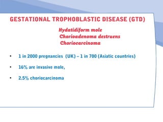
- 1. GESTATIONAL TROPHOBLASTIC DISEASE (GTD) Hydatidiform mole Chorioadenoma destruens Choriocarcinoma • 1 in 2000 pregnancies (UK) – 1 in 700 (Asiatic countries) • 16% are invasive mole, • 2.5% choriocarcinoma
- 2. GESTATIONAL TROPHOBLASTIC DISEASE (GTD) Features are large-for-dates uterus, PV bleeding and high hCG Curettage should be repeated after one month if bleeding don't stopped or hCG remains elevated Follow-up is by hCG and advise not to get pregnant for 12 months Choriocarcinoma blood-borne metastases, treatment with methotrexate
- 3. GESTATIONAL TROPHOBLASTIC DISEASE (GTD) Abnormality of early trophoblast may arise as a develepment anomaly of placental tissue and results in the formation of a mass of oedematous and avascular villi. There is usually no fetus but the condition can be found in the presence of a fetus. The placenta is replaced by a mass of grapelike vesicles known as a hydatidiform mole. Invasion of the myometrium without systemic spread occurs in about 16% of cases of benign mole and is known as invasive mole or chorioadenoma destruens. Malignant change occurs in 2.5% and is known as choriocarcinoma.
- 4. GESTATIONAL TROPHOBLASTIC DISEASE (GTD) Pathology Benign mole remains confined to the uterine cavity and decidua. The histopathology exhibits a villous pattern, which is also found in the invasive mole, the tissue penetrates the myometrium deeply and may result in serious haemorrhage. Choriocarcinoma comprises plexiform columns of trophoblastic cells without villous patterns. Widespread blood-borne metastases are a feature of this disease which, until recent years, carried a very high mortality rate. Metastases may occur locally in the vagina but most commonly appear in the lungs. Theca lutein cysts occur in about one-third of all cases as a result of high circulating levels of hCG. These regress spontaneously with removal of the molar tissue.
- 5. GESTATIONAL TROPHOBLASTIC DISEASE (GTD) Clinical presentation Molar pregnancy most commonly presents as bleeding in the first half of pregnancy and spontaneous abortion often occurs at about 20 weeks' gestation. Occasionally, the passage of a grape-like villus heralds the presence of a mole. The uterus is larger than dates in about half the cases but this is not a reliable sign as it may sometimes be small for dates. Severe hyperemesis, pre-eclampsia and unexplained anaemia are all factors suggestive of this disorder. The diagnosis can be confirmed by ultrasound scan and by the presence of very high levels of hCG in the blood or urine.
- 6. Sponsored Medical Lecture Notes – All Subjects USMLE Exam (America) – Practice
- 7. GESTATIONAL TROPHOBLASTIC DISEASE (GTD) Management Once the diagnosis is established, the pregnancy is terminated by suction curettage. Adequate replacement of blood loss is essential and the procedure is accompanied by the infusion of oxytocin to ensure that the uterus remains well contracted. All cases of molar pregnancy should be followed for 1 year and further pregnancy during this time is con-traindicated. Serial estimations of subunit hCG are followed; where these remain elevated for more than 1 month after uterine evacuation, or where persistent haemorrhage occurs, repeat curettage should be performed.
- 8. GESTATIONAL TROPHOBLASTIC DISEASE (GTD) If the histological evidence shows malignant change, chemotherapy with methotrexate and actinomycin D is employed and produces good results. Management of these cases is concentrated in specialised centres. It may occasionally be necessary to remove the uterus where haemorrhage becomes life-threatening. It must be remembered that choriocarcinoma sometimes can occur following an abortion or a normal term intra-uterine pregnancy.
- 9. GESTATIONAL TROPHOBLASTIC DISEASE (GTD) Hydatidiform moles Risk factors include: maternal age >35 years (>2 x increase), prior molar pregnancy (10 x increase), long-term use of oral contraceptives (2 x increase), dietary deficiency ((3-carotene, vitamin A). Clinical presentation. Partial moles usually present as a missed abortion during the 1 st or early 2nd trimester. Normal or marginally elevated þ-hCG levels are common. Complete moles typically have abnormal vaginal bleeding (85%). 10% of women will have anaemia, hyperemesis gravidarum or preeclampsia.
- 10. GESTATIONAL TROPHOBLASTIC DISEASE (GTD) Sonographic findings. Partial moles may be suspected by visualizing a fetus with focal cystic spaces in the placenta and an increase in the transverse diameter of the gestational sac. Complete moles classically have a 'snowstorm' appearance of diffuse hydropic swelling without a fetus. Diagnosis of hydatidiform moles is made by histopathological analysis. Partial moles have a non-viable fetus with malformations (syndactyly, hydrocephalus, growth restriction), variably hydropic (swollen) villi and minimal trophoblastic hyperplasia. Complete moles have no fetal tissue and consist of diffusely hydropic villi (grape-like vesicles) with widespread trophoblastic hyperplasia.
- 11. GESTATIONAL TROPHOBLASTIC DISEASE (GTD) Treatment. Dilatation and evacuation is the most common initial treatment for molar pregnancy. Hysterectomy is an alternative in selected patients who desire surgical sterilization. Prophylaxis. Anti-D immunoglobulin should be administered to appropriate Rh-negative patients. Surveillance. þ-hCG levels should be monitored until they are undetectable. Hormonal contraception should be encouraged to prevent pregnancy and reduce the potential for complicating þ-hCG interpretation. ·Future pregnancies. Patients may expect normal reproductive outcome of subsequent conceptions. The risk of developing another hydatidiform mole
- 12. GESTATIONAL TROPHOBLASTIC DISEASE (GTD) Gestational trophoblastic neoplasia (GTN) • Antecedent gestation: most commonly occurs following a molar pregnancy, but may occur after any gestational event (termination or spontaneous miscarriage , ectopic pregnancy , term pregnancy). • Diagnosis is not uniform worldwide, but includes one of these criteria: þ-hCG plateau of 4 measurements over a period of at last 3 weeks þ-hCG rise of 3 measurements over a period of at last 2 weeks þ-hCG level remains elevated for more than 6 months • Histological diagnosis of choriocarcinoma.
- 13. GESTATIONAL TROPHOBLASTIC DISEASE (GTD) Choriocarcinoma consists of sheets of anaplastic cytotrophoblast and syncytiotrophoblast cells without chorionic villi. Invasive moles may have the histological features of either choriocarcinoma or hydatidiform mole, but metastases are always choriocarcinoma.
- 14. GESTATIONAL TROPHOBLASTIC DISEASE (GTD) GTN is anatomically staged. The combination of a chest X-ray, abdominal-pelvic CT scan and pelvic examination is an effective strategy to determine the extent of disease. Chest and head CT are indicated if the chest X-ray is abnormal. Biopsy of suspected metastatic lesions is not recommended and may cause haemorrhage. The World Health Organization (WHO) prognostic scoring system (opposite) is used to categorize patients with GTN into low-risk (score: 0-6) or high-risk (score: 7 or higher) groups.
- 15. GESTATIONAL TROPHOBLASTIC DISEASE (GTD) Treatment Low-risk GTN is most frequently treated by methotrexate. If the tumour is resistant, the patient may be switched to dactinomycin. High-risk GTN is usually best managed by combination chemotherapy (etoposide, methotrexate, dactinomycin, Cytoxan, vincristine) due to the increased risk of tumour resistance to a single agent. Surveillance. þ-hCG levels are measured until therapy is completed. Follow-up should continue for 12 (stage I— III) to 24 months (stage IV). Prognosis. 98-100% of stage I—III patients and 75-80% of stage IV patients will be cured.