Recomendados

Recomendados
Más contenido relacionado
La actualidad más candente
La actualidad más candente (20)
Similar a Poster SVF
Similar a Poster SVF (20)
Poster SVF
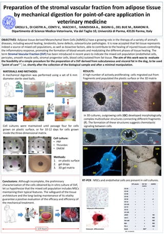
- 1. RESULTS: A high number of actively proliferating cells migrated out from fragments and populated the plastic surface or the 3D matrix In 3D cultures, outgrowing cells (OC) developed morphologically complex multicellular structures connecting different fragments (F). The formation of these structures suggests chemotactic signaling between cells. RT-PCR : MSCs and endothelial cells are present in cell cultures. 4X 20X 10X 2cm OC F OC F F OC F F OC Adipocytic differentiation Osteocytic differentiation ND*: not detected Preparation of the stromal vascular fraction from adipose tissue by mechanical digestion for point-of-care application in veterinary medicine GROLLI S., DI CASTRI A., CONTI V., MACCHI F., IVANOVSKA A., BASINI G., DEL BUE M., RAMONI R. Dipartimento di Scienze Medico-Veterinarie, Via del Taglio 10, Università di Parma, 43126 Parma, Italy OBJECTIVES: Adipose tissue derived Mesenchymal Stem Cells (AdMSCs) have a growing role in the therapy of a variety of animal’s diseases, including wound healing, tendonitis, bone defects, osteoarticular pathologies. It is now accepted that fat tissue represents indeed a source of mixed cell populations, as well as bioactive factors, able to contribute to the healing of injured tissues controlling the inflammatory response, promoting the formation of blood vessels and modulating the different phases of tissue healing. The term Stromal Vascular Fraction (SVF) has been introduced in recent years to indicate the mixed cell population (endothelial cells, pericytes, smooth muscle cells, stromal progenitor cells, blood cells) isolated from fat tissue. The aim of this work was to evaluate the feasibility of a simple procedure for the preparation of a SVF derived from subcutaneous and visceral fat in the dog, to be used “point of care” ”, i.e. shortly after the collection of the biological sample and after a minimal manipulation. MATERIALS AND METHODS: A mechanical digestion was performed using a set of 6 mm diameter sterile steel balls. Cell cultures were maintained until passage four for cells grown on plastic surface, or for 10-12 days for cells grown inside the three-dimensional matrix. Cell culture: • PRP • Thrombin • DMEM Methods: 1. on plastic surface 2. gel drops 3. 3D gel matrix Conclusions: Although incomplete, the preliminary characterization of the cells obtained by in-vitro culture of SVF, let us hypothesize that the mixed cell population includes MSCs maintaining their typical features. The safeguard of the tissue architecture and the long-lasting maintenance of its vitality guarantee a positive evaluation of the efficacy and efficiency of the mechanical treatment. ≈ 2/3 hours +PRP