
Recomendados
Más contenido relacionado
La actualidad más candente
La actualidad más candente (20)
Destacado
Destacado (19)
Similar a Spinal Cord Injury Evaluation and Management
Similar a Spinal Cord Injury Evaluation and Management (20)
Último
Último (20)
Spinal Cord Injury Evaluation and Management
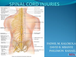
- 1. FADHIL M. KALOKOLA DAVID B. MBANYE PHILEMON RAHAEL (MD5)
- 2. Outlines Introduction Aetiology & Epidemiology Mechanism of injury classification Neurological evaluation Work up Management Complications
- 3. 31 pairs of spinal nerves: 8 cervical 12 thoracic 5 lumbar 5 sacral 1 coccygeal Spinal cord: Extends from medulla oblongata – L1 Lower part tapered to form conus medullaris Enclosed within 33 vertebrae Anatomy :
- 5. On the surface : Deep anterior median fissure Shallower posterior median sulcus Spinal cord segment : Section of the cord from which a pair of spinal nerves are given off
- 6. Dorsal root – sensory fibres Ventral root – motor fibres Dorsal and ventral roots join at intervertebral foramen to form the spinal nerve
- 8. Physiology and function Grey matter – sensory and motor nerve cells White matter – spinal tracts - Ascending, descending and intersegmental tracts
- 9. Tracts :
- 10. Posterior column and lateral corticospinal tract crosses over at medulla oblongata Spinothalamic tract crosses in the spinal cord and ascends on the opposite side NB Understanding this helps to reveal the clinical features of injury patterns and the neurological deficit
- 11. Dermatomes Area of skin innervated by sensory axons within a particular segmental nerve root Knowledge is essential in determining level of injury Useful in assessing improvement or deterioration
- 12. Downloaded from: Rosen's Emergency Medicine (on 29 April 2009 06:34 PM) © 2007 Elsevier
- 13. Myotomes : Segmental nerve root innervating a muscle Again important in determining level of injury Upper limbs: C5 - Deltoid C6 - Wrist extensors C7 - Elbow extensors C8 - Long finger flexors T 1 - Small hand muscles Lower Limbs : L2 - Hip flexors L3,4 - Knee extensors L4,5 – S1 - Kneeflexion L5 - Ankle dorsiflexion S1 - Ankle plantar flexion
- 14. Definition Insult to spinal cord resulting in a change, in the normal motor, sensory or autonomic function. This change is either temporary or permanent.
- 15. Terminologies Plegia = complete lesion Paresis = some muscle strength is preserved Tetraplegia (or quadriplegia) Injury of the cervical spinal cord Patient can usually still move his arms using the segments above the injury (e.g., in a C7 injury, the patient can still flex his forearms, using the C5 segment) Paraplegia – paralysis of both LL Injury of the thoracic or lumbo-sacral cord, or cauda equina Hemiplegia Paralysis of one half of the body Usually in brain injuries (e.g., stroke) Monoplegia is a paralysis of one limb only. Diplegia is a paralysis of two corresponding limbs (i.e., arms or legs).
- 16. Causes of SCI
- 17. CAUSES MVA Falls Violence Sports injuries Gunshot Injuries Diving accidents Blunt Assault Stab Wounds Sport Injuries 55% cases occur in 16 – 30yrs of age > 80% are male!
- 18. Other causes: Vascular disorders Tumours Infectious conditions Spondylosis Iatrogenic Vertebral fractures secondary to osteoporosis Development disorders
- 19. Mechanisms: i) Direct trauma ii) Compression by bone fragments / haematoma / disc material iii) Ischemia from damage / impingement on the spinal arteries
- 20. Patterns of injury Fracture Dislocation Fracture dislocation SCIWORA
- 22. ASIA – American Spinal Injury Association Based on neurological responses, touch & pinprick sensation (dermatome), +muscle strength. A – Complete: no sensory or motor function preserved in sacral segments S4– S5 B – Incomplete: sensory, but no motor function in sacral segments C – Incomplete: motor function preserved below level and power graded < 3 D – Incomplete: motor function preserved below level and power graded 3 or more E – Normal: sensory and motor function normal
- 23. Can be 1) Complete 2) Incomplete Complete: i) Loss of voluntary movement of parts innervated by segment, this is irreversible ii) Loss of sensation iii) Spinal shock
- 24. Incomplete: i) Some function is present below site of injury ii) More favourable prognosis overall iii) Are recognisable patterns of injury, although they are rarely pure and variations occur
- 25. Incomplete injury syndromes i) Central Cord Syndrome ii) Anterior Cord Syndrome iii) Posterior Cord Syndrome iv) Brown – Sequard Syndrome v) Cauda Equina Syndrome
- 26. i) Central Cord Syndrome : Typically in older patients Hyperextension injury Compression of the cord anteriorly by osteophytes and posteriorly by ligamentum flavum
- 27. Also associated with fracture dislocation and compression fractures More centrally situated cervical tracts tend to be more involved hence flaccid weakness of arms > legs Perianal sensation & some lower extremity movement and sensation may be preserved
- 30. ii) Anterior cord Syndrome: Due to flexion / rotation Anterior dislocation / compression fracture of a vertebral body encroaching the ventral canal Corticospinal and spinothalamic tracts are damaged either by direct trauma or ischemia of blood supply (anterior spinal arteries)
- 32. Clinically: Loss of power Decrease in pain and sensation below lesion Dorsal columns remain intact
- 33. ii) Posterior Cord Syndrome: Hyperextension injuries with fractures of the posterior elements of the vertebrae Clinically: Proprioception affected – ataxia and faltering gait Usually good power and sensation
- 36. v) Cauda Equina Syndrome: Due to bony compression or disc protrusions in lumbar or sacral region Clinically Non specific symptoms – back pain - bowel and bladder dysfunction - leg numbness and weakness
- 37. Spinal Shock Transient reflex depression of cord function below level of injury Initially hypertension due to release of catecholamines Followed by hypotension Flaccid paralysis Bowel and bladder involved Sometimes priaprism develops Symptoms last several hours to days
- 38. Asessment Spinal shock Bulbocavernosus reflex Complete VS incomplete cord injury spinal shock Sacral sparing Voluntary anal sphincter control Toe flexor Perianal sensation Anal wink reflex
- 39. Neurogenic shock: Triad of - i) hypotension ii) bradycardia iii) hypothermia More commonly in injuries above T6 Secondaryto disruption of sympathetic outflow from T1 – L2
- 40. Loss of vasomotor tone – pooling of blood Loss of cardiac sympathetic tone – bradycardia Blood pressure will not be restored by fluid infusion alone Massive fluid administration may lead to overload and pulmonary edema Vasopressors may be indicated
- 41. Autonomic dysreflexia (AD) A syndrome of massive imbalanced reflex sympathetic discharge Occurring in patients with spinal cord injury (SCI) 48-90% in patients with SCI above T6 Above the splanchnic sympathetic outflow (T5-T6). Primarily a male phenomenon; M:F=4:1 About 66% in females in labour
- 42. Pathophysiology Occurs after the phase of spinal shock in which reflexes return. Below the injury, intact peripheral sensory nerves transmit impulses That ascend in the spinothalamic and posterior columns Stimulate sympathetic neurons located in the intermediolateral gray matter of the spinal cord.
- 43. Inhibitory outflow above the SCI from vasomotor centres is increased, Unable to pass below the block of the SCI. Release of various neurotransmitters (dopamine-b-hydroxylase, norepinephrine, dopamine), Causing piloerection, skin pallor, and severe vasoconstriction in arterial vasculature. Sudden elevation in blood pressure + vasodilatation above the level of injury. Headache caused by vasodilation of pain sensitive intracranial vessels.
- 44. Increase in parasympathetic vagal tone by Vassomotor centers compensatory bradycardia. Parasympathetic nerves prevail above the level of injury sweating and vasodilatation with skin flushing.
- 45. Clinically Headaches, and a sense of anxiety sudden rise in both systolic and diastolic blood pressures, usually with bradycardia profuse sweating above the level of lesion, flushing of the skin nasal congestion is common.
- 46. Neurological evaluation Motor: how to test each segment?
- 47. Sensory: how to determine the level?
- 48. Spinal assessment Palpate entire spine Deformity Grating / crepitus Tenderness Gapping interspinous spaces Bogginess Lacerations DRE, perineal sensation + tone
- 49. Imaging modalities CT scan MRI X-ray - standard trauma series is composed of 5 x-ray views: cross-table lateral, swimmer's, oblique, odontoid, Anteroposterior Radiographic level = the level of fracture on plain XRays / CT scan / MRI NB: spine level does not correspond to spinal cord level below the cervical region
- 50. Radiolographic evaluation X-ray Guidelines (cervical) AABBCDS Adequacy, Alignment Bone abnormality, Base of skull Cartilage Disc space Soft tissue
- 51. Alignment • The anterior vertebral line, posterior vertebral line, and spinolaminar line should have a smooth curve with no steps or discontinuities • Malalignment of the posterior vertebral bodies is more significant than that anteriorly, which may be due to rotation • A step-off of >3.5mm is significant anywhere
- 52. Bones
- 53. Disc Disc Spaces Should be uniform Assess spaces between the spinous processes
- 54. AP C-spine Films Spinous processes should line up Disc space should be uniform Vertebral body height should be uniform. Check for oblique fractures.
- 55. Swimmer’s view
- 56. Open mouth view Adequacy: all of the: all of the dens and lateraldens and lateral borders of C1 & C2borders of C1 & C2 Alignment: lateral: lateral masses of C1 and C2masses of C1 and C2 Bone: Inspect dens for lucent fracture lines
- 57. Management
- 58. Management…. Advance Trauma Life Support (ATLS) guidelines Primary survey; ABCDE -Adequate airway and ventilation are the most important factors Supplemental oxygenation Early intubation is critical to limit secondary injury from hypoxia Secondary surveys (e.g. Hx) Initial Management
- 59. Immobilization Entire spine until when the x-ray are available Supine, no rotation no bending It protect further damage -Beware of decubitus ulcers
- 60. Methods of immobilization Rigid collar (Philadelphia hard collar)>>> Sandbags and straps Spine board Braces Log-roll to turn
- 62. Logrolling At least 4 people 1 Maintain inline manual immobilization 1 For the torso (trunk) 1 For the pelvis and legs 1 To direct the move Move the pt towards the attendants as a unit Maintain neutral position of the spine Children have proportionally large heads
- 64. IV fluids Persistent hypotension after 2 liters neurogenic shock Use of vasopressors Dopamine / Adrenalin Invasive monitoring; CVP and urethral catheters Steroids Methylprednisolone - 30mg/kg in the first 15min Then 5.4mg/kg/hr for 24-48hrs Exclusion criteria Cauda equina syndrome Pregnancy Age <13 years Patient on maintenance steroids
- 65. Mnx … cont NGT Prevents aspiration Decompresses the abdomen (paralytic ileus is common in the first days) Foley catheter Urinary retention is common and For monitoring - Spinal assessment
- 66. Care of paraplegics Skin care Bowel and bladder Psychological support Wheelchair rehabilitation
- 67. Surgical Mnx Indications for surgery Instability maintaining alignment to allow development of solid bone fusion; preventing progression of deformity; alleviating pain progressive neurological deficit ???? For early rehabilitation????
- 69. complications Hypotension Neurogenic pain Spinal shock hypothermia paraplegia Sexually dysfunction Bladder + bowel dysfunction -Incontinence -paralytic ileus -urinary problems (UTI) Weight loss or gain Depression Autonomic dysreflexia Pressure sores (bed sores) Pneumonia and asthma CVS diseases Spasm syringomyelia Deep vein thrombosis -pulmonary embolism
- 70. References: 1. ATLS, et al. Student Course Manual. 7th Edition 2004;7:177-204 2. Keith L Moore et al. Clinically Orientated Anatomy. 3rd Edition1992;4:359 3. Snell.Clinical.Neuroanatomy.7th.2009 4. Essential of Orthopedics
Notas del editor
- Pathophysiology: This phenomenon occurs after the phase of spinal shock in which reflexes return. Individuals with injury above the major splanchnic outflow may develop AD. Below the injury, intact peripheral sensory nerves transmit impulses that ascend in the spinothalamic and posterior columns to stimulate sympathetic neurons located in the intermediolateral gray matter of the spinal cord. The inhibitory outflow above the SCI from cerebral vasomotor centers is increased, but it is unable to pass below the block of the SCI. This large sympathetic outflow causes release of various neurotransmitters (norepinephrine, dopamine-b-hydroxylase, dopamine), causing piloerection, skin pallor, and severe vasoconstriction in arterial vasculature. The result is sudden elevation in blood pressure and vasodilation above the level of injury. Patients commonly have a headache caused by vasodilation of pain sensitive intracranial vessels. Vasomotor brainstem reflexes attempt to lower blood pressure by increasing parasympathetic stimulation to the heart through the vagus nerve to cause compensatory bradycardia. This reflex action cannot compensate for severe vasoconstriction, explained by the Poiseuille formula, where pressure in a tube is affected to the fourth power by change in radius (vasoconstriction) and only linearly by change in flow rate (bradycardia). Parasympathetic nerves prevail above the level of injury, which may be characterized by profuse sweating and vasodilation with skin flushing. Cameron and colleagues have found that site-directed genetic manipulation of fiber sprouting in the spinal dorsal horns in a cord compression rat model could alter the extent of hyperreflexia after bowel distention, indicating that endogenous spinal cord circuitry/neural sprouting plays a role in the pathophysiology of AD (Cameron, 2006).
- The patient generally gives a history of blurry vision, headaches, and a sense of anxiety. Feelings of apprehension or anxiety over an impending physical problem commonly are exhibited Physical: A patient may have one or more of the following findings on physical examination: A sudden significant rise in both systolic and diastolic blood pressures, usually associated with bradycardia, can appear. The normal systolic blood pressure for SCI above T6 is 90-110 mm Hg. Blood pressure 20-40 mm Hg above the reference range for such patients may be a sign of AD. Profuse sweating above the level of lesion, especially in the face, neck, and shoulders, may be noted, but it rarely occurs below the level of the lesion because of sympathetic cholinergic activity. Goose bumps above, or possibly below, the level of the lesion may be observed. Flushing of the skin above the level of the lesion, especially in the face, neck, and shoulders, frequently is noted. The patient may report blurred vision. Spots may appear in the patient&apos;s visual fields. Nasal congestion is common. No symptoms may be observed, despite elevated blood pressure.